Anti-myostatin antibodies and methods of use

a technology of myostatin and antibodies, applied in the field of anti-myostatin antibodies, can solve the problems of limited treatment options for these disorders, and achieve the effects of increasing muscle tissue mass, and increasing strength of muscle tissu

Active Publication Date: 2019-01-03
CHUGAI PHARMA CO LTD
View PDF2 Cites 8 Cited by
  • Summary
  • Abstract
  • Description
  • Claims
  • Application Information

AI Technical Summary

Benefits of technology

[0042]Anti-myostatin antibodies of the present invention may be for use as a medicament. Anti-myostatin antibodies of the present invention may be for use in treating a muscle wasting disease. Anti-myostatin antibodies of the present invention may be for use in increasing mass of muscle tissue. Anti-myostatin antibodies of the present invention may be for use in increasing strength of muscle tissue.
[0043]Anti-myostatin antibodies of the present invention may be used in the manufacture of a medicament. In some embodiments, the medicament is for treatment of a muscle wasting disease. In some embodiments, the medicament is for increasing mass of muscle tissue. In some embodiments, the medicament is for increasing strength of muscle tissue.
[0044]The invention also provid

Problems solved by technology

Patients will benefit from an increase in muscle mass and/or muscle strength;

Method used


Image

  • Anti-myostatin antibodies and methods of use
  • Anti-myostatin antibodies and methods of use
  • Anti-myostatin antibodies and methods of use

Examples


example 1

Expression and Purification of Human, Cynomolgus Monkey, and Mouse Myostatin Mature Form

[0265]Human latent myostatin (also described herein as myostatin latent form) (SEQ ID NO:1) was expressed transiently using FreeStyle293-F cell line (Thermo Fisher, Carlsbad, Calif., USA). Conditioned media containing expressed human myostatin latent form was acidified to pH 6.8 and diluted with ½ vol of milliQ water, followed by application to a Q-sepharose FF anion exchange column (GE healthcare, Uppsala, Sweden). The flow-through fraction was adjusted to pH 5.0 and applied to a SP-sepharose HP cation exchange column (GE healthcare, Uppsala, Sweden), and then eluted with a NaCl gradient. Fractions containing the human myostatin latent form were collected and subsequently subjected to a Superdex 200 gel filtration column (GE healthcare, Uppsala, Sweden) equilibrated with 1×PBS. Fractions containing the human myostatin latent form were then pooled and stored at −80 degrees C.

[0266]Human mature my...

example 2

Identification of Anti-Mature Myostatin Antibody

[0269]Anti-mature myostatin antibodies were prepared, selected, and assayed as follows.

[0270]Twelve to sixteen week old NZW rabbits were immunized intradermally with human mature myostatin, human latent myostatin or mature myostatin conjugated with KLH (50-100 micro g / dose / rabbit). This dose was repeated 4-5 times. One week after the final immunization, the spleen and blood from immunized rabbit was collected. Antigen-specific B-cells were stained with labelled antigen, sorted with FCM cell sorter (FACS aria III, BD), and plated in 96-well plates at one cell / well density together with 25,000 cells / well of EL4 cells (European Collection of Cell Cultures) and with rabbit T-cell conditioned medium diluted 20 times, and were cultured for 7-12 days. EL4 cells were treated with mitomycin C (Sigma) for 2 hours and washed 3 times in advance. The rabbit T-cell conditioned medium was prepared by culturing rabbit thymocytes in RPMI-1640 containin...

example 3

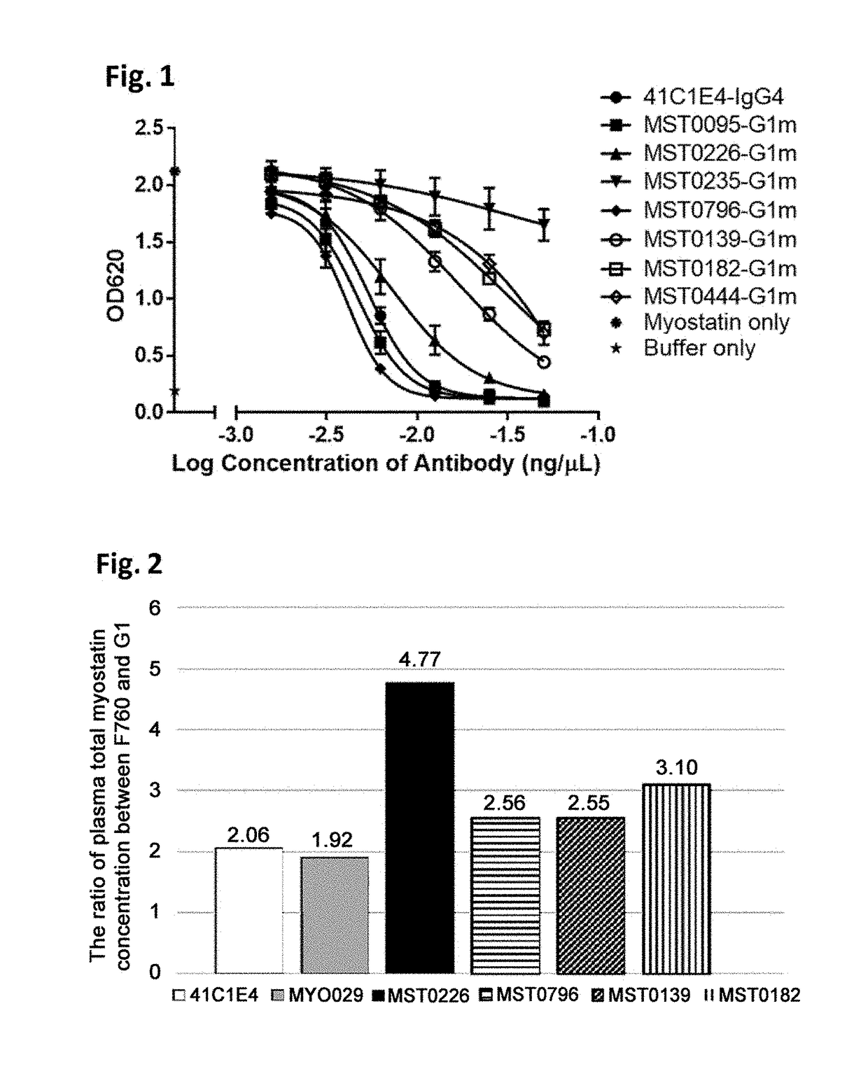
Characterization of Anti-Mature Myostatin Antibody (HEK Blue Assay (BMP-1 Activation))

[0274]Reporter gene assay was used to assess the biological activity of active myostatin in vitro. HEK-Blue™ TGF-beta cells (Invivogen), which express a Smad3 / 4-binding elements (SBE)-inducible SEAP (Secreted embryonic alkaline phosphatase) reporter genes, allow the detection of bioactive myostatin by monitoring the activation of the activin type 1 and type 2 receptors. Myostatin mature form stimulates the production of SEAP into cell supernatant by activating Smad3 / 4 signal through the binding to its receptor. The quantity of SEAP secreted is then assessed using QUANTIBlue™ (Invivogen).

[0275]HEK-Blue™ TGF-beta cells were maintained in DMEM medium (Gibco) supplemented with 10% fetal bovine serum, 50 micro g / mL streptomycin, 50 U / mL penicillin, 100 micro g / mL Normocin™, 30 micro g / mL of Blasticidin, 200 micro g / mL of HygroGold™ and 100 micro g / mL of Zeocin™. During functional assay, cells were chang...


PUM

PropertyMeasurementUnit
Fractionaaaaaaaaaa
Acidityaaaaaaaaaa
Acidityaaaaaaaaaa

Abstract

The invention provides anti-myostatin antibodies and methods of using the same. In some embodiments, an isolated anti-myostatin antibody of the present invention binds to mature myostatin, and uptake of the antibody into cells is enhanced when complexed with the antigen. The invention also provides isolated nucleic acids encoding an anti-myostatin antibody of the present invention. The invention also provides host cells comprising a nucleic acid of the present invention. The invention also provides a method of producing an antibody comprising culturing a host cell of the present invention so that the antibody is produced. Anti-myostatin antibodies of the present invention may be for use as a medicament. Anti-myostatin antibodies of the present invention may be for use in treating a muscle wasting disease. Anti-myostatin antibodies of the present invention may be for use in increasing mass of muscle tissue. Anti-myostatin antibodies of the present invention may be for use in increasing strength of muscle tissue.

Description

TECHNICAL FIELD[0001]The present invention relates to anti-myostatin antibodies and methods of using the same.BACKGROUND ART[0002]Myostatin, also referred to as growth differentiation factor-8 (GDF-8), is a secreted protein and is a member of the transforming growth factor-beta (TGF-beta) superfamily of proteins. Members of this superfamily possess growth-regulatory and morphogenetic properties (See, e.g., NPL 1, NPL 2, and PTL 1). Myostatin is expressed primarily in the developing and adult skeletal muscle and functions as a negative regulator of muscle growth. Systemic overexpression of myostatin in adult mice leads to muscle wasting (See, e.g., NPL 3) while, conversely, a myostatin knockout mouse is characterized by hypertrophy and hyperplasia of the skeletal muscle resulting in two- to threefold greater muscle mass than their wild type littermates (See, e.g., NPL 4).[0003]Like other members of the TGF-beta family, myostatin is synthesized as a large precursor protein containing ...

Claims


Application Information

Patent Timeline
IPC IPC(8): C07K16/22A61P21/00
CPCC07K16/22A61P21/00C07K2317/52C07K2317/76C07K2317/92C07K2317/21C07K2317/24C07K2317/567C07K2317/56C07K2317/77A61K2039/505C07K2317/94
Inventor RUIKE, YOSHINAOKURAMOCHI, TAICHI
Owner CHUGAI PHARMA CO LTD
Why Eureka
Try Eureka