Systems and methods for automated image recognition of implants and compositions with long-lasting echogenicity

an image recognition and composition technology, applied in the field of image processing and imageable compositions, can solve the problems of other implants degrading and their length/width changing, and achieve the effects of increasing visibility, identifying and analysing implants more accurately and efficiently, and computationally more efficien

Inactive Publication Date: 2019-02-21
CONTRALINE INC
View PDF0 Cites 19 Cited by
  • Summary
  • Abstract
  • Description
  • Claims
  • Application Information

AI Technical Summary

Benefits of technology

[0010]Bodily implants are typically engineered medical devices consisting of non-biological or biological materials. Previous work has shown that implants may be made more visible across the imaging modalities by adding contrast agents, such as microbubbles or radioactive dye. Regardless of the homogeneity of the contrast agent throughout the implant (which can impact the visible texture of the implant in medical media), the systems and methods of the present invention include one or more algorithm(s) which are capable of extracting Regions of Interest (ROIs) with or without increased visibility as a result of said contrast agents. In one embodiment, the one or more algorithms are capable of extracting and analyzing the ROI based on morphological features, which is computationally more efficient than previously existing methods. Advantageously, the one or more algorithms require no additional, manual input for such analysis. Furthermore, machine learning algorithms may be applied to identify and analyze the implants more accurately and efficiently, especially if algorithms are used more frequently or the number of images inputted into the algorithm are increased.

Problems solved by technology

Other implants may degrade over time, and as such, their length / width may change.

Method used


Image

  • Systems and methods for automated image recognition of implants and compositions with long-lasting echogenicity
  • Systems and methods for automated image recognition of implants and compositions with long-lasting echogenicity
  • Systems and methods for automated image recognition of implants and compositions with long-lasting echogenicity

Examples


examples

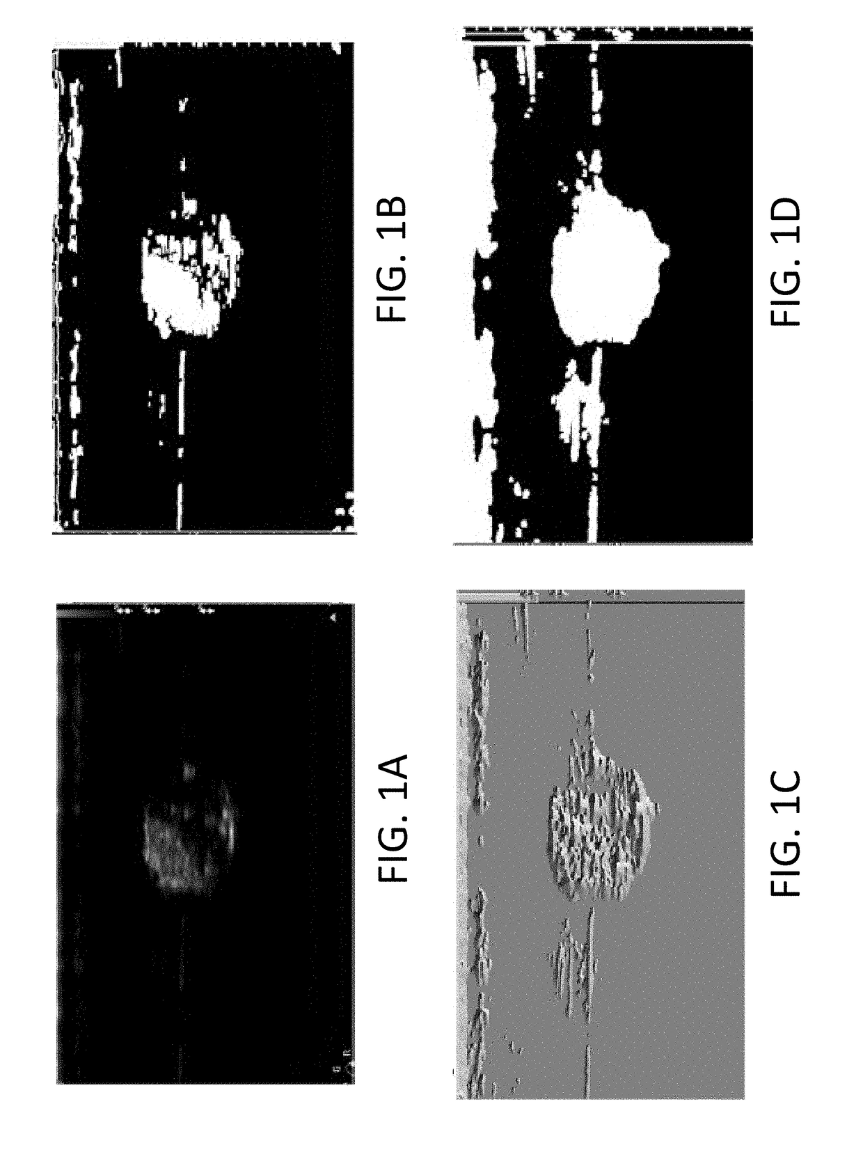
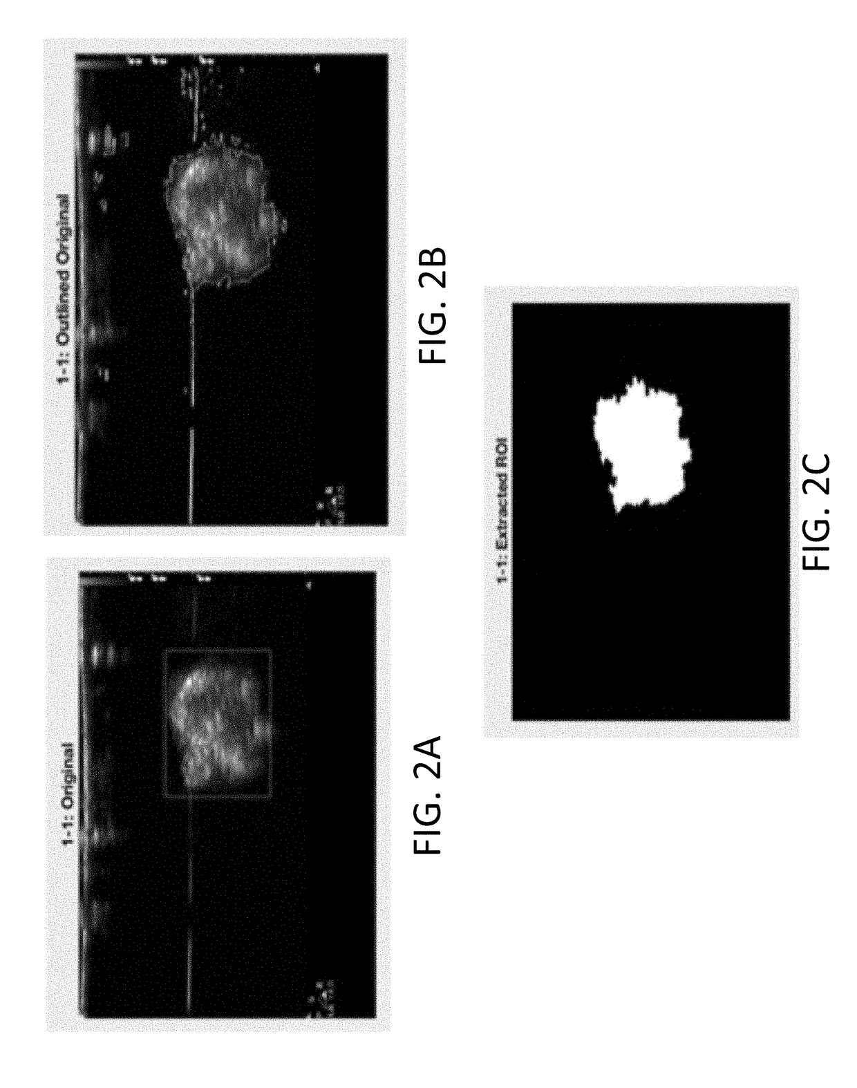
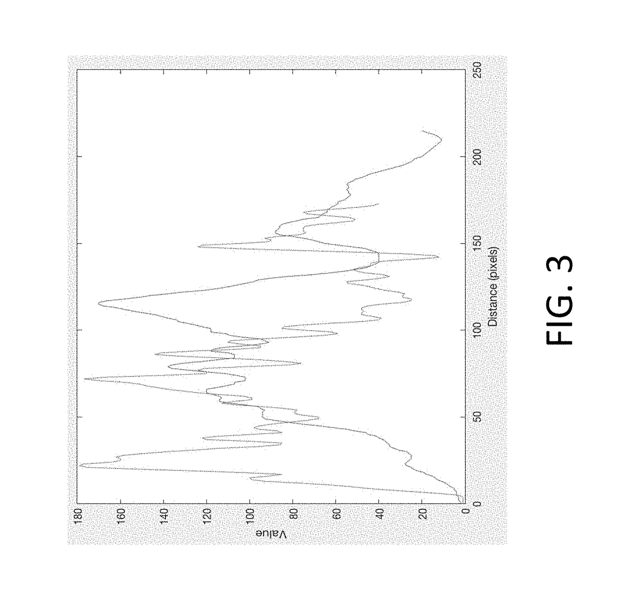
[0102]1. An algorithm was designed to automatically detect soft materials (specifically, hydrogels) in ultrasound images. The gels comprised various ultrasound contrast agents. The objective was to analyze the echogenicity and homogeneity of various ultrasound contrast agents. Gels were prepared consisting of the following UCA's: argon microbubbles (n=6), chitosan (n=5), and glass microspheres (n=4). The algorithm was run on the images and showed an accuracy of 96.47% (p-value<0.0001). After converting the images to binary images and performing morphological techniques such as smoothing, the Sobel edge detection method was used to find the location parameters of each gel. The extracted ROI was then used to determine the echogenicity and homogeneity. The results showed that glass microspheres were more echogenic than chitosan, which was more echogenic than argon microbubbles. Chitosan had the greatest homogeneity of the three UCA's, but did have aggregation in certain areas of the ge...


PUM

PropertyMeasurementUnit
diameteraaaaaaaaaa
diameteraaaaaaaaaa
diameteraaaaaaaaaa

Abstract

Systems and methods for imaging an object that are capable of capturing an image or images of the object using an imaging modality, automatically detecting and analyzing the image or images by way of converting the image or images to at least one binary image, and analyzing the at least one binary image to extract and / or segment regions-of-interest (ROIs) from the at least one binary image. The object can be or include an implantation, occlusion, medical device, body lumen, tissue, organ, duct, and / or vessel. The imaging modality can be or include X-ray, CT, MRI, PET, and / or ultrasound, or any combination thereof. Also included are compositions of soft, implantable materials with one or more carbon-based material, nanomaterial, and / or allotrope present in an amount sufficient as an ultrasound contrast agent effective for days, months, or years and which compositions are useful in the automated imaging methods of the invention.

Description

CROSS-REFERENCE TO RELATED APPLICATIONS[0001]The present application relies on the disclosure of and claims priority to and the benefit of the filing date of U.S. Provisional Application No. 62 / 546,718, filed Aug. 17, 2017, the disclosure of which is hereby incorporated by reference herein in its entirety.BACKGROUND OF THE INVENTIONField of the Invention[0002]The invention relates to the field of image processing and imageable compositions. Specifically, embodiments are directed to automated identification and analyzation of bodily implants by way of one or more medical imaging modality. Embodiments are also directed to determining information regarding an implant's brightness, echogenicity, location, density, volume, and / or homogeneity, along with other measurements and properties, as well as directed to compositions and methods for implanting soft materials that are echogenic, or ultrasound visible, and where the echogenicity of the material lasts as long as the implant itself.Des...

Claims


Application Information

Patent Timeline
Patent Type & Authority Applications(United States)
IPC IPC(8): A61B8/08A61B8/00A61F6/22A61F2/02A61L31/02A61L31/14A61L31/04A61L31/06
CPCA61B8/5223A61B8/469A61B8/0841A61B8/085A61B8/481A61F6/22A61F2/02A61L31/024A61L31/145A61L31/146A61L31/048A61L31/06A61L31/042A61L31/044A61L31/045A61L31/046A61L31/047A61L31/148A61F2250/0096A61L2400/12A61L31/18A61L31/126A61L27/443G06T7/0012G06T2207/10132G06T2207/30052G06T2207/20036G06T7/11G06T7/136G06T7/155G06T7/194G06T2207/20081G16H50/30
Inventor GROVER, GREGORYEISENFRATS, KEVINPATEL, SUCHI
Owner CONTRALINE INC
Why Eureka
Try Eureka