
Multiplexed single molecule RNA visualization with a two-probe proximity ligation system

a proximity ligation and multi-molecule technology, applied in the field of multi-molecule rna visualization with a two-probe proximity ligation system, can solve the problems of limited sensitivity, low efficiency of reverse transcriptase, limited analysis of both abundance and spatial distribution of mrnas, etc., and achieve the effect of minimizing the chance that the probes will enable “ligation”

Active Publication Date: 2019-02-21
THE BOARD OF TRUSTEES OF THE LELAND STANFORD JUNIOR UNIV
View PDF0 Cites 98 Cited by
  • Summary
  • Abstract
  • Description
  • Claims
  • Application Information

AI Technical Summary

Benefits of technology

The patent describes a method for amplifying nucleic acid sequences in single cells using a process called rolling circle amplification. This method requires a circular molecule, which can be created by a specific enzyme. The method is highly specific and produces low background noise. Using a small number of probes, the method allows for cost-efficient detection of specific nucleic acids in single cells and can be combined with other techniques such as flow cytometry or mass cytometry for simultaneous analysis of multiple nucleic acids and other cellular parameters. The method is also applicable to different platforms such as microscopy or scanning mass spectrometry. The patent also discusses the selection of probes based on their Tm (the temperature at which they bind to their DNA target) to minimize the likelihood of unwanted ligation. Overall, the method provides a reliable and efficient tool for studying nucleic acids in single cells.

Problems solved by technology

Analyzing both the abundance and the spatial distribution of mRNAs is often limited by either the number of fluorophores that can be simultaneously detected by a conventional microscope or by laborious, time consuming and expensive methods.
Detection of specific mRNA molecules in single cells usually involves production of cDNA, for example the FISSEQ technique, or padlock probe ligation on cDNA followed by rolling circle amplification, whereby the sensitivity is limited by the low efficiency of reverse transcriptase.
These methods have a disadvantage that multiple probes must be synthesized; and such probes generally need to be targeted to open reading frames.
The problem is, however, that PLAYR involves a complex four-probe system that requires two-step hybridization and features intermediate hybridization specificity sequences that complicate the probe design process.
Additionally, each gene requires a different intermediate hybridization sequence, and each new sequence has to be tested independently to ensure the efficiency of ligation and lack of cross-talk, which makes the design of highly multiplexed experiments a laborious task.
While flow cytometry provides an excellent platform for the detection of proteins in single cells using antibodies, no comparable solution exists for the detection of nucleic acids.
Microfluidic technologies for the detection and quantification of mRNA in single cells are very costly and their throughput is several orders of magnitude lower compared with what can be achieved for proteins using flow cytometry.
This technology enables the detection of only few RNA copy numbers in millions of single cells but is currently limited to the simultaneous detection of small numbers of measured transcripts.
Furthermore, the protocol is long and laborious and the buffers used are not compatible with some fluorophores commonly used in flow cytometry and cannot be used at all in mass cytometry.
Therefore, it introduces additional variability in the assay and requires the design and optimization of both probes and primers.
This is currently the only genome-wide solution but the very limited throughput (96 cells per run) makes it unsuitable for the analysis of samples with multiple cell populations such as blood samples or tumors.
Additionally, the technology is expensive compared to the other approaches, and does not allow for the simultaneous detection of proteins and mRNAs in the same cell.

Method used


Image

  • Multiplexed single molecule RNA visualization with a two-probe proximity ligation system
  • Multiplexed single molecule RNA visualization with a two-probe proximity ligation system
  • Multiplexed single molecule RNA visualization with a two-probe proximity ligation system

Examples


example 1

Multiplexed Single Molecule RNA Visualization with a Simplified Two-Probe Proximity Ligation System

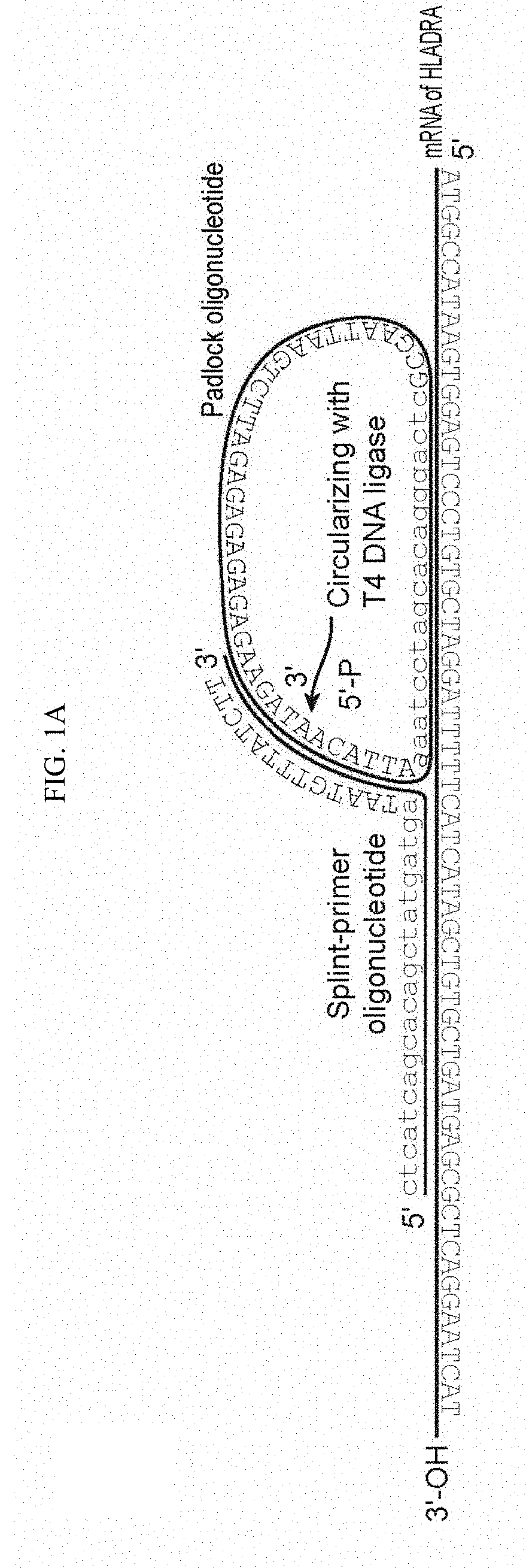
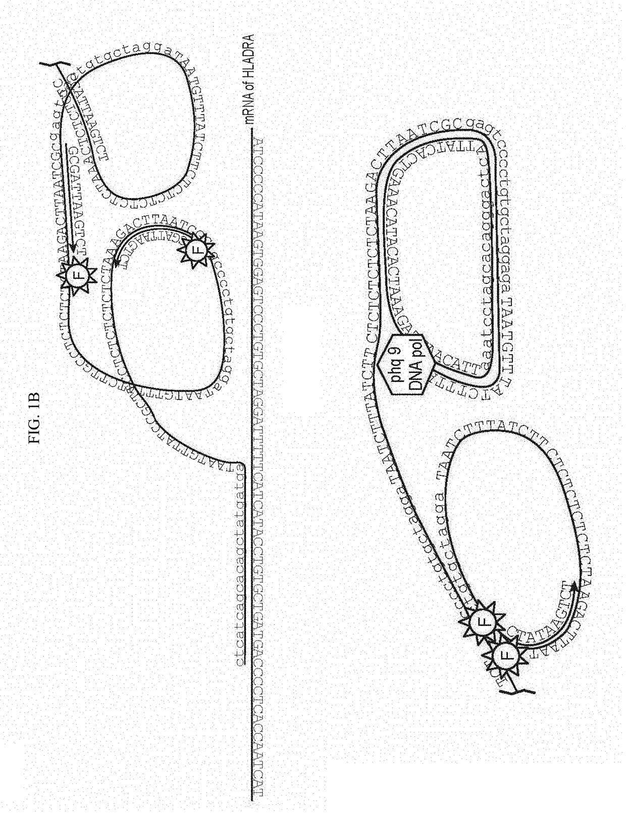
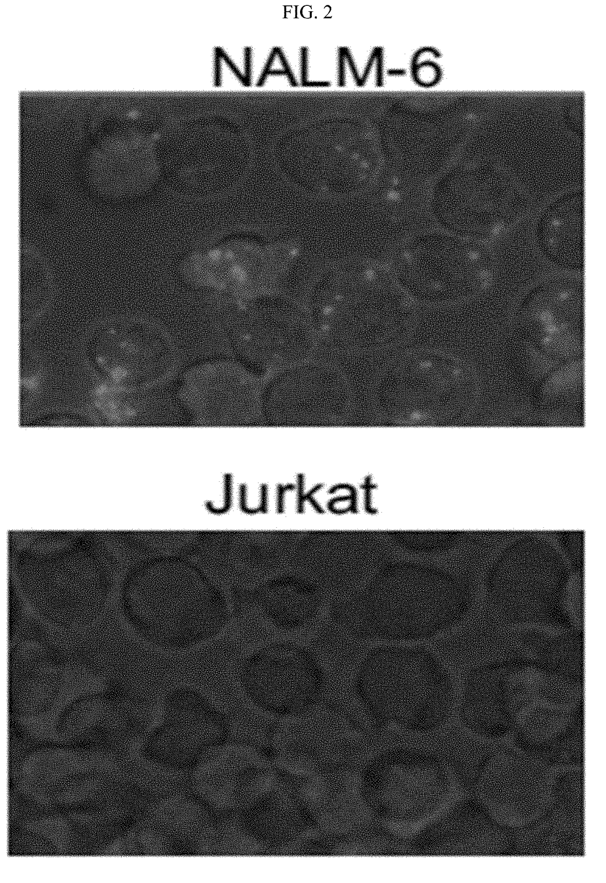
[0146]Quantifying the gene transcriptional activity on a single-cell level is key to studying cell phenotypic heterogeneity, differentiation processes and gene regulatory networks. Most modern single-cell expression profiling methods require cDNA production which limits the efficiency and introduces sequence bias. An alternative method of smRNA-FISH is limited to long transcripts. We created a simple two-probe proximity ligation system termed SNAIL-RCA that enables in situ amplification, detection and visualization of genes. RCA products are detected via hybridization of unlabeled detection probes coupled with single-nucleotide extension with fluorescent nucleotide analogs. Fluorescent imaging and automatic image analysis enable precise quantification of expression levels. Multiplexing is enabled through re-hybridization, which, combined with parse barcoding strategy enables simultaneo...

example 2

[0152]Multiplexed visualization of single RNA molecules in cells and tissues typically relies on smRNA-FISH, which uses multiple fluorescently labelled probes that are directly hybridized to mRNAs. However, prior approaches typically uses 48 20-nt probes that need to be hybridized to each mRNA, which could limit the approach to large mRNAs. Also, creating large libraries representing a large fraction of the genome might be prohibitively expensive. bDNA technology can detect short RNAs and even miRNAs, but its multiplexing is limited by the difficulty of finding orthogonal bDNA sequences. Alternatively, cDNA can be produced in situ and then hybridized with padlock probes, which are ligated and amplified via RCA, but low efficiency and sequence bias of reverse transcription pose a bottleneck to this approach.

[0153]We conceived of a simplified design termed SNAIL-RCA (Specific Amplification of Nucleic Acids via Intramolecular Ligation and Rolling Circle Amplification) that uses two pro...


PUM

PropertyMeasurementUnit
Massaaaaaaaaaa

Abstract

SNAIL provides cost-efficient detection of specific nucleic acids in single cells, and may be combined with flow cytometry to simultaneously analyze large numbers of cells for a plurality of nucleic acids, e.g. at least one, to up to 5, up to 10, up to 15, up to 20 or more transcripts can be simultaneously analyzed, at a rate of up to about 50, 100, 250, 500 or more cells / second. The methods require only two primers for amplification, and may further include a detection primer.

Description

CROSS REFERENCE[0001]This application is a 371 application and claims benefit of PCT Application No. PCT / US2017 / 019443, filed Feb. 24, 2017, which claims benefit of U.S. Provisional Patent Application No. 62 / 300,596, filed Feb. 26, 2016, which applications are incorporated herein by reference in their entirety.FEDERALLY SPONSORED RESEARCH AND DEVELOPMENT[0002]This invention was made with Government support under contract HHSF223201210194C awarded by the Food and Drug Administration and under contract A1100627 awarded by the National Institutes of Health. The Government has certain rights in the invention.BACKGROUND OF THE INVENTION[0003]Studying the mechanisms of gene expression regulation is necessary to understand how their dysregulation can lead to disease states. Spatial distribution of messenger RNAs (mRNAs) is tightly regulated both at the cellular and tissue levels. Analyzing both the abundance and the spatial distribution of mRNAs is often limited by either the number of flu...

Claims


Application Information

Patent Timeline
IPC IPC(8): C12Q1/6841G01N33/542C12Q1/682
CPCC12Q2543/10C12Q2525/307C12Q1/6841C12Q2533/107C12Q1/682C12Q2600/16C12Q2531/125G01N33/542
Inventor SAMUSIK, NIKOLAYBAVA, FELICE ALESSIOGOLTSEV, YURYNOLAN, GARRY P.
Owner THE BOARD OF TRUSTEES OF THE LELAND STANFORD JUNIOR UNIV
Learn More