[0004]In one aspect, disclosed herein are engineered, living, three-dimensional connective tissue constructs comprising: connective tissue cells cohered to one another to provide a living, three-dimensional connective tissue construct; wherein the construct is substantially free of pre-formed scaffold. In some embodiments, the construct is substantially free of any pre-formed scaffold at the time of use. In some embodiments, the construct is non-innervated. In some embodiments, the connective tissue cells comprise connective tissue cells derived in vitro from multi-potent cells. In some embodiments, the multi-potent cells comprise one or more of: tissue-specific progenitors, mesenchymal stem / stromal cells, induced pluripotent stem cells, and embryonic stem cells. In some embodiments, the multi-potent cells are derived from mammalian adipose tissue. In other embodiments, the multi-potent cells are derived from mammalian bone marrow. In yet other embodiments, the multi-potent cells are derived from a non-adipose, non-bone marrow tissue source. In some embodiments, the multi-potent cells were exposed to one or more differentiation signals before fabrication of the construct. In some embodiments, the multi-potent cells were exposed to one or more differentiation signals during fabrication of the construct. In some embodiments, the multi-potent cells were exposed to one or more differentiation signals after fabrication of the construct. In some embodiments, the construct was bioprinted. In further embodiments, the construct further comprises an extrusion compound, the extrusion compound improving the suitability of the cells for bioprinting. In some embodiments, the connective tissue is selected from the group consisting of: bone, cartilage, tendon, and ligament. In some embodiments, the construct further comprises one or more of the following cell types: vascular, endothelial, fibroblasts, pericytes, stem / progenitor cells, immune cells. In some embodiments, the construct is substantially in the form of a sheet, patch, ring, tube, cube, polyhedron, or sphere. In some embodiments, the construct is substantially in the form of a shape that mimics the shape or architecture of a native human connective tissue in vivo. In some embodiments, the construct is for implantation in a subject at a site of injury, disease, or degeneration. In some embodiments, the construct further comprises one or more of discrete filler bodies, each filler body comprising a biocompatible material, wherein the one or more filler body creates a gap or space in the cohered cells. In further embodiments, each filler body substantially resists migration and ingrowth of cells.
[0005]In another aspect, disclosed herein are arrays of engineered, living, three-dimensional connective tissue constructs, each construct fabricated by a process comprising: exposing multi-potent cells to one or more differentiation signals to provide a living, three-dimensional connective tissue construct; wherein each connective tissue construct is substantially free of pre-formed scaffold; wherein each connective tissue construct is maintained in culture. In some embodiments, each construct is substantially free of any pre-formed scaffold at the time of use. In some embodiments, each construct is non-innervated. In some embodiments, the multi-potent cells comprise one or more of: tissue-specific progenitors, mesenchymal stem / stromal cells, induced pluripotent stem cells, and embryonic stem cells. In some embodiments, the multi-potent cells are derived from mammalian adipose tissue. In other embodiments, the multi-potent cells are derived from mammalian bone marrow. In yet other embodiments, the multi-potent cells are derived from a non-adipose, non-bone marrow tissue source. In some embodiments, the multi-potent cells were exposed to the one or more differentiation signals before fabrication of the construct. In some embodiments, the multi-potent cells were exposed to the one or more differentiation signals during fabrication of the construct. In some embodiments, the multi-potent cells were exposed to the one or more differentiation signals after fabrication of the construct. In some embodiments, each construct was bioprinted. In some embodiments, the connective tissue is selected from the group consisting of: bone, cartilage, tendon, and ligament. In some embodiments, one or more connective tissue constructs further comprises one or more of the following cell types: endothelial cells, fibroblasts, stem / progenitor cells, pericytes, satellite cells, or vascular cells. In some embodiments, one or more connective tissue constructs are compound tissue constructs comprising one or more connective tissues. In further embodiments, one or more connective tissue constructs are compound tissue constructs comprising connective tissue and a non-connective tissue. In still further embodiments, one or more connective tissue constructs are compound tissue constructs comprising bone tissue and a non-connective tissue. In some embodiments, the arrays are for use in in vitro assays. In further embodiments, the arrays are for use in one or more of: drug discovery, drug testing, toxicology testing, disease modeling, three-dimensional biology studies, and cell screening. In some embodiments, the one or more differentiation signals comprise mechanical, biomechanical, soluble, or physical signals, or combinations thereof. In some embodiments, one or more constructs further comprises one or more discrete filler bodies, each filler body comprising a biocompatible material, wherein the one or more filler body creates a gap or space in the cohered cells. In further embodiments, each filler body substantially resists migration and ingrowth of cells.
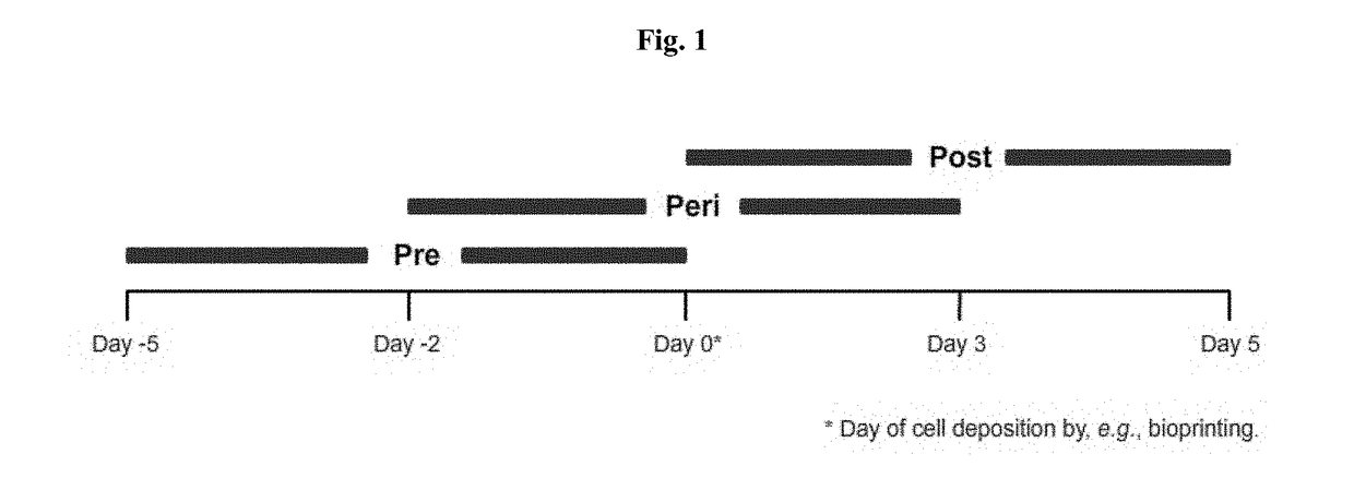
[0006]In another aspect, disclosed herein are methods of fabricating a living, three-dimensional connective tissue construct comprising: incubating a bio-ink, comprising multi-potent cells that have been deposited on a support and exposed to one or more differentiation signals, to allow the bio-ink to cohere and to form a living, three-dimensional connective tissue construct, wherein said incubation has a duration of about 1 hour to about 30 days. In some embodiments, the multi-potent cells comprise one or more of: mesenchymal stem / stromal cells, induced pluripotent stem cells, and embryonic stem cells. In some embodiments, the multi-potent cells are derived from mammalian adipose tissue. In other embodiments, the multi-potent cells are derived from mammalian bone marrow. In yet other embodiments, the multi-potent cells are derived from a non-adipose, non-bone marrow tissue source. In some embodiments, the connective tissue cells are exposed to one or more differentiation signals at one or more time intervals between about 1-21 days before depositing the bio-ink onto the support to about 1-21 days after depositing the bio-ink onto the support. In some embodiments, the bio-ink is deposited by bioprinting. In some embodiments, the construct is substantially free of any pre-formed scaffold at the time of use. In some embodiments, the construct is non-innervated. In some embodiments, the connective tissue is selected from the group consisting of: bone, cartilage, tendon, and ligament. In some embodiments, the bio-ink further comprises one or more of the following cell types: vascular, endothelial, fibroblasts, pericytes, stem / progenitor cells, immune cells. In some embodiments, the bio-ink further comprises an extrusion compound. In some embodiments, the one or more differentiation signals comprise mechanical, biomechanical, soluble, or physical signals, or combinations thereof. In some embodiments, the method further comprises the step of depositing one or more discrete filler bodies, each filler body comprising a biocompatible material, wherein the one or more filler body creates a gap or space in the cohered cells. In further embodiments, each filler body substantially resists migration and ingrowth of cells. In some embodiments, the method further comprises the step of assembling a plurality of living, three-dimensional connective tissue constructs into an array by spatially confining the constructs onto or within a biocompatible surface. In some embodiments, the construct is suitable for implantation in a subject at a site of injury, disease, or degeneration.
[0007]In another aspect, disclosed herein are methods of fabricating a living, three-dimensional connective tissue construct comprising the steps of: preparing bio-ink comprising multi-potent cells; depositing the bio-ink onto a support; and incubating the bio-ink to allow the bio-ink to cohere and to form a living, three-dimensional connective tissue construct, wherein said incubation has a duration of about 1 hour to about 30 days; with the proviso that the multi-potent cells are exposed to one or more differentiation signals. In some embodiments, the multi-potent cells comprise one or more of: mesenchymal stem / stromal cells, induced pluripotent stem cells, and embryonic stem cells. In some embodiments, the multi-potent cells are derived from mammalian adipose tissue. In other embodiments, the multi-potent cells are derived from mammalian bone marrow. In yet other embodiments, the multi-potent cells are derived from a non-adipose, non-bone marrow tissue source. In some embodiments, the connective tissue cells are exposed to one or more differentiation signals at one or more time intervals between about 1-21 days before depositing the bio-ink onto the support to about 1-21 days after depositing the bio-ink onto the support. In some embodiments, the bio-ink is deposited by bioprinting. In some embodiments, the construct is substantially free of any pre-formed scaffold at the time of use. In some embodiments, the construct is non-innervated. In some embodiments, the connective tissue is selected from the group consisting of: bone, cartilage, tendon, and ligament. In some embodiments, the bio-ink further comprises one or more of the following cell types: vascular, endothelial, fibroblasts, pericytes, stem / progenitor cells, immune cells. In some embodiments, the bio-ink further comprises an extrusion compound. In some embodiments, the one or more differentiation signals comprise mechanical, biomechanical, soluble, or physical signals, or combinations thereof. In some embodiments, the method further comprises the step of depositing one or more discrete filler bodies, each filler body comprising a biocompatible material, wherein the one or more filler body creates a gap or space in the cohered cells. In further embodiments, each filler body substantially resists migration and ingrowth of cells. In some embodiments, the method further comprises the step of assembling a plurality of living, three-dimensional connective tissue constructs into an array by spatially confining the constructs onto or within a biocompatible surface. In some embodiments, the construct is suitable for implantation in a subject at a site of injury, disease, or degeneration.