

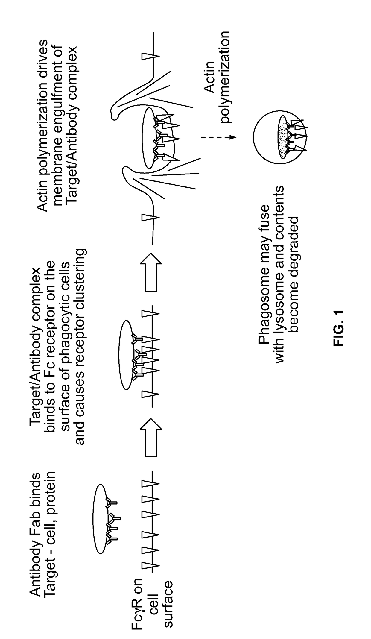
Antibody-dependent cell-mediated phagocytosis assay for reliably measuring uptake of aggregated proteins
a phagocytosis and antibody technology, applied in the field of antibodies, can solve the problems of inconvenient assays, inconvenient assays, and insufficient assays, and achieve the effects of allowing phagocytosis, high throughput analysis, and consistent data analysis
- Summary
- Abstract
- Description
- Claims
- Application Information
AI Technical Summary
Benefits of technology
Problems solved by technology
Method used
Image

Examples
example 1
Evaluating ADCP Assay Formats
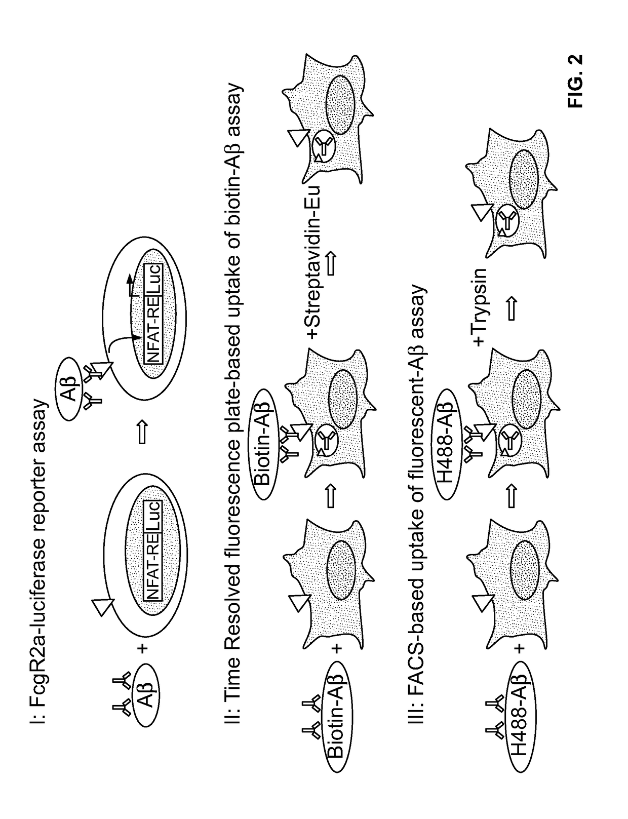
[0103]In order to develop a robust and accurate ADCP assay, several different methods were evaluated. First, an FcγR2a / CD32A reporter gene cell line from Promega was assessed (FIG. 2, I). Fcy receptor reporter cell lines have been shown to be robust, accurate and precise surrogate assay systems to measure Fab and Fc activities of antibodies (Parekh et al., 2012). A fully human monoclonal IgG1 antibody, Aducanumab, was used in the ADCP assays. Aducanumab targets aggregated forms of Aβ with high affinity. Aducanumab / Aβ complexes were added to FcγR2a-expressing Jurkat cells which contain an NFAT-luciferase (luc) reporter gene. Aducanumab / Aβ binds to FcγR2a and activates the NFAT-Luc pathway. After 5-24 h, luminescence is read on a plate reader. The signal to noise measured was very low compared to a positive control (Promega), and therefore, this format was not used for further analysis.
[0104]Second, a time-resolved fluorescence assay format that measures i...
example 2
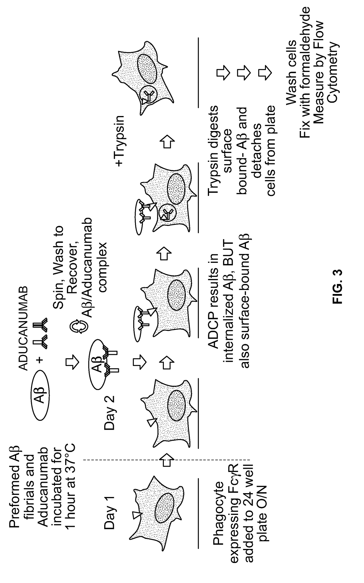
[0106]Several reagents used in the assay were characterized. 10% HiLyte488 Aβ monomers were mixed with 90% unlabeled Aβ monomers and polymerized overnight while shaking at 37° C. to make Aβ fibrils. First, it was demonstrated that fluorescent (HiLyte488) Aβ fibrils could be bound similarly by Aducanumab as unlabeled Aβ fibrils (data not shown). Second, flow cytometry was used to demonstrate that the BV-2 microglial cell line express Fc receptors (data not shown).
[0107]Microglial BV-2 cells were first allowed to adhere to tissue culture plates overnight (FIG. 3). Next, Aducanumab was incubated with 10% HiLyte488 Aβ
[0108]fibrils. After this incubation, Aducanumab / Aβ fibril complexes were isolated with several centrifugation and wash steps and added to the adherent BV-2 cells. Following ADCP, trypsin was added to detach adherent cells and digest any cell surface-bound Aβ fibrils. Following several washes to remove extracellular Aβ, cells containing internalized fluorescen...
example 3
Evaluating Cell Lines
[0114]Several cell lines were assessed for their ability to promote Aducanumab-mediated ADCP of Aβ fibrils. The BV-2 cell line is a murine microglial cell line used to measure Aducanumab's phagocytosis of Aβ fibril activity. Microglial cells are presumed to be the main effector cells in the brain. The BV-2 cell line has been used to measure IgG-dependent uptake of Aβ fibrils (Webster et al., 2001). However, one potential challenge to using the BV-2 cell line may be the use of a murine cell line and murine Fc gamma receptors to assess the biological activity of a human antibody. A second challenge may be the lack of suitable reagents to assess murine Fc gamma receptors. A third challenge may be the consistency of ADCP activity, because the BV-2 cell line expresses multiple Fc receptors.
[0115]One alternative approach to the murine BV-2 cell line is the use of a surrogate cell line that stably expresses a human Fc receptor. Stable cell lines have been used in other...
PUM
| Property | Measurement | Unit |
|---|---|---|
| time | aaaaa | aaaaa |
| time | aaaaa | aaaaa |
| intracellular fluorescence | aaaaa | aaaaa |
Abstract
Description
Claims
Application Information