Microbeads for cell culture and method of monitoring cell culture using the same

Active Publication Date: 2019-05-16
UNIV IND COOP GRP OF KYUNG HEE UNIV
View PDF1 Cites 3 Cited by
  • Summary
  • Abstract
  • Description
  • Claims
  • Application Information

AI Technical Summary

Benefits of technology

[0029]As apparent from the foregoing, a hydrophilic surface formed by surface modification of each of microbeads for cell culture according to an embodiment of the present invention, induces adsorption of a medium solution onto surfaces of the microbeads when the microbeads are immersed in the medium solution. Accordingly, cell may adhere to surfaces of the microbeads and proliferate thereon.
[0030]In addition, since the microbeads for cell culture according to an embodiment of the present invention have a lower specific gravity than a medium solution, the microbeads float in an upper layer (surface layer) of the medium solution at the beginning of cell culture. Subsequently, the specific gravities of the microbeads increase as cells adhere to surfaces of the microbeads and proliferate threreon, whereby an exhibited phenomenon that the microbeads sink in a lower part (lower layer) of the medium solution indicate the proliferation state of cells enabling intuitive monitoring.
[0031]In addition, by applying a cell culture method using the micro

Problems solved by technology

Accordingly, cells may be damaged, and cell loss may occur due to unclear separation between microbeads and cells.
In addition, to culture cells in safe and efficient way, materials of microbeads are limited to bio-compatible polymers.
Because the specific gravities of the polymers are similar with those of cell- or human-derived solid materials, it is not easy to separate the polymers from the cell- or human-derived solid materials by centrifugation.
Meanwhile, when the cells are three-dimensionally cultured using microbeads made of glass or plastic, it is difficult to monitor the density of the cells.
In

Method used


Image

  • Microbeads for cell culture and method of monitoring cell culture using the same
  • Microbeads for cell culture and method of monitoring cell culture using the same
  • Microbeads for cell culture and method of monitoring cell culture using the same

Examples


example 2

ring of Microbeads for Cell Culture (Diameter: 200 μm)

[0150]Spherical microbeads surface-modified with methoxyaminopropylsilane were manufactured in the same manner as in Example 1, except that polyethylene cores having a diameter of about 180 μm were used to prepare microbeads having a diameter of about 200 μm.

example 3

ure Using Microbeads with Low Specific Gravity (Diameter: 500 μm) of Example 1 (for Two Hours)

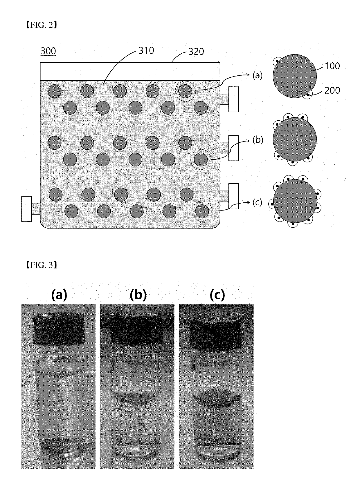
[0151]340 Surface-modified microbeads with a diameter of 500 μm of Example 1 and 3.5×10² mesodermal stem cells were mixed in a plastic tube. Temperature was maintained at 37° C. such that the cells were bound to the surfaces of the microbeads. Cell culture was performed for two hours while shaking every 10 minutes over a period of two hours such that the microbeads and the cells were dispersed.

[0152]FIG. 4 is a view illustrating culture period-dependent behavior of microbeads in a culture medium according to an example of the present invention.

[0153]Referring to (a) to (c) of FIG. 4, as a result of the cell culture according to Example 3, the number of sunken microbeads tended to increase as cell culture time increased (from (a) to (c)).

example 4

ure Using Microbeads with Low Specific Gravity (Diameter: 500 μm) of Example 1 (for Five Days)

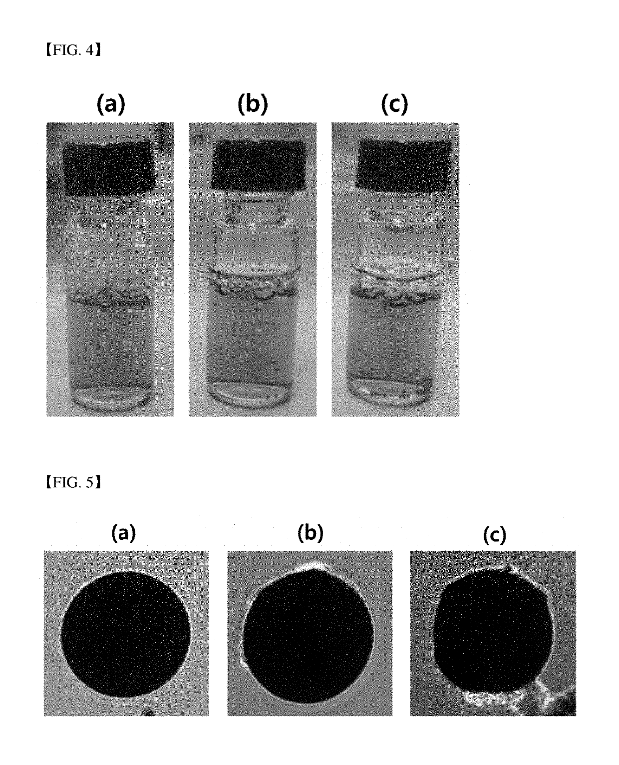
[0154]Cell culture was carried out using the microbeads in the same manner as in Example 3, except that a cell culture time was extended to five days.

[0155]FIG. 5 is a view illustrating an adhesion degree of cells to microbeads depending upon position thereof in a culture medium according to an example of the present invention.

[0156]Referring to (a) to (c) of FIG. 5, after culturing cells in a plastic tube for five days according to Example 4, microbeads distributed in (a) an upper layer (a surface layer). (b) an middle layer, and (c) a lower layer (bottom layer) of a culture medium were fractionized and adhesion degrees of cells on surfaces of the microbeads were observed by means of an inverted microscope. As a result, it was confirmed that the number of cells attached to each of the microbeads increased in proportion to sinking degrees of the microbeads.

[0157]FIG. 6 illustrates an MTT as...


PUM


Abstract

Disclosed are microbeads for cell culture and a method of monitoring cell culture using the same. More particularly, each of the microbeads for cell culture according to an embodiment of the present invention include a core and a surface modification layer formed on a surface of the core. By using the method of monitoring cell culture with the microbeads for cell culture according to an embodiment of the present invention, cell culture may be carried out in highly scaled-up dimension and easily monitored.

Description

TECHNICAL FIELD[0001]The present invention relates to microbeads for cell culture and a method of monitoring cell culture using the same.BACKGROUND ART[0002]Recently, biotechnology in the field of regenerative medicine, involving replacing or regenerating human tissues and organs using stem cells or progenitor cells such that the tissues and organs perform original functions thereof, is actively underway.[0003]Most cells derived from solid tissues adhere to the surface of glass, plastic, or nylon as a single layer and proliferate thereon. To maximize stem cell yield, various bioreactors and incubators are being developed.[0004]For effective stem cell yield, a surface area, to which the cells are attached, needs to be increased within limited volume increase using a biocompatible micro-bead, as a support for cell culture. Microbeads, providing a large surface area for cell proliferation, enable culturing adherent cells in suspension state and this method can effectively decrease cult...

Claims


Application Information

Patent Timeline
IPC IPC(8): C12N5/00G01N33/50
CPCC12N5/0075G01N33/5005C12N2531/00C12N2537/00C12N2533/30C12N2533/40C12M23/20C12M41/46C12N5/0068C12N11/00G01N15/06G01N27/02C12N2533/12C12N5/00
Inventor OH, TONG INKIMLEE, EUN AHKIM, TAE WOO
Owner UNIV IND COOP GRP OF KYUNG HEE UNIV
Why Eureka
Try Eureka