
Treating optic neuritis with induced pluripotent stem cell-derived oligodendrocyte precursor cells

a technology of oligodendrocyte precursor cells and optic neuritis, which is applied in the field of treating optic neuritis with induced pluripotent stem cell-derived oligodendrocyte precursor cells, can solve the problems of reduced contrast sensitivity, persistent visual impairment of a third of affected eyes, and poor visual function of patients without a clinical history of optic neuritis, so as to improve the maturation or myelination efficiency

Pending Publication Date: 2019-05-23
MAYO FOUND FOR MEDICAL EDUCATION & RES
View PDF0 Cites 2 Cited by
  • Summary
  • Abstract
  • Description
  • Claims
  • Application Information

AI Technical Summary

Benefits of technology

The patent text describes methods for treating a damaged optic nerve in a mammal by using induced pluripotent stem cell-derived oligodendrocyte precursor cells. These cells have a high remyelination potential, meaning they can effectively treat the damaged nerve. The patent also describes a method for screening factors that enhance the maturation or myelination efficiency of these cells. The technical effects of this patent are the development of effective treatments for optic nerve damage and the identification of important factors for the maturation and myelination of oligodendrocyte precursor cells.

Problems solved by technology

While some patients recover central vision, one third of affected eyes exhibit persistent visual impairment that includes reduced contrast sensitivity and problems with motion processing and depth perception.
Moreover, the prevalence of persistent visual complaints in MS patients has been estimated at around 35%, and even MS patients without a clinical history of optic neuritis exhibit poor visual function, including diffuse visual field defects, reduced low-contrast acuity, impaired depth and motion perception, and decreased color-sensitivity.

Method used


Image

  • Treating optic neuritis with induced pluripotent stem cell-derived oligodendrocyte precursor cells
  • Treating optic neuritis with induced pluripotent stem cell-derived oligodendrocyte precursor cells
  • Treating optic neuritis with induced pluripotent stem cell-derived oligodendrocyte precursor cells

Examples


example 1 — experimental procedures

Example 1—Experimental Procedures

Preparation of Microfluidic Chambers

[0053]MFCs were prepared as described elsewhere (Sauer et al., Neurobiol. Dis., 59:194-205 (2013)). Briefly, silicone elastomer (Sylgard 184) base and curing agent (mixed 10:1) were poured over etched fused silica molds. MFCs were placed in a vacuum chamber and incubated at 37° C. before being cut out from the molds and sterilized for use. In a sterile hood, acid washed and sterile coverslips (22×22 mm) were placed in 6-well tissue culture plates and coated with 0.5 mg / mL poly-ornithine overnight at 37° C. Prior to plating cells, the printed surface of the sterilized MFCs was adhered to glass cover slips to achieve a leak-proof chamber.

Primary Cortical Neurons Grown in MFCs

[0054]Primary murine cortical neuron cultures were prepared as described elsewhere (Sauer et al., Neurobiol. Dis., 59:194-205 (2013)). Briefly, cells were obtained from embryonic day 15 B6 mouse cortices and plated at 1.5×10⁵ cells per microfluid...

example 2

dic Chamber Design

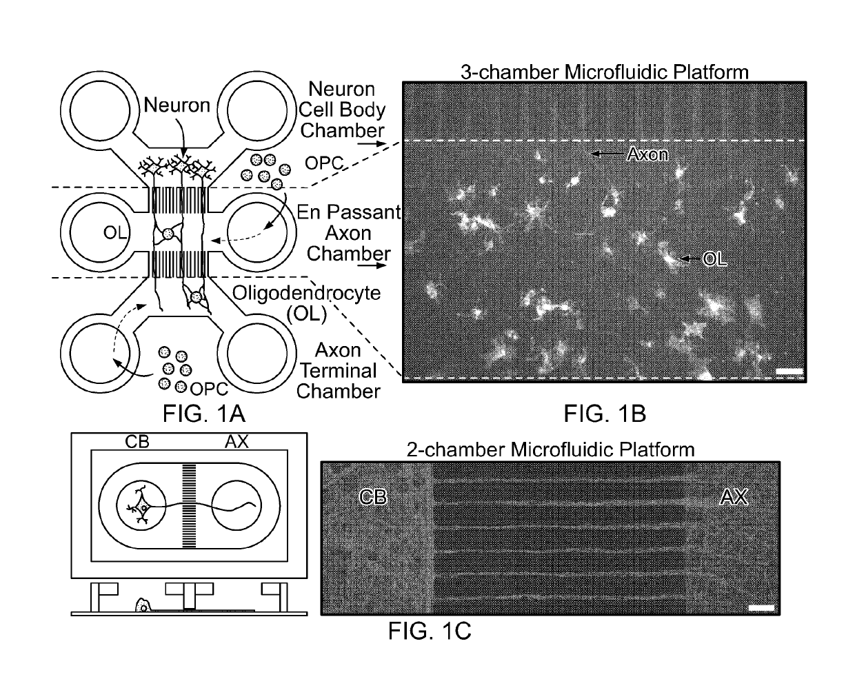
[0064]Murine cortical neurons were successful cultured in microfluidic chambers in order to isolate the neuronal cell bodies from their axons. Microgrooves within a microfluidic device prevented cell bodies from entering the axonal chamber, thereby allowing easier visualization and manipulation of axons without the interference of neuronal cell bodies and other cells types (such as oligodendrocytes and astrocytes) that are present at plating (FIG. 1C). A three-chamber microfluidic device was designed that not only allowed the separation of neuronal cell bodies and axons but permitted the addition of OPCs to a middle chamber that provided en passant access to the axons while minimizing fluidic shear stress (FIGS. 1A and 1B), thus allowing for easier quantification of in vitro myelination. The design shown in FIGS. 1A and 1B also permits the use of different combinations of growth and differentiation factors in order to optimize survival and maturation of OPCs and ol...

example 3

Differentiation Protocol for Generating OPCs from Induced Pluripotent Stem Cells

[0065]Cell populations enriched for OPCs were created from murine lines as described elsewhere (Terzic et al., Cell Transplantation, 25(2):411-424 (2016)).


PUM


Abstract

This document provides materials and methods for treating a damaged optic nerve in a mammal to restore visual function comprising administering a population of induced pluripotent stem cell-derived oligodendrocyte precursor cells. This document also provides materials and methods for determining a remyelination potential quotient of a population of induced pluripotent stem cell-derived oligodendrocyte precursor cells. This document also provides materials and methods for screening factors that enhance maturation or myelination efficiency of an induced pluripotent stem cell-derived oligodendrocyte precursor cell or cells.

Description

CROSS-REFERENCE TO RELATED APPLICATIONS[0001]This application claims priority to U.S. Application Ser. No. 62 / 317,839, filed on Apr. 4, 2016. The disclosure of the prior application is considered part of the disclosure of this application, and is incorporated in its entirety into this application.BACKGROUND1. Technical Field[0002]This document provides materials and methods for treating a damaged optic nerve in a mammal comprising administering a population of induced pluripotent stem cell-derived oligodendrocyte precursor cells, materials and methods for determining a remyelination potential quotient of a population of induced pluripotent stem cell-derived oligodendrocyte precursor cells, and materials and methods for screening for factors that enhance maturation or myelination efficiency of an induced pluripotent stem cell-derived oligodendrocyte precursor cell or cells.2. Background Information[0003]Across the United States, approximately 400,000 people have multiple sclerosis (M...

Claims


Application Information

Patent Timeline
IPC IPC(8): A61K35/30C12N5/079A61K9/00G01N33/50
CPCA61K35/30C12N5/0622A61K9/0048A61K9/0019G01N33/5058C12N2502/08A61P25/28
Inventor HOWE, CHARLES L.STANDIFORD, MIRANDA M.MIRCHIA, KANISH
Owner MAYO FOUND FOR MEDICAL EDUCATION & RES
Learn More