System and method for guiding invasive medical treatment procedures based upon enhanced contrast-mode ultrasound imaging

Inactive Publication Date: 2019-06-27
CHARLES STARK DRAPER LABORATORY +1
View PDF0 Cites 6 Cited by
  • Summary
  • Abstract
  • Description
  • Claims
  • Application Information

AI Technical Summary

Benefits of technology

[0007]This invention overcomes disadvantages of the prior art by providing a system and method for removal of various features from images acquired by an ultrasound scanner in the presence of molecularly bound contrast agent. The removed features can include background features, camouflaging features, confounding artifacts, and/or other features. The system and method e

Problems solved by technology

However, existing approaches to detection of the accumulation of targeted contrast agents in living tissue using ultrasound are not sufficient to achieve this potential.
Existing techniques are either not suitable for widespread use in humans, due to techniques that necessitate microbubble destruction using a high burst of acoustic energy that risks damage to blood vessels, or due to measurement challenges in which ultrasound signals from other sources are confounded with the signal from the accumulating microbubbles, leading to low-confidence measurements.
Thus, existing techniques are generally incompatible with the real

Method used


Image

  • System and method for guiding invasive medical treatment procedures based upon enhanced contrast-mode ultrasound imaging
  • System and method for guiding invasive medical treatment procedures based upon enhanced contrast-mode ultrasound imaging
  • System and method for guiding invasive medical treatment procedures based upon enhanced contrast-mode ultrasound imaging

Examples


Embodiment Construction

[0042]I. System Overview

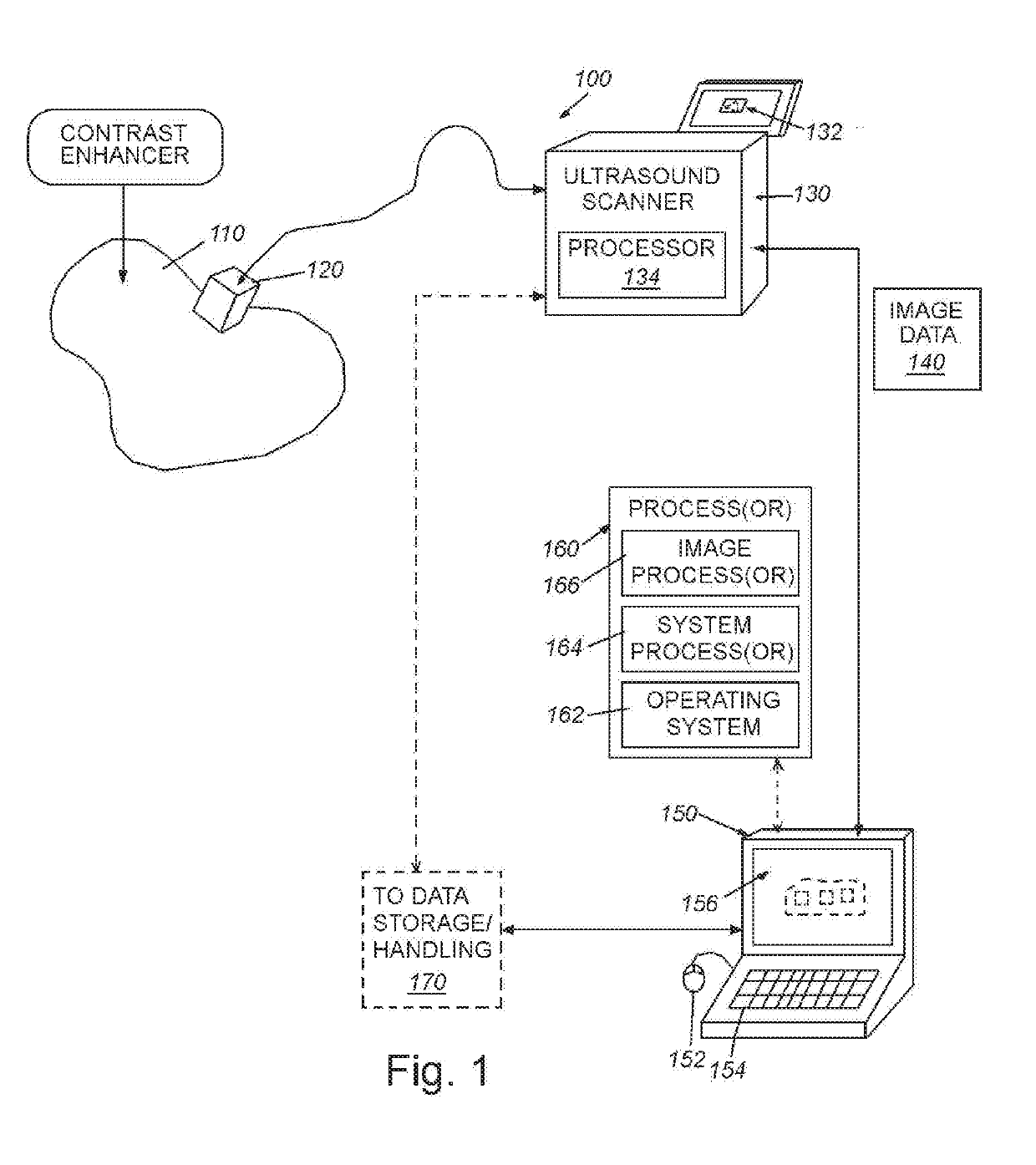
[0043]FIG. 1 shows a diagram of a generalized system 100 for scanning tissue 110 (e.g. human or mammalian) using ultrasound energy. The exemplary system 100 includes a transducer / probe 120, which is shown held against the tissue in an appropriate orientation using freehand guidance or a mechanical device (e.g. a robotic manipulator, such as the da Vinci® surgical robot, available from Intuitive Surgical, Inc. of Sunnyvale, Calif.). The probe 120 defines a transceiver that transmits ultrasound energy to the tissue, and receives echoes / reflections that are converted into electromagnetic signals. These signals are received by the base scanner unit 130, which can be any acceptable manufacturer and model—for example, Philips, Siemens, HP, General Electric, etc. The exemplary base scanner unit 130 includes an onboard display 132 that allows for local viewing and control of images acquired by the probe. It can include touch screen functions to allow a user to interf...


PUM


Abstract

This invention provides a system and method for guiding a surgical instrument based on information obtained using enhanced contrast-mode ultrasound. The enhanced information can be added to information or images obtained in one or more imaging modes. Various pieces of information can be combined and composited, including information regarding tumors, blood vessels, location information, confidence levels, and other information, and can be composited into operative imaging.

Description

RELATED APPLICATION[0001]This application claims the benefit of co-pending U.S. Provisional Application Ser. No. 62 / 589,491, entitled SYSTEM AND METHOD FOR DYNAMIC BACKGROUND SIGNAL REMOVAL AND RESOLVING REGIONS OF INTEREST IN CONTRAST-ENHANCED ULTRASOUND IMAGES, filed Nov. 21, 2017, the teachings of which are expressly incorporated herein by reference.FIELD OF THE INVENTION[0002]This invention relates to medical imaging and more particularly to processing and analysis of contrast-enhanced ultrasound imagesBACKGROUND OF THE INVENTION[0003]Ultrasound is sound waves with frequencies which are significantly higher than those audible to humans (>20,000 Hz). Ultrasonic images also known as sonograms are made by sending pulses of ultrasound into tissue using a transducer (also termed a probe). The sound echoes off the tissue, with different tissues reflecting varying degrees of sound. These echoes are recorded and displayed as an image to the operator.[0004]A recent development in medi...

Claims


Application Information

Patent Timeline
IPC IPC(8): A61B34/20A61B8/08A61B5/055A61B6/03A61B8/06
CPCA61B34/20A61B8/481A61B8/0841A61B8/5246A61B8/5261A61B8/488A61B5/055A61B6/032A61B8/06A61B8/5269A61B2034/2051A61B2034/2063A61B2505/05
Inventor BERLIN, ANDREW A.MD, JUERGEN K.YOUNG, MON Y.
Owner CHARLES STARK DRAPER LABORATORY
Why Eureka
Try Eureka