Methods for diagnosing pancreatic cancer

Inactive Publication Date: 2019-07-18
THE TRUSTEES OF THE UNIV OF PENNSYLVANIA
View PDF0 Cites 0 Cited by
  • Summary
  • Abstract
  • Description
  • Claims
  • Application Information

AI Technical Summary

Benefits of technology

The patent text explains that the biological samples used in this patent can be taken from a subjects' blood, stool, or plasma. Using stool or plasma samples can reduce the invasiveness of the diagnostic or prognostic procedure and make the examination more comfortable for the subject. Additionally, the patent allows for the detection of biomarkers in these samples, which can aid in the diagnosis and monitoring of various health conditions.

Problems solved by technology

However, no iPS cell lines from solid primary human cancers have been reported.
However, the study observed very few EMT-derived stromal cells at the PanIN stages.
There has been an absence of live human cell models of PDAC progression and consequently little information about proteins that could serve as released biomarkers and pathway indicators for early stages of the disease.

Method used


Image

  • Methods for diagnosing pancreatic cancer
  • Methods for diagnosing pancreatic cancer
  • Methods for diagnosing pancreatic cancer

Examples


example 1

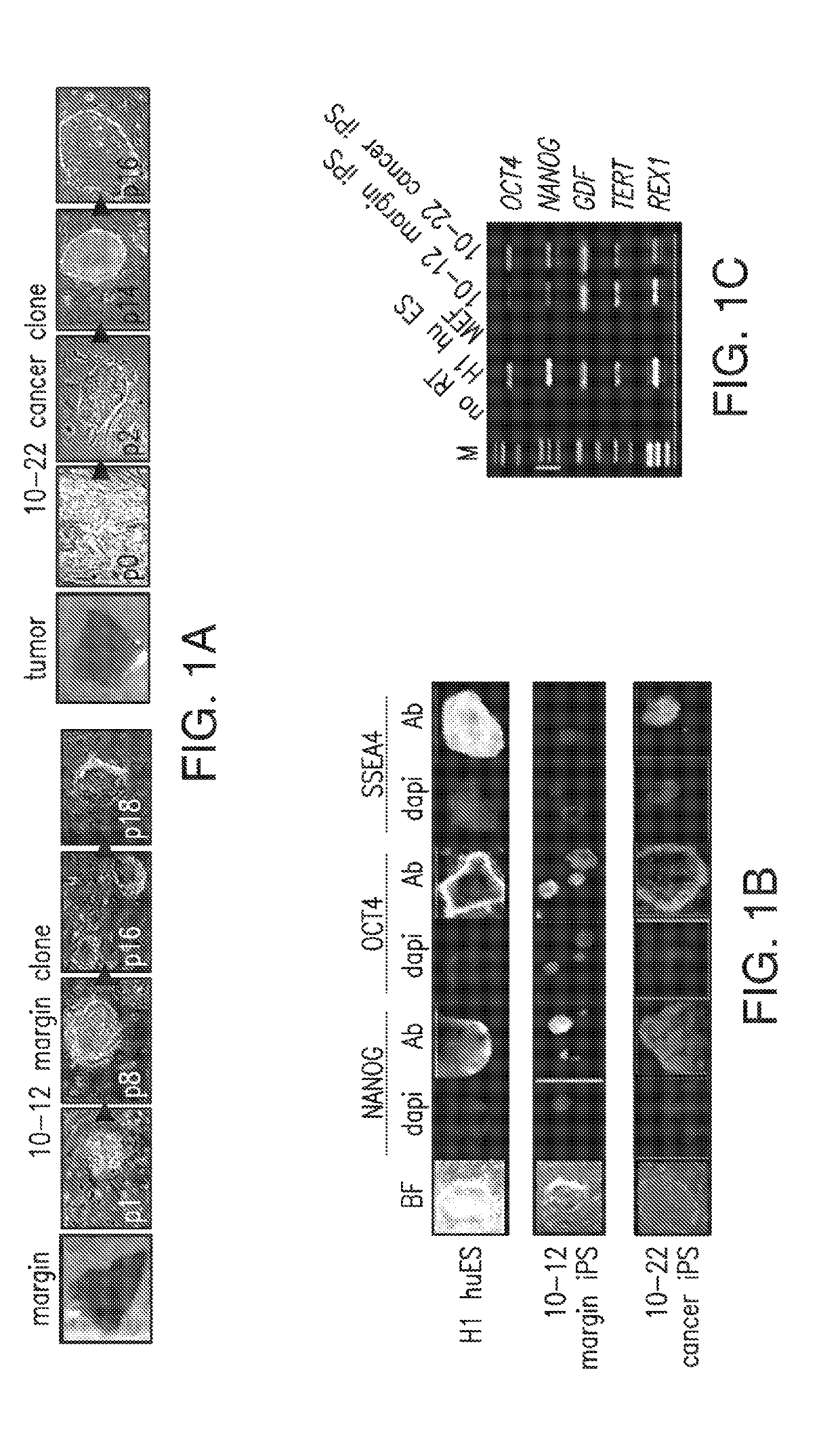
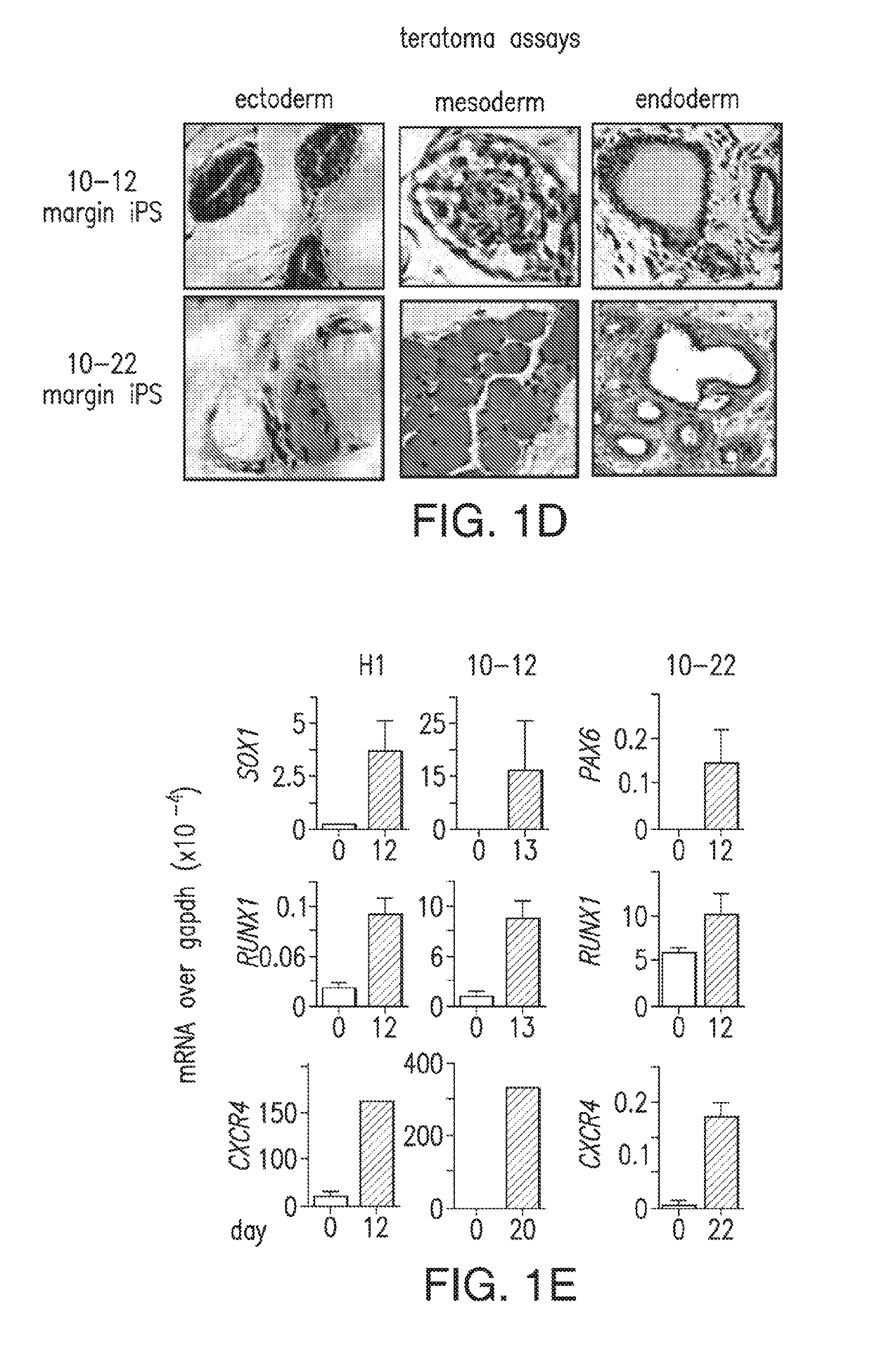
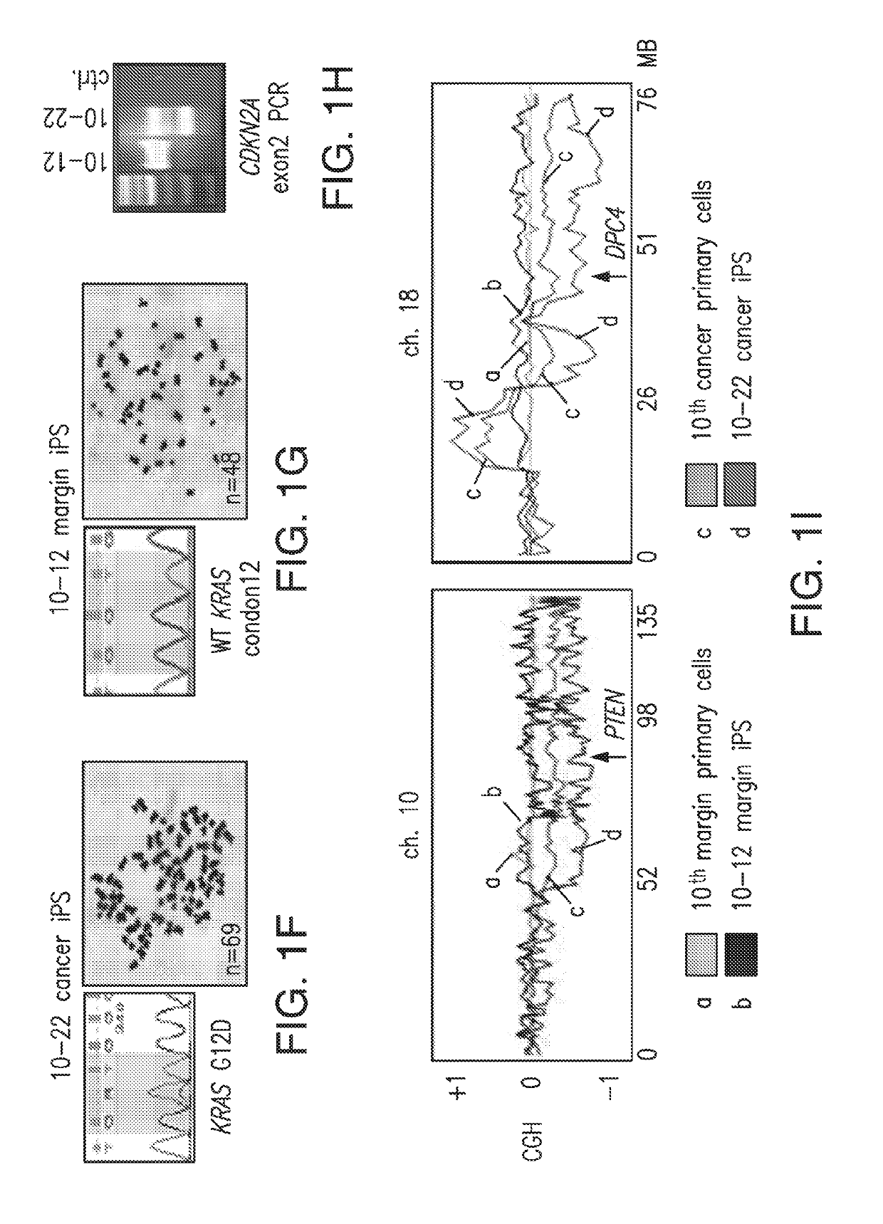
An iPS Cell Line from Human Pancreatic Ductal Adenocarcinoma Undergoes Early to Invasive Stages of Pancreatic Cancer Progression

MATERIALS AND METHODS

General Cell Culture

[0181]293T cells and human pancreatic ductal carcinoma cell lines PanC-1 and MIAPaCa-2 were maintained in 90% Dulbecco's modified essential medium (Invitrogen, Carlsbad, Calif.) supplemented with 10% FBS (Hyclone, Logan, Utah) and fed every other day. Irradiated mouse embryonic fibroblast (MEF) cells were purchased from R&D systems (Minneapolis, Minn.) and maintained in 85% DMEM (Invitrogen) supplemented with 15% FBS (Hyclone) on 0.1% gelatin (Millipore, Billerica, Mass.) pre-treated tissue culture dishes. Plated irradiated MEFs were used within 5 days.

H1 huES / human iPS Culture

[0182]H1 huES (Thomson et al., 1998) and iPS-like clones were maintained in 80% Dulbecco's modified essential medium (DMEM) / F12 supplemented with 20% KNOCKOUT serum replacement, 0.1 mM nonessential amino acids (Invitrogen), 0.1 mM -mercaptoetha...

example 2

Detection of Biomarkers in Subjects with Pancreatic Cancer by ELISA

[0218]In this Example, validation of the disclosed biomarkers for pancreatic cancer was performed in plasma samples of subjects that had pancreatic cancer using enzyme-linked immunosorbent assays (ELISA).

[0219]Validation of the disclosed biomarkers was performed by analyzing plasma samples of 10 patients with pancreatic cancer at various stages and analyzing plasma samples of 10 control subjects that did not have pancreatic cancer. Of the 10 subjects with pancreatic cancer, 7 had resectable and locally advanced cancer and 5 had resectable pancreatic cancer that was not as far advanced (Table 15).

[0220]Thirty three of the identified biomarkers described in Example 1, and one control, were analyzed in the plasma of the subjects by ELISA (Table 15). Human plasma samples were prepared from blood drawn from patients or controls by treating with EDTA or heparin as an anticoagulant. The mixture was centrifuged for 15 min at...

example 3

Detection of Biomarkers in Subjects with Pancreatic Cancer by Mass Spectrometry

[0223]In this Example, detection of the disclosed biomarkers for pancreatic cancer was performed in plasma samples of subjects that had pancreatic cancer using mass spectrometry.

[0224]Two control plasmas (M1, M2) and two metastatic PDAC case plasmas (M3, M4) were selected and coded to allow blind testing. Each plasma sample was subjected to a serum albumin removal gel (“pull-down”), and the superntants were collected. The supernatant of each plasma sample was subjected to a mixture of protein A beads and protein G beads to remove IgG, and adjusted with 6M Urea, 10 mM DTT and 50 mM IAA to block free sulfhydryl groups. The supernatants were subsequently treated with trypsin at 1:50 and subjected to a C18 cartridge desalting step. 30 μg were aliquoted from each supernatant sample for each LC-MS / MS run. pFind was used to analyze the data and to identify peptide IDs. The reverse peptide decoys were compared wi...


PUM


Abstract

The present disclosure relates to the use of one or more biomarkers to determine the presence of pancreatic cancer precursor lesions or pancreatic cancer in a subject. This disclosure is based, at least in part, on the discovery that early to invasive stages of pancreatic cancer release or secrete biomarkers that can be detected in biological samples of a subject. Accordingly, in certain non-limiting embodiments, the present disclosure provides for methods and kits for determining the presence of one or more biomarkers in a biological sample of a subject, and methods of using such determinations in selecting a therapeutic regimen for a cancer subject and in methods of treating cancer subjects.

Description

PRIORITY CLAIM[0001]This application is a divisional of U.S. patent application Ser. No. 14 / 976,275, filed Dec. 21, 2015, which is a continuation of PCT Application No. PCT / US2014 / 043457, filed Jun. 20, 2014, and claims priority to U.S. Provisional Application No. 61 / 837,358, filed Jun. 20, 2013, the contents of all of which are incorporated by reference herein in their entirety.GRANT INFORMATION[0002]This invention was made with government support under Grant Number GM036477 awarded by the National Institutes of Health. The government has certain rights in the invention.SEQUENCE LISTING[0003]The instant application contains a Sequence Listing which has been submitted electronically in ASCII format and is hereby incorporated by reference in its entirety. Said ASCII copy, created on Jan. 14, 2019, is named 046483-7171US2_Sequence_Listing and is 20,457 bytes in size.BACKGROUND[0004]Pancreatic ductal adenocarcinoma (PDAC) carries a dismal prognosis, with less than a 5% survival rate (H...

Claims


Application Information

Patent Timeline
IPC IPC(8): G01N33/574C12N5/074C12N5/09C12N5/071
CPCG01N33/57438C12N5/0696C12N5/0693C12N5/0676G01N2800/60
Inventor ZARET, KENNETH S.KIM, JUNGSUN
Owner THE TRUSTEES OF THE UNIV OF PENNSYLVANIA
Why Eureka
Try Eureka