Adoptive transfer of plasmacytoid dendritic cells to prevent or treat ocular diseases and conditions
a plasmacytoid dendritic cell and optimal technology, applied in the field of ocular diseases and conditions, can solve problems such as corneal nv loss and other problems
- Summary
- Abstract
- Description
- Claims
- Application Information
AI Technical Summary
Benefits of technology
Problems solved by technology
Method used
Image
Examples
example 1
arization
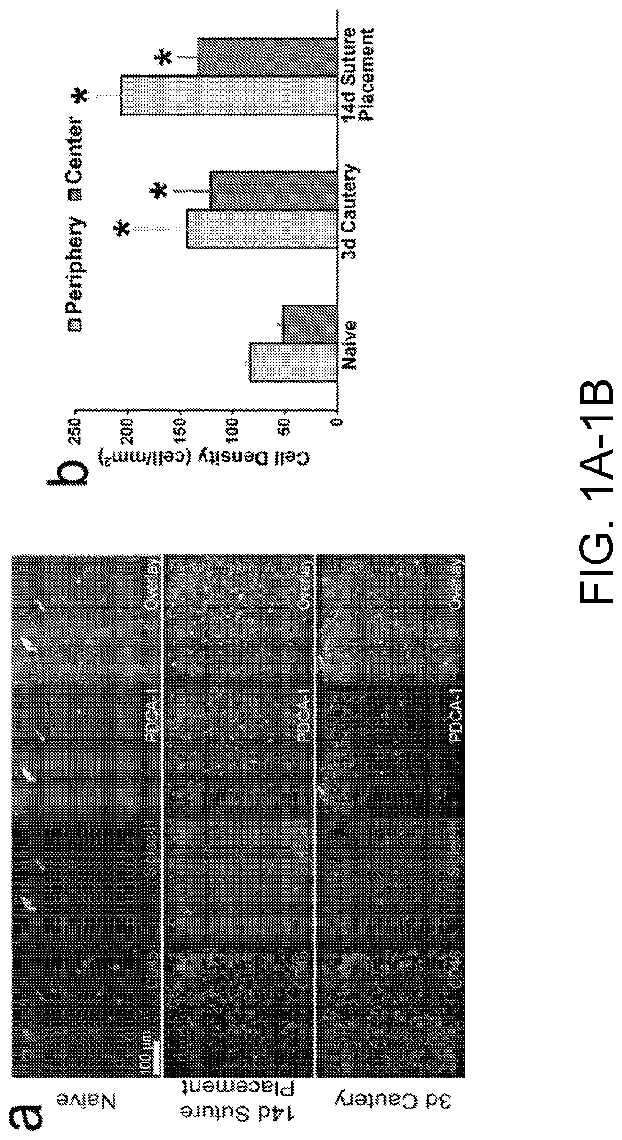
[0056]Presence of Resident Plasmacytoid Dendritic Cells in the Naïve Murine Cornea and their Significant Increase in Density Following Induction of Inflammation
[0057]To demonstrate the presence of corneal pDCs in steady state, we performed immunofluorescence (IF) staining on wild-type (WT) C57BL / 6 (B6) mice corneal whole-mounts with fluorochrome-conjugated antibodies against Siglec-H (eBioscience, San Diego, Calif.), PDCA-1 (Miltenyi Biotec Inc., San Diego, Calif.; two specific murine pDC markers), and CD45 (pan-leukocyte marker; Biolegend, San Diego, Calif.). Briefly, corneas were excised (n=3-5), fixed for 15 minutes in chilled acetone, blocked for 60 minutes with 2% bovine serum albumin+1% FC block at room temperature (RT), incubated with antibodies overnight at 4° C. and, after washing, mounted and imaged by a Leica TCS Spectral photometric SP5 laser confocal microscope.
[0058]To assess whether pDCs increased during inflammation, we used two well-established models of co...
example 2
eneration
Results
Plasmacytoid Dendritic Cells Reside in Close Proximity to Sub-Basal Nerve Plexus in Normal Cornea
[0073]As noted above, the cornea hosts resident pDCs under steady state. In order to study potential communication of pDCs with corneal nerves, we first assessed the spatial relation of pDCs and corneal nerves. As shown in FIG. 12, whole mount immunofluorescence (IF) staining of naïve wild-type (WT) C57BL / 6 cornea with 13111-tubulin (pan-neuronal marker; white), CD45 (pan-leukocyte marker; red), and PDCA-1 (pDC marker; green), reveals that pDCs lay in anterior stroma in close proximity to corneal sub-basal nerve plexus.
Local Depletion of Corneal Plasmacytoid Dendritic Cells is Accompanied by Abrupt Corneal Nerve Loss
[0074]Next, we depleted resident corneal pDCs by subconjunctival injection of 30 ng DT in transgenic BDCA2-DTR (pDC-DTR) mice. As previously mentioned, in these mice, diphtheria toxin receptor is expressed under transcriptional control of human BDCA2, a specif...
example 3
ce Nerve Regeneration after Corneal Nerve Damage
Results
Local Adoptive Transfer of Plasmacytoid Dendritic Cells as a Therapeutic Approach for Corneal Nerve Regeneration
[0097]Corneas of 6-8-week-old male wildtype (WT) C57BL / 6 mice underwent deep stromal trephination with a 2 mm trephine to sever corneal nerves. Splenic GFP+ pDCs from DPE-GFP×RAG1− / − mice and WT CD11b myeloid cells were isolated. After trephination, 104 pDCs, CD11b cells, or PBS control were locally applied onto the corneas using Tisseel tissue glue. On day 3, corneas underwent flow cytometry to assess protein expression of NGF. On day 14, corneas were stained for 13111-tubulin (pan-neuronal marker), CD45 (pan-leukocyte marker), and MHC-II (maturation marker). Total length of corneal nerves was quantified via NeuronJ and densities of MHC-II cells were measured by ImageJ. ANOVA with LSD post hoc test was used to assess statistical significance. p<0.05 was considered significant.
[0098]Confocal microscopy confirmed succes...
PUM
 Abstract
Description
Claims
Application Information
 - R&D
- Intellectual Property
- Life Sciences
- Materials
- Tech Scout
- Unparalleled Data Quality
- Higher Quality Content
- 60% Fewer Hallucinations
Browse by: Latest US Patents, China's latest patents, Technical Efficacy Thesaurus, Application Domain, Technology Topic, Popular Technical Reports.
© 2025 PatSnap. All rights reserved.Legal|Privacy policy|Modern Slavery Act Transparency Statement|Sitemap|About US| Contact US: help@patsnap.com


