Anti-angiogenesis fusion protein and uses thereof

Active Publication Date: 2020-02-20
TRICAN BIOTECH CO LTD
0 Cites 3 Cited by

AI-Extracted Technical Summary

Problems solved by technology

Suppression of any one of these steps would inhibit the formation of new...

Benefits of technology

[0007]Accordingly, the present invention provides a fusion protein that inhibits an ANG-TIE signaling pathway and a VEGF-VEGFR signaling pathway, wherein the fusion protein contains an ANG inhibiting ...

Abstract

The present invention provides a bi-functional fusion protein, (AT)a-Fc-(VT)b, simultaneously targeting vascular endothelial growth factor (VEGF) and angiopoietins (ANGs), wherein AT is an ANG binding motif; and VT is a VEGF binding motif, Fc is an N-terminal of IgG; each of a and b is an integer from 1 to 10. The bi-functional fusion proteins contain two or more domains of human proteins and are of all human sequences, and thus are expected to be non-immunogenic, and potentially can be used therapeutically in human targeting angiogenesis-associated diseases.

Application Domain

Hybrid immunoglobulinsPeptide/protein ingredients +7

Technology Topic

Protein proteinMolecular biology +12

Image

  • Anti-angiogenesis fusion protein and uses thereof
  • Anti-angiogenesis fusion protein and uses thereof
  • Anti-angiogenesis fusion protein and uses thereof

Examples

  • Experimental program(10)

Example

Example 1
Expression and Purification of Angiogenesis Blockers
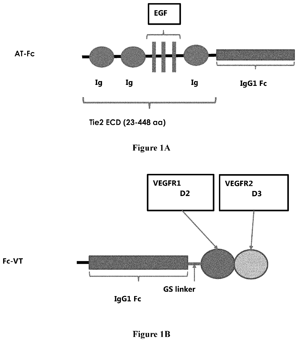
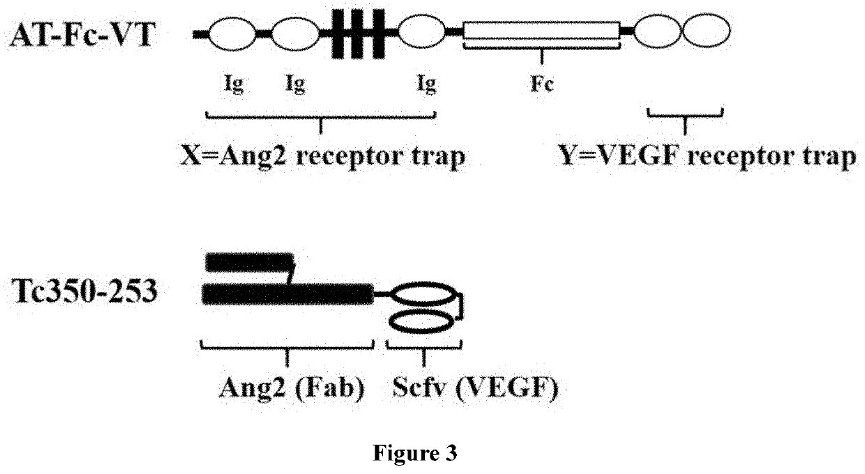
[0071]The TIE2 ECD amino acid residues from 23-448 was used as ANG binding motif, and the chimeric domains with VEGFR1 D2 and VEGFR2 D3 were used as VEGF binding trap. The corresponding DNAs were synthesized and fused with IgG1 Fc fragment gene at C-terminal for ANG blocker (AT-Fc) (FIG. 1A) or at N-terminal for VEGF blocker (Fc-VT) (FIG. 1B) with a short flexible GS linker was placed in between. To construct bi-functional AT-Fc-VT expression vector, AT-Fc was fused with GS linker and the VEGF motif, sequentially, at C-terminal (FIG. 3). These fusion constructs were cloned to pCI-neo (Promega, Inc.) expression vector between the EcoR I and Not I restriction sites under the control of a CMV promoter. The vector contains a Neomycin resistant gene and an Ampicillin resistant gene for mammalian cell selection and E. coli propagation. All fusion constructs contained a signal peptide at the N-terminal for the secretion of soluble fusion proteins. These expression constructs were used to transfect 50 ml of HEK293 cells at density of 1×10⁶ cells per ml transiently. The culture media was harvested after 96 hours of transfection and fusion proteins were purified via Protein G chromatography.
[0072]As shown in FIGS. 2 and 4, about 3 mg fusion proteins (per transfection) having a purity greater than 90% was obtained.
[0073]To construct bi-functional protein Tc350-253 expression vector (FIG. 3), Anti-Ang2 Fab HC was sequentially fused with Lucentis Vh and Lucentis Vk separated by GS linker. Tc350-253 protein was transiently expressed by HEK293 cells and purified from the transfected cell culture supernatant via nickel affinity chromatography. The purified protein (5 μg/lane) was gel-analyzed under reducing or non-reducing condition. The Tc350-253 bi-functional protein was properly expressed with a molecular weight about 75 kD, and greater than 90% purity was obtained (FIG. 5).

Example

Example 2
In Vitro Binding of Angiogenesis Blockers
[0074]Overnight ligand pre-coated (100 ng/well) wells were washed 3 times with 1× PBS and blocked with 400 ul 5% non-fat skin milk in PBS for 2 hr. After blocking, 100 ul of 3-fold serially diluted purified protein started from 30 nM were added to each corresponding well in duplicate and incubated for 1 hr at room temperature on shaker. After binding, wells were washed 3 times with PBST (1× PBS with 0.1% Tween-20) before adding 100 ul of 1:2500 diluted goat anti-human IgG Fe HRP conjugates (Jackson ImmunoResearch Inc.) to each well and incubated on shaker for another hour. After final washing, TMB reagent (Invitrogen, Inc.) was added and OD absorption at 450 nm was measured. The data analyzed by sigmoidal curve fitting using Prism 4. Binding of AT-Fc, Fc-VT, and AT-Fc-VT to ANG or VEGF were assessed by similar assays.
[0075]As shown in FIG. 6, the calculated EC50 for AT-Fc against ANG1 and ANG2 are 4.26 nM and 2.01 nM, respectively. The results collected herein indicated the soluble extracellular domain (amino acid residues 23-448) is functionally active in angiopoietin binding and could modulate the activity of the membrane-bound form by competing for angiopoietins.
[0076]As shown in FIG. 7, the Fc-VT had the anti-VEGF efficacy in the VEGF-A binding assessment. The calculated EC50 against VEGF-A was 0.33 nM. As shown in the results as collected, the C-terminally localized VEGFR1 D2 and VEGFR2 D3 chimeric VEGF trap was functional with high affinity and could neutralize VEGF's biological activities.
[0077]As shown ion FIGS. 8 and 9, the binding activities to ANG2 and VEGF of the purified AT-Fc-VT bi-functional fusion protein were assessed and the results indicated that the AT-Fc-VT fusion protein was able to bind ANG2 and VEGF-A with EC50 of 2.57 nM and 0.35 nM, respectively, and provided significantly improved binding activities as compared with the AT-Fc against ANG2, and the Fc-VT against VEGF.
[0078]In conclusion, the AT-Fc-VT fusion protein were confirmed to have bi-functions to modulate the ANG-TIE and VEGF-VEGFR pathways through the inhibition of ligand binding and receptor activation.

Example

Example 3
Inhibition of VEGF-Dependent HUVEC Proliferation Assay by Angiogenesis Blockers
[0079]The Fc-VT and AT-Fc-VT were used to inhibit VEGF activities in a cell base assay. Human Umbilical Vein Endothelial Cells (HUVEC cells, Lonza, Inc.) were used to demonstrate VEGF-dependent cell proliferation which can be inhibited by VEGF blocker. In the experiment, HUVECs were maintained in the Endothelial Cell Growth Medium (Lonza, Inc.) with 2% FBS. Collagen pre-coated wells were loaded with 50 ml of 1 nM of VEGF-A (R&D systems, Inc.) and various concentrations of Fc-VT or AT-Fc-VT per well for 1 hour at 37° C. before adding 50 ml of HUVECs at 1×10⁵ cells/ml in Medium-199 (10% FBS, Hyclone, Inc.) to each well. After 72 hours incubation at 37C with 5% CO2, the cell growth was assayed by adding 10 ml MTS detection reagent (Promega Inc.) to each well and then measuring OD absorption at 450/650 nm.
[0080]As shown in FIG. 10, the 50 percent inhibitory activity (IC50) of AT-Fc-VT on VEGF-dependent HUVEC cell proliferation assay was 0.26 nM, similar to that of Fc-VT (0.20 nM) in this example.

PUM

PropertyMeasurementUnit
Flexibility

Description & Claims & Application Information

We can also present the details of the Description, Claims and Application information to help users get a comprehensive understanding of the technical details of the patent, such as background art, summary of invention, brief description of drawings, description of embodiments, and other original content. On the other hand, users can also determine the specific scope of protection of the technology through the list of claims; as well as understand the changes in the life cycle of the technology with the presentation of the patent timeline. Login to view more.

Similar technology patents

Honeysuckle-scutellaria baicalensis-forsythia superfine powder and production method thereof

InactiveCN103463203AFast crushingImprove efficacy
Owner:SICHUAN LONGHAIYANG ANIMAL PHARMA CO LTD

Novel Skin Remodeling Strategy

InactiveUS20160243023A1improve efficacy
Owner:ELC MANAGEMENT LLC

Classification and recommendation of technical efficacy words

  • improve efficacy
Why Eureka
Try Eureka