Anatomical measurements from ultrasound data

anatomical measurement and ultrasound data technology, applied in the field of anatomical measurement from ultrasound data, can solve the problems of insufficient acquisition frame rate, inability to access 3-d ultrasound image data, and inability to acquire 3-d ultrasound images, so as to reduce the uncertainty in the 3-d, increase the reliability of correlation function, and increase the accuracy of acquisition.

Active Publication Date: 2020-03-05
KONINKLJIJKE PHILIPS NV
View PDF0 Cites 8 Cited by
  • Summary
  • Abstract
  • Description
  • Claims
  • Application Information

AI Technical Summary

Benefits of technology

[0024]This obviates the need to provide separate 2-D ultrasound images for the training purpose of the machine learning algorithm and has the further advantage that typical slicing directions of the 3-D ultrasound images, e.g. 2-chamber view and 4-chamber view directions in case of echocardiography images, can be easily obtained, which may be used to strengthen the reliability of the correlation function developed by the machine learning algorithm for such commonly used views. Hence, in an example embodiment, providing a set of 2-D image planes comprises providing a first set of 2-D image planes providing a 2-chamber view of a human heart and a second set of 2-D image planes providing a 4-chamber view of the human heart.
[0025]Preferably, the computer-implemented method further comprises training the machine-learning algorithm to generate a further estimator function to estimate an uncertainty in the estimated anatomical body measurement value using the obtained ground truth values, extrapolated values and at least one of the outline contour measurements and the cross-sectional measurements as inputs of said machine-learning algorithm. It is particularly useful to provide an indication of the uncertainty in the 3-D anatomical body measurement value obtained with a model, such that a user of the model can determine if the obtained value can be relied upon. This furthermore may provide the user with an indication of whether the 2-D ultrasound image acquisition should be repeated alo...

Problems solved by technology

Or, the operator may not have access to 3-D ultrasound image data, in which case the operator needs to rely on 2-D ultrasound images to obtain the desired anatomical measurement.
In yet another scenario, the acquisition of 3-D ultrasound images may be practically unfeasible, for example in the field of contrast imaging, where the acquisition frame rate may be insufficient to capture usef...

Method used


Image

  • Anatomical measurements from ultrasound data
  • Anatomical measurements from ultrasound data
  • Anatomical measurements from ultrasound data

Examples


Embodiment Construction

[0047]It should be understood that the Figures are merely schematic and are not drawn to scale. It should also be understood that the same reference numerals are used throughout the Figures to indicate the same or similar parts.

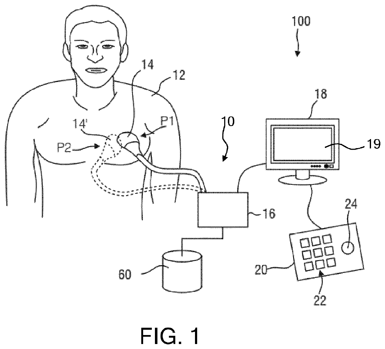
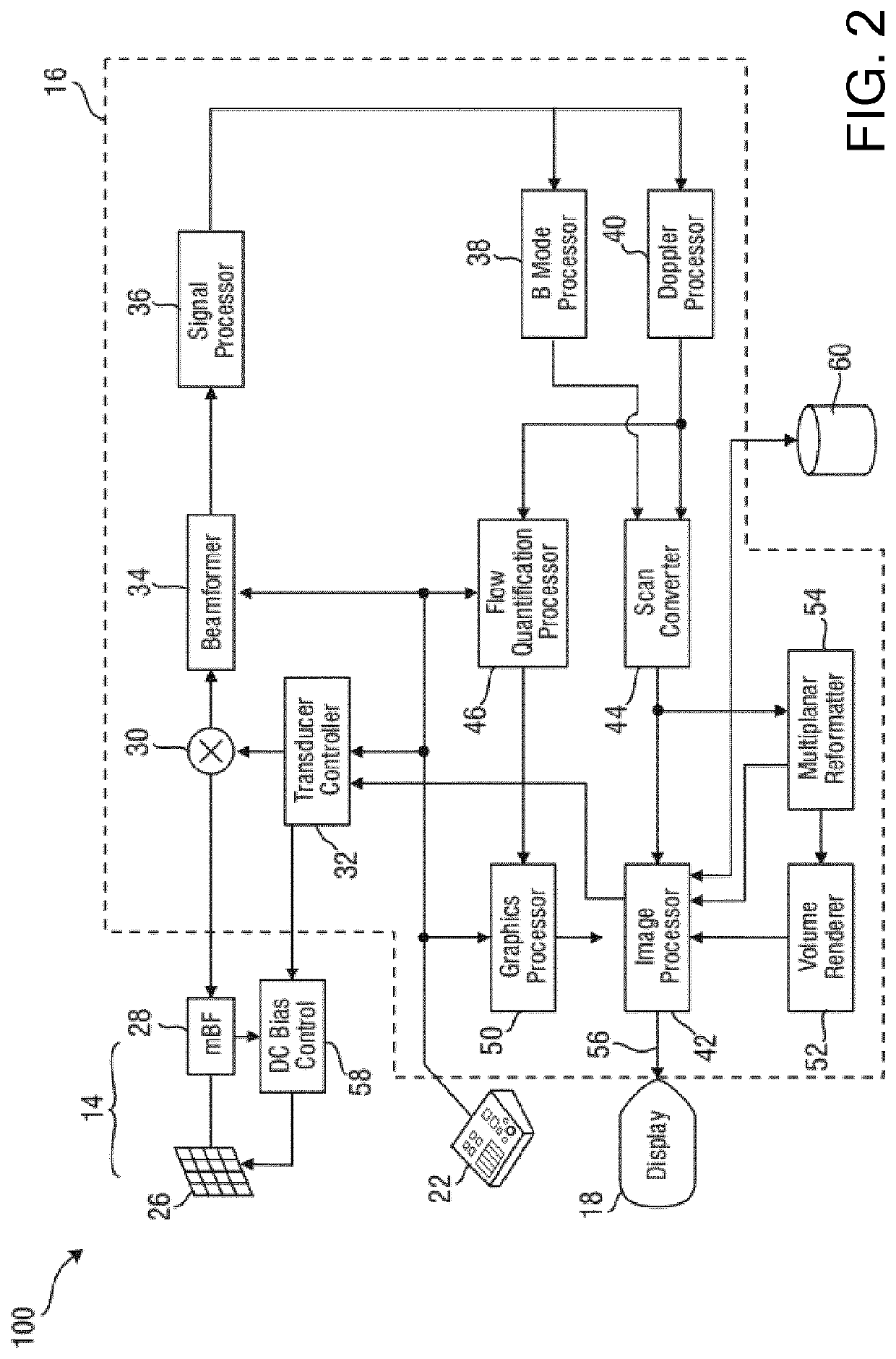
[0048]In the present application, where reference is made to a set of ultrasound images, it should be understood that such a set contains at least one ultrasound image. FIG. 1 shows a schematic illustration of an ultrasound system 100, in particular a medical two-dimensional (2-D) or three-dimensional (3-D) ultrasound imaging system. The ultrasound system 100 may be applied to inspect a volume of an anatomical site, in particular an anatomical site of a patient 12, such as the patient's heart. This for example may involve monitoring the anatomical site over a period of time to track progress of a condition affecting the anatomical site. The ultrasound system 100 comprises an ultrasound probe 14 having at least one transducer array having a multitude of transd...


PUM


Abstract

The application discloses a computer-implemented method (100) of providing a model for estimating an anatomical body measurement value from at least one 2-D ultrasound image including a contour of the anatomical body, the method comprising providing (110) a set of 3-D ultrasound images of the anatomical body; and, for each of said 3-D images, determining (120) a ground truth value of the anatomical body measurement; generating (130) a set of 2-D ultrasound image planes each including a contour of the anatomical body, and for each of the 2-D ultrasound image planes, extrapolating (140) a value of the anatomical body measurement from at least one of an outline contour measurement and a cross-sectional measurement of the anatomical body in the 2-D ultrasound image plane; and generating (150) said model by training a machine-learning algorithm to generate an estimator function of the anatomical body measurement value from at least one of a determined outline contour measurement and a determined cross-sectional measurement of a contour of the anatomical body within a 2-D ultrasound image using the obtained ground truth values, extrapolated values and at least one of the outline contour measurements and the cross-sectional measurements as inputs of said machine-learning algorithm. A computer-implemented method of deploying such a model, a computer program product, an ultrasound image processing apparatus and an ultrasound imaging system adapted to implement such methods are also disclosed.

Description

FIELD OF THE INVENTION[0001]The present invention relates to a computer-implemented method of providing a model for estimating an anatomical body measurement value from at least one 2-D ultrasound image including a contour of the anatomical body.[0002]The present invention further relates to a computer-implemented method of estimating an anatomical body measurement value from at least one 2-D ultrasound image including a contour of the anatomical body with such a model.[0003]The present invention further relates to a computer program product for implementing such methods on an ultrasound image processing apparatus.[0004]The present invention further relates to an ultrasound image processing apparatus implementing such methods.[0005]The present invention further relates to an ultrasound imaging system comprising such an ultrasound image processing apparatus.BACKGROUND OF THE INVENTION[0006]Ultrasound plays an essential role in many diagnostic imaging techniques including but not limi...

Claims


Application Information

Patent Timeline
IPC IPC(8): G06T7/62G06T7/149G06T17/00A61B8/08
CPCG06T7/62G06T17/00G06T2207/20081G06T2207/10136G06T7/149G06T2207/30048A61B8/5223G06T2207/30044
Inventor WEBER, FRANK MICHAELWAECHTER-STEHLE, IRINABUERGER, CHRISTIAN
Owner KONINKLJIJKE PHILIPS NV
Why Eureka
Try Eureka