Flexible device and its application for bio-cell in-vitro electrical and mechanical stimulation and characterization

a bio-cell and in-vitro electrical and mechanical stimulation technology, applied in the direction of skeletal/connective tissue cells, prosthesis, specific use bioreactors/fermenters, etc., can solve the problems of patch failure and the need for reoperation, the number of autologous cardiomyocytes that can be isolated from discarded heart tissue is insufficient to allow for the construction of effectively sized cell sheets, and the inability to meet the tissue requirements of the hear

Pending Publication Date: 2020-06-25
ROBERTS ERIN GLENN
View PDF6 Cites 3 Cited by
  • Summary
  • Abstract
  • Description
  • Claims
  • Application Information

AI Technical Summary

Benefits of technology

[0020]FIG. 2 Design of the device: cross-section of wall profile, groove / ridge architecture, cell locations and alignment of cells perpendicular to wall direction are shown. Top down view of the device shows orientation of cells in relationship to wall and groove / ridge directions. The benefit of the positive sloped groove / ridge profile during gold deposition is depicted; it allows for continuous gold deposition along the top of the wall. The benefit of the negative sloped side walls during gold deposition is depicted; this wall profile prevents electrical shorting between walls after deposition.
[0021]FIG. 3 Two configurations for electrical stimulation. In both of these configurations, the user is able to manipulate voltage, frequency, and pulse duration and shape. On the left, in the direct contact to contact stimulation, the user can directly stimulate individual cells, readout the current through the device, and analyze effects of conditioning on cells by measuring alignment, protein content, contractility, etc. of cells before and after stimulation. On the right, in the contact to common ground stimulation, the user can readout the time for signal to travel from one gold coated side of the device to the other, allowing for measurements of conduction velocity.

Problems solved by technology

However, these patches are unable to grow and electromechanically incorporate upon implantation within the resident heart tissue, often resulting in patch failure and the need for re-operation.
The number of autologous cardiomyocytes that can be isolated from discarded heart tissue is insufficient to allow for the construction of effectively sized cell sheets.
Therefore, cardiomyocytes are produced in vitro from various stem cell sources, but these are usually of an immature phenotype, not able to meet the tissue requirements of the heart.
Implanting immature myocyte cells into a mature cardiac environment can result in electromechanical mismatch and arrhythmia.
However it does not have cell sheet capabilities cell sheets being single layered or otherwise much thinner constructs that allow for more specific measurements across the sheet.
This limits the ability of the device to track cell duster properties as they mature and form full sheets.
The design and resulting capability for electrical stimulation and mechanical measurement is at the tissue level, rather than the cell level, limiting the specificity of measurement / readout.
Again, these platforms have not been designed to couple co-stimulation with measurement of electromechanical properties at the cellular-level.
The previously described devices and techniques can measure electrical and mechanical properties of an entire piece of tissue as well as condition these tissues, but provide no greater level of detail on a cellular level.

Method used


Image

  • Flexible device and its application for bio-cell in-vitro electrical and mechanical stimulation and characterization
  • Flexible device and its application for bio-cell in-vitro electrical and mechanical stimulation and characterization
  • Flexible device and its application for bio-cell in-vitro electrical and mechanical stimulation and characterization

Examples


Embodiment Construction

Design

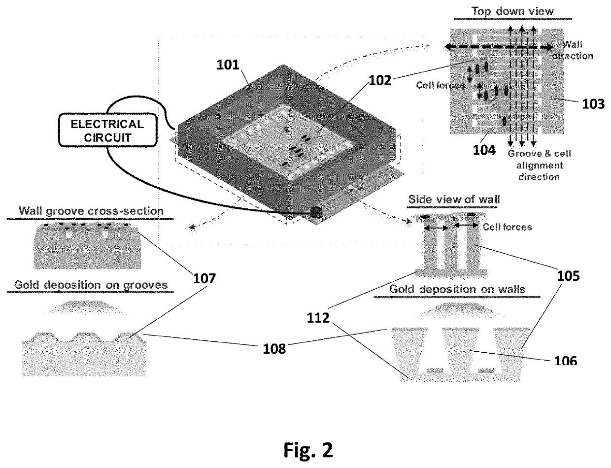
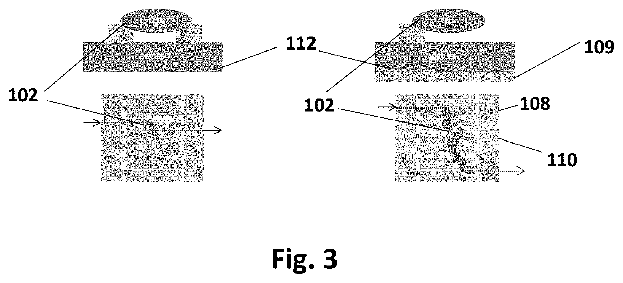
[0023]This device has been designed to achieve the following: cellular alignment, electrical stimulation, mechanical stimulation, conduction velocity readout, contraction force readout, and upon characterization, cell sheet release. In brief, the device achieves these design criteria through the following setup. The platform is based on a set of interdigitated comb electrical contacts 104. These are three-dimensional walls made of polydimethylsiloxane (PDMS) 105 coated with electrically conductive films of titanium and gold 108. Not only do the walls serve as a method for stimulating cells that are sitting on top of them, but their geometry and physical properties (namely stiffness) can be tailored to make them flexible (or rigid) enough to be bent by the range of forces applied by cells during contraction. In this way, they can be used to measure force throughout the cell culture, at the cellular or cell-group level. The walls 105 also play the role of a substrate that has an...


PUM

PropertyMeasurementUnit
angleaaaaaaaaaa
angleaaaaaaaaaa
aspect ratioaaaaaaaaaa

Abstract

Disclosed below is a device comprising a base 112, polymer walls 105, comb fingers 104, groove/ridge architecture 107, metal film 108, comb buses 103, electrical ground electrode 109, and polymer well 101 for electrical and mechanical stimulation as well as for measuring of contractile force and conduction velocity of the cellular sheet/cluster 102 in response to mechanical and electrical stimulation of the cell source as it grows into a multilayer cellular sheet on the device. This allows cell sheets to be cultured and conditioned to be compatible with the patient's cardiac environment in vitro, prior to sheet release and implantation. Major innovative elements of this device include: real-time data for rich understanding of engineered tissues as they are grown, ability to expose engineered tissues to patient-derived stimuli (specifically localized electrical stimuli, mechanical stimuli, and micro-architecture), and the option to implant after characterization.

Description

CROSS REFERENCE TO RELATED APPLICATIONS[0001]This application is a continuation of U.S. provisional patent application No. 62 / 625,049, filed Feb. 1, 2018.STATEMENT REGARDING FEDERALLY SPONSORED RESEARCH OR DEVELOPMENT[0002]Not applicable.REFERENCE TO SEQUENCE LISTING, A TABLE, OR A COMPUTER PROGRAM LISTING COMPACT DISC APPENDIX[0003]Not applicable.BACKGROUND OF THE INVENTION[0004]Cardiovascular diseases (CVDs) are the most common cause of death worldwide with an estimated 17.7 million people dying from them in 2015, 31% of all global deaths. Ischaemic heart disease and stroke are the primary conditions responsible for these deaths and have been so for the past 15 years.[0005]Currently the most widely used surgical patch solutions for repairing damaged myocardium include synthetic materials such as Dacron, Gore-Tex, and ePTFE as well as biological materials such as autologous, allogenic, and xenogenic pericardium. However, these patches are unable to grow and electromechanically inco...

Claims


Application Information

Patent Timeline
Patent Type & Authority Applications(United States)
IPC IPC(8): C12M1/42C12N5/077A61F2/00
CPCC12N5/0657C12M35/02A61F2/0077C12M35/04A61F2002/0086C12M23/20C12M21/08A61K35/12A61L2430/20A61L27/3826C12M41/36
Inventor ROBERTS, ERIN GLENN
Owner ROBERTS ERIN GLENN
Why Eureka
Try Eureka