
Systems and methods for platform agnostic whole body image segmentation

Active Publication Date: 2020-08-06
EXINI DIAGNOSTICS
View PDF0 Cites 26 Cited by
  • Summary
  • Abstract
  • Description
  • Claims
  • Application Information

AI Technical Summary

Benefits of technology

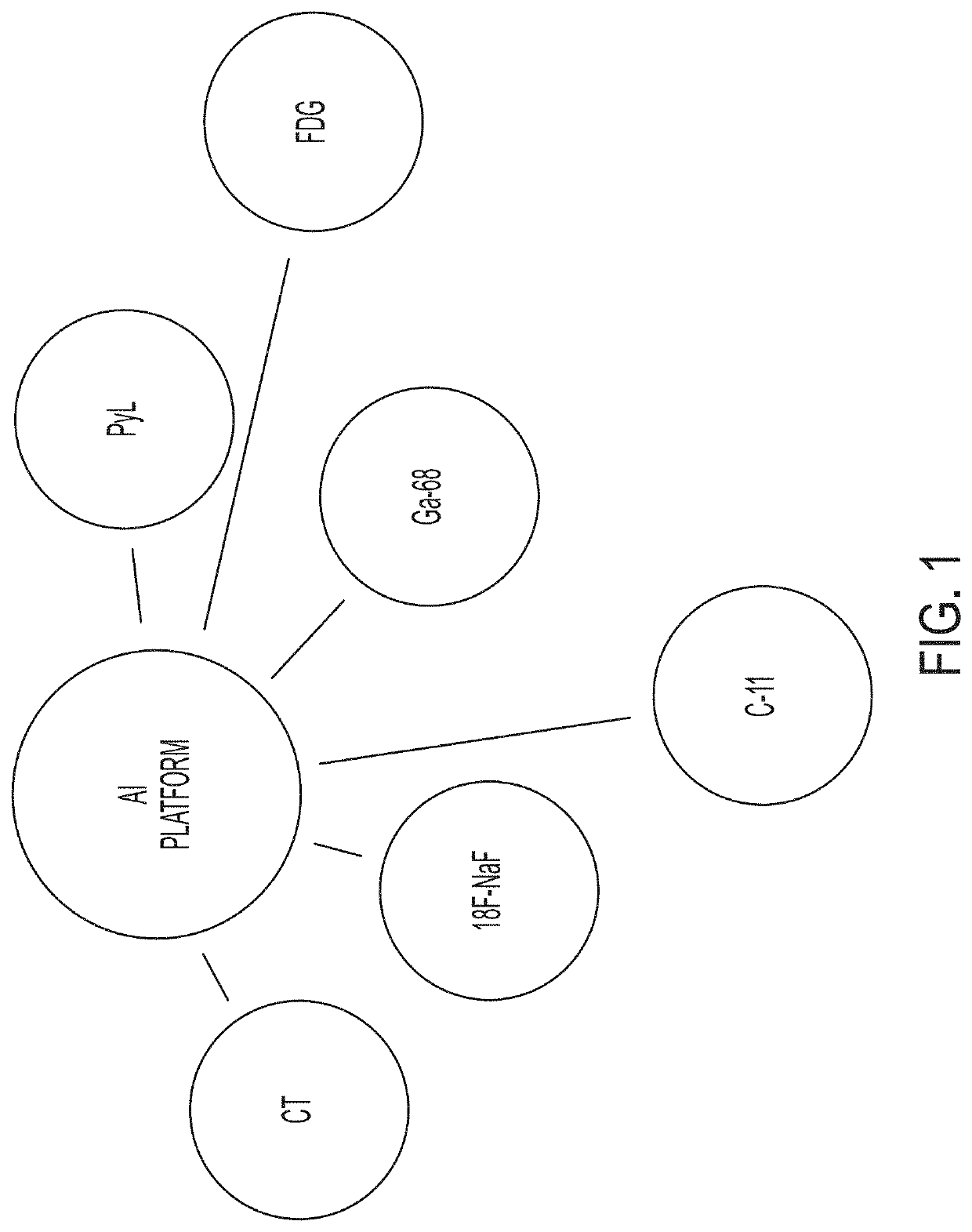
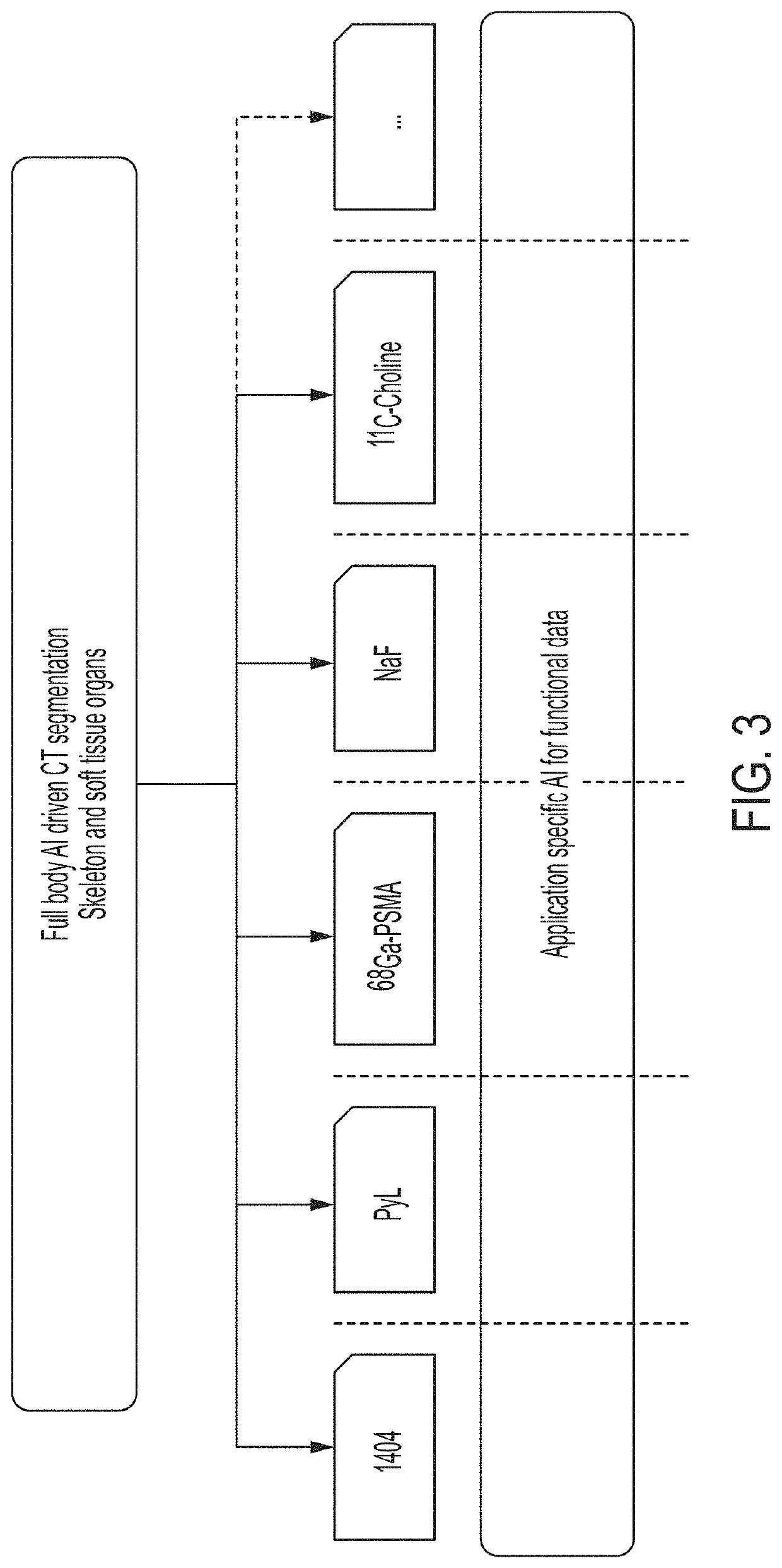
The patent describes a system and method for automatically analyzing medical images of a person's body to identify specific parts like organs and tissue. This analysis can help determine how well a treatment is working or if a person has cancer. The system works well with different types of imaging agents and can be used with various detection modalities. This makes it easier to assess the function of organs and structures in the body and improve cancer diagnosis and treatment.

Problems solved by technology

Although CT and MRI provide detailed anatomical information, conventional (e.g., manual) identification of organs and structures is difficult, subjective and time consuming, making certain assessment infeasible without computer support.

Method used


Image

  • Systems and methods for platform agnostic whole body image segmentation
  • Systems and methods for platform agnostic whole body image segmentation
  • Systems and methods for platform agnostic whole body image segmentation

Examples


example 3

iii. Automated Whole Body Segmentation for PyL™-PET Image Analysis and Lesion Detection

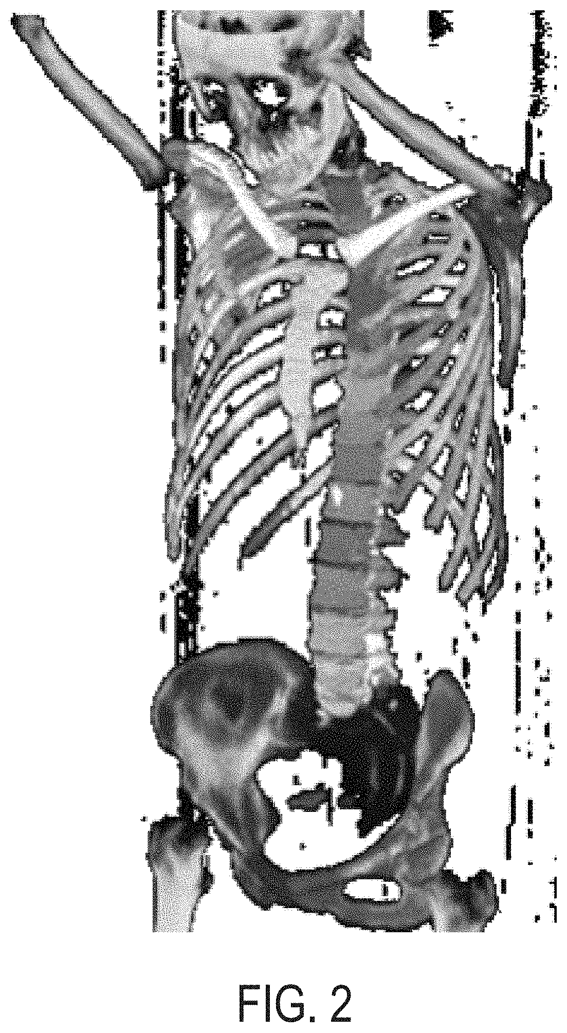
[0280]This example demonstrates automated segmentation of 49 bones and 27 soft-tissue regions in whole body CT images using deep learning approaches in accordance with the whole body segmentation technology described herein. This example also demonstrates how the anatomical information gained via such segmentation can be used to create a fully automated lesion detection algorithm in [18F]DCFPyL (PyL™-PSMA) PET / CT images. This example also shows how the segmentation can be used to remove background signal from PET images to facilitate observation and detection of lesions in which PyL™ has accumulated.

[0281]FIG. 6A shows a block flow diagram illustrating an embodiment of the segmentation processes described herein that is used in the PyL™-PET / CT image analysis described in this example. As in process 500, shown in FIG. 5, in process 600 an anatomical image is received. In particular, in process 600...

example 6

vi. Automated Hotspot Detection and Uptake Quantification in Bone and Local Lymph

[0303]PyL™-PSMA PET / CT hybrid imaging (e.g., images PET / CT images acquired for a patient after administering PyL™ to the patient) is a promising tool for detection of metastatic prostate cancer. Image segmentation, hotspot detection, and quantification technologies of the present disclosure can be used as a basis for providing automated quantitative assessment of abnormal PyL™-PSMA uptake in bone and local lymph (i.e., lymph nodes localized within and / or in substantial proximity to a pelvic region of a patient). In particular, as shown in this example, image segmentation and hotspot detection techniques in accordance with the systems and methods described herein can be used to automatically analyze in PET images to detect hotspots that a physician might identify as malignant lesions.

[0304]In this example, PET / CT scans were evaluated to automatically identify, within the PET images of the scans, hotspot...

example 8

vii. Improved Performance in AI-Assisted Image Analysis for Patients with Low or Intermediate Risk Prostate Cancer

[0316]99mTc MIP-1404 (1404) is a PSMA targeted imaging agent for the detection and staging of clinically significant prostate cancer. Manual assessment of tracer uptake in SPECT / CT images introduces inherent limitations in inter- and intra-reader standardization. This example describes a study that evaluated the performance of PSMA-AI assisted reads, wherein automated segmentation of prostate volumes and other target tissue regions are performed in accordance with the embodiments described herein, over manual assessment and known clinical predictors.

[0317]The study analyzed 464 evaluable patients with very low-, low-, or intermediate-risk prostate cancer, whose diagnostic biopsy indicated a Gleason grade of ≤3+4 and / or who were candidates for active surveillance (1404-3301). All subjects received an IV injection of 1404 and SPECT / CT imaging was performed 3-6 hours postd...


PUM

PropertyMeasurementUnit
Fractionaaaaaaaaaa
Capacitanceaaaaaaaaaa
Sizeaaaaaaaaaa

Abstract

Presented herein are systems and methods that provide for automated analysis of three-dimensional (3D) medical images of a subject in order to automatically identify specific 3D volumes within the 3D images that correspond to specific anatomical regions (e.g., organs and / or tissue). Notably, the image analysis approaches described herein are not limited to a single particular organ or portion of the body. Instead, they are robust and widely applicable, providing for consistent, efficient, and accurate detection of anatomical regions, including soft tissue organs, in the entire body. In certain embodiments, the accurate identification of one or more such volumes is used to automatically determine quantitative metrics that represent uptake of radiopharmaceuticals in particular organs and / or tissue regions. These uptake metrics can be used to assess disease state in a subject, determine a prognosis for a subject, and / or determine efficacy of a treatment modality.

Description

CROSS-REFERENCE TO RELATED APPLICATIONS[0001]This application claims priority to and benefit of U.S. Provisional Application No. 62 / 789,155, filed Jan. 7, 2019, U.S. Provisional Application No. 62 / 837,941, filed Apr. 24, 2019, U.S. Provisional Application No. 62 / 863,608, filed Jun. 19, 2019, U.S. Provisional Application No. 62 / 870,210, filed Jul. 3, 2019, U.S. Provisional Application No. 62 / 907,158, filed Sep. 27, 2019, U.S. Provisional Application No. 62 / 934,305, filed Nov. 12, 2019, the content of each of which are hereby incorporated by reference in their entirety.FIELD OF THE INVENTION[0002]This invention relates generally to methods, systems, and architectures for automated analysis of medical images. More particularly, in certain embodiments, the invention relates to automated identification of one or more particular regions of interest (e.g., corresponding to specific organs or tissue) within images of a subject.BACKGROUND OF THE INVENTION[0003]Targeted image analysis involve...

Claims


Application Information

Patent Timeline
IPC IPC(8): A61B6/00A61B6/03A61K51/04G06K9/00G06K9/68G06K9/62G16H30/20G16H30/40G16H50/20G16H50/50G16H50/30
CPCG16H50/20A61B6/481G16H30/20A61B6/5247A61B6/032A61B6/463A61B6/505G16H50/50A61B6/507G16H50/30A61B6/037G16H30/40A61K51/0455G06K9/6256A61B6/5205G06K9/00201G06K2209/055G06K9/6857G06K2209/051A61B6/466A61B6/5241G06K9/0014G06K9/00147G06T7/11G06T2207/10104G06T2207/10108G06T2207/20084G06V20/64G06V20/695G06V20/698G06V30/2504G06V2201/031G06V2201/033G06F18/214
Inventor RICHTER, JENS FILIP ANDREASJOHNSSON, KERSTIN ELSA MARIAGJERTSSON, ERIK KONRADANAND, ASEEM UNDVALL
Owner EXINI DIAGNOSTICS
Learn More