Rapid diagnosis of peritonitis in peritoneal dialysis patients

a technology for peritoneal dialysis and rapid diagnosis, applied in the field of medical devices, can solve the problems of peritonitis being a major cause of morbidity and mortality in peritoneal dialysis, increased risk of drug-resistant bacteria, and simple device not available to at-home peritoneal dialysis patients or dialysis clinic clinicians, so as to improve the management of peritoneal dialysis-related peritonitis

Active Publication Date: 2020-08-06
FRESENIUS MEDICAL CARE HLDG INC
View PDF0 Cites 5 Cited by
  • Summary
  • Abstract
  • Description
  • Claims
  • Application Information

AI Technical Summary

Benefits of technology

[0026]Sensitivity may be enhanced by concentrating the PDE, particularly for gram-positive and gram-negative antigens / markers. In certain embodiments, a syringe is used to filter and concentrate the PDE rather than a centrifuge.
[0033]In a particularly preferred embodiment, a paper device that utilizes lateral flow assay technology and / or dry chemistry test technology is disclosed to simultaneously detect the presence of cell wall components and / or surface antigen(s) from contaminating microorganisms and / or inflammatory biomarkers in a patient's PDE. In some embodiments, lateral flow technology and / or dry chemistry test technology is used to rapidly assist clinicians in diagnosing an infection in the peritoneal dialysis patient when the patient presents in the clinic with cloudy peritoneal dialysis effluent and / or other symptoms of peritonitis. In certain embodiments, detection is based upon a lateral flow test (also known as a lateral flow immunochromatographic assay), which is a simple, paper-based device used to detect the presence of a target analyte in a liquid sample without the need for specialized and costly equipment. The device aids in the determination of the contaminating microorganism(s) so that, for example, appropriate antibiotics therapy can be promptly prescribed.
[0034]The device and associated methods for using it assist clinicians (and patients and caregivers) in quickly determining the existence of an infection in a peritoneal dialysis patient and aid in providing treatment therefore so as to improve the management of peritoneal dialysis-related peritonitis. A goal is to thus rapidly and simultaneously detect the presence of multiple molecular markers that are released from the patient's host immune system and / or pathogens as soon as the invasion of pathogens is thought to have occurred in the patient.

Problems solved by technology

Peritonitis is a major cause of morbidity and mortality in peritoneal dialysis (“PD”) patients globally.
However, in practice, when a patient presents in the clinic with symptoms of peritonitis, it typically takes clinicians from a couple of hours to five days to get the results of a white blood cell count and peritoneal dialysis effluent cell culture.
The long-term use of broad spectrum antibiotics is however known to be associated with the risk of developing drug-resistant bacteria and an increased risk of subsequent fungal infection.
At present, a simple device is not available for at-home peritoneal dialysis patients or for clinicians in dialysis clinics to confirm the presence of infection as well as to identify the putative causative agents or class of causative agents.
However, the product does not specifically identify the causative agent(s) of infection, which would assist in, e.g., prescribing antibiotics.
In such cases, inflammatory status alone does not provide sufficient information for a clinician to make a timely and specific decision about treatment.
However, the described method is not specific for causative agent(s) of infection, and requires incubation times of at least two hours, and more typically 12 hours or more.
Another limitation of the method is that it may not distinguish between Gram negative infections and infection with both Gram positive and Gram negative bacteria.
Other devices may be useful to determine gram status of bacteria in bodily fluids, but they cannot simultaneously detect inflammation, and / or they cannot indicate specific bacterial species known that may be associated with peritonitis.
Furthermore, the sensitivity of a turbidity-based WBC test may be inadequate for use with many continuous ambulatory peritoneal dialysis (“CAPD”) patients.
This technology is intended to provide an accurate WBC count in a short time, however, it does not provide any information regarding causative agents and the device is sophisticated and relatively expensive.
While these may be sensitive, they typically require bulky and expensive equipment, specialized training, and relatively long incubation periods (2+ hours), so may not be practical in many point-of-care applications where a determination is desired in minutes or less than an hour, particularly for use in the home or remote clinic setting
Accordingly, despite a long-term need for rapid, early detection of inflammation and specific Gram / microbial status of peritonitis in PD patients, no suitable solution has yet been identified.

Method used


Image

  • Rapid diagnosis of peritonitis in peritoneal dialysis patients
  • Rapid diagnosis of peritonitis in peritoneal dialysis patients
  • Rapid diagnosis of peritonitis in peritoneal dialysis patients

Examples


example i

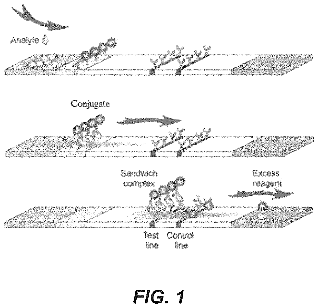
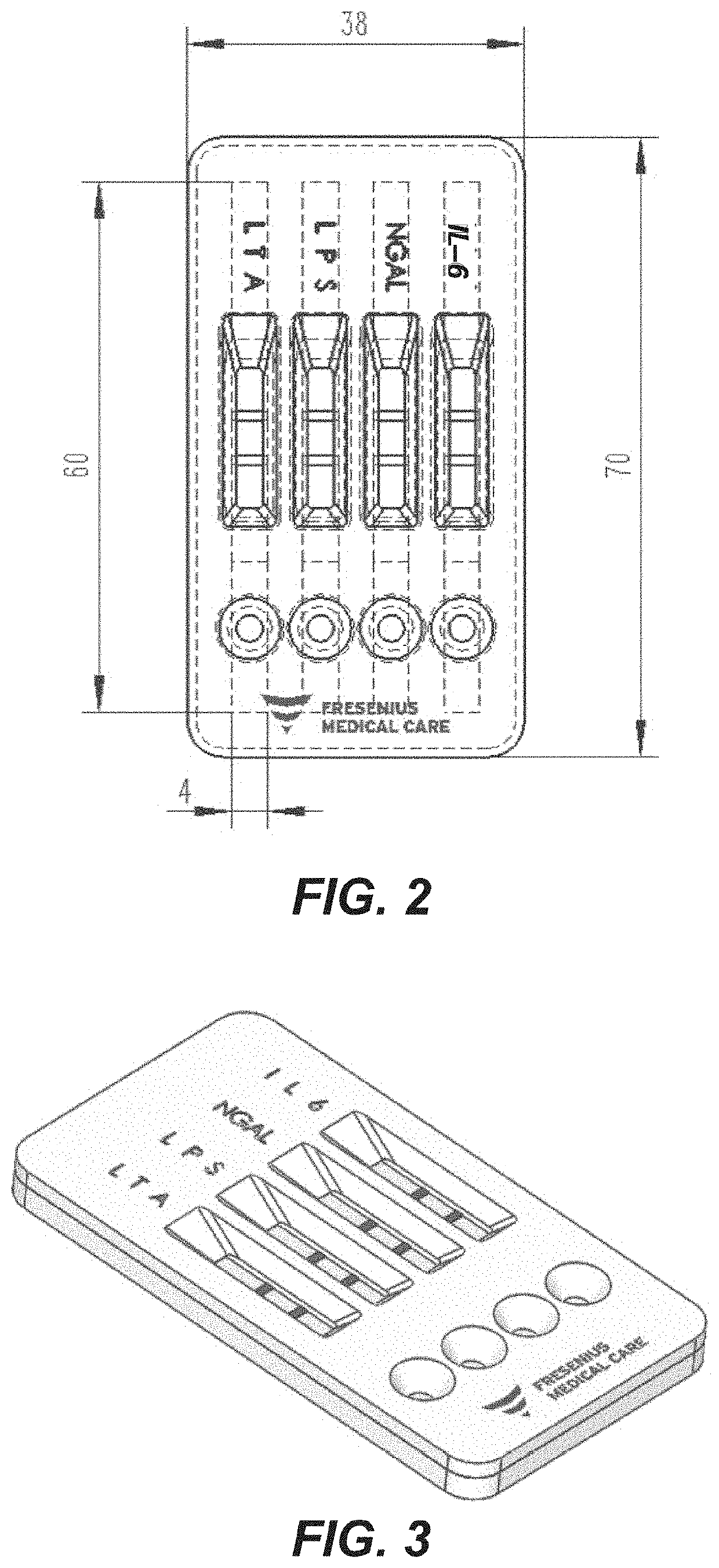
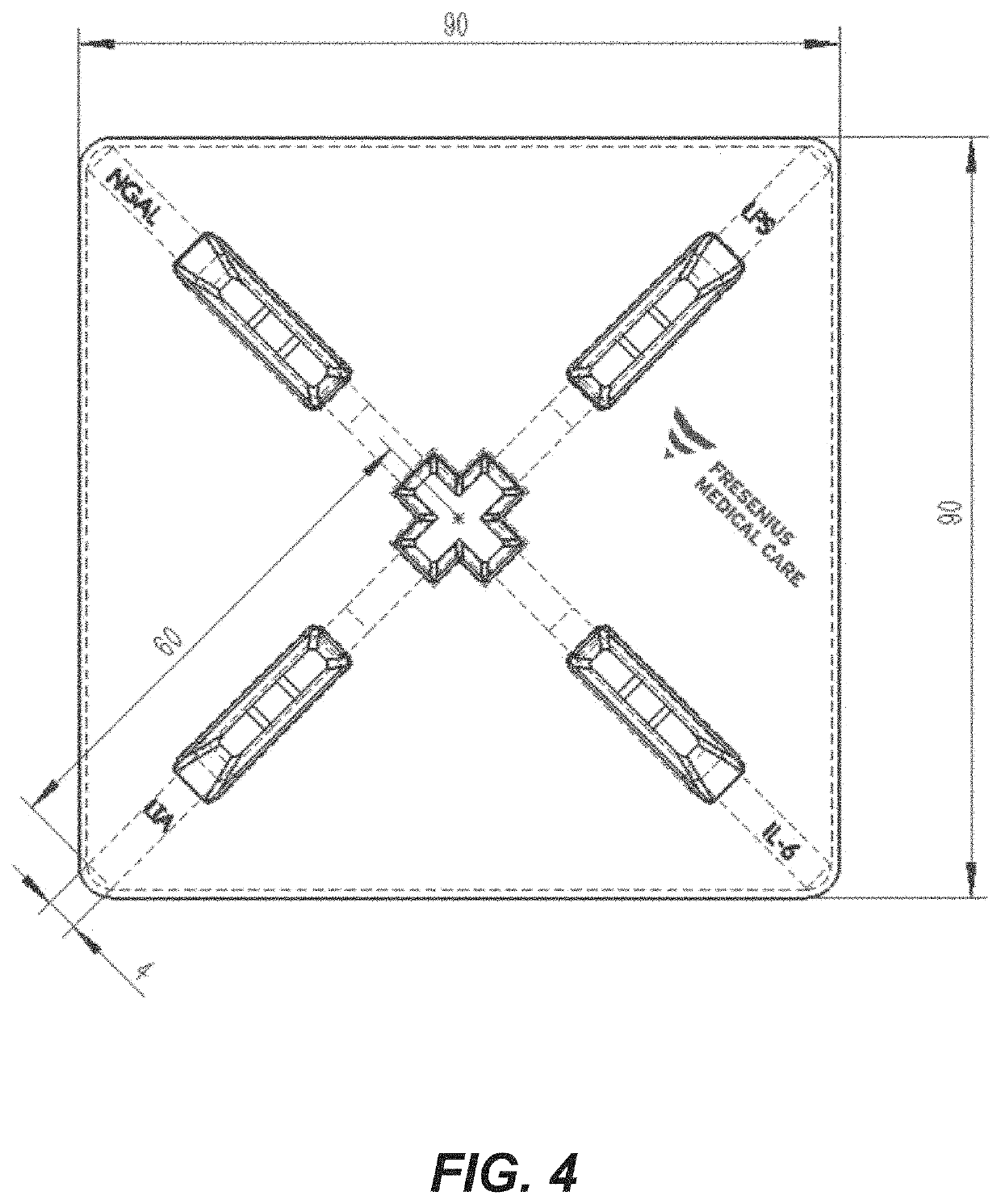
[0119]The assay of FIGS. 1-3 is made with the following antibody components incorporated into a paper-based (e.g., nitrocellulose) LFD:

BindingMolecule(s)SupplierProduct No.ReferenceAnti-IL-6R&D SystemsMAB206 (Capture)Cell. Physiol.614 McKinley Place NEBiochem., 2017;Minneapolis, MN 5541342(5): 1961-1972R&D SystemsBAF206 (Detection)Cell. Physiol.614 McKinley Place NEBiochem., 2017;Minneapolis, MN 5541342(5): 1961-1972Abcamab48478 (Pair)1 Kendall Square, Suite B2304Cambridge, MA 02139-1517Anti-NGALR&D SystemsMAB17571 (Capture)614 McKinley Place NEMinneapolis, MN 55413R&D SystemsBAF1757 (Detection)J. Neurosci. 2016;614 McKinley Place NE36(20): 5608-22Minneapolis, MN 55413Abcamab220126 (Pair)1 Kendall Square, Suite B2304Cambridge, MA 02139-1517Anti-LPSMeridian BioscienceC55308M3471 River Hills DriveCincinnati, OH 45244Anti-LTAMeridian BioscienceC65380M *3471 River Hills DriveCincinnati, OH 45244Meridian BioscienceC65813M *3471 River Hills DriveCincinnati, OH 45244Anti-Staph.Abcamab37644...

example ii

[0122]Protocol—Rapid PD-related peritonitis diagnosis at dialysis clinic or home (following the flow diagram of FIG. 6).

[0123]Background: When a PD patient displays any sign of peritonitis (e.g., cloudy PDE, abdominal pain, and / or fever), he or she is to visit a dialysis clinic or doctor as soon as possible. Upon presentation at the dialysis clinic, the nurse or other health care professional drains the PD effluent into a waste bag and collects samples for routine testing such as cell count, gram stain, and culture. Alternatively, a PD patient may collect and test samples using the disclosed assay and device before going to a dialysis clinic or doctor. What follows is rapid testing of the same PDE sample before it is sent for routine testing (e.g., cell culture, Gram stain, cell count, etc.)[0124]1. Process[0125]1.1 Equipment and reagents[0126]1. PPE (“Personal Protective Equipment”)[0127]2. Testing kit contains:[0128]A sterile 50 mL syringe with a needle pre-attached[0129]A sterile...

example iii

Enrichment by UF Membrane

[0154]25 ml of PDE is placed into a container for centrifugation, which is separated into an upper chamber and a lower chamber by an ultrafiltration (“UF”) membrane having a molecular weight cutoff of 29,000. The container is capped and placed into a centrifuge and spun (×4000 g). The PDE is filtered through the UF membrane and pushed from the upper chamber to the lower chamber, creating an enriched PDE.

[0155]A few drops of the enriched PDE are placed onto the sample port of the IL-6 / NGAL panel of the device of EXAMPLE I, and any reaction is determined.

[0156]The UF membrane is mixed with elution buffer, and a few drops are placed onto the sample port of the LPS / LTA panel of the device of EXAMPLE I, and any reaction determined.


PUM

PropertyMeasurementUnit
heightaaaaaaaaaa
heightaaaaaaaaaa
heightaaaaaaaaaa

Abstract

Described is an assay for diagnosing an infection such as peritonitis in a subject. The assay includes a binding molecule, such as an antibody that specifically binds to an inflammatory marker in a sample from the subject, and a second binding molecule that binds to a marker indicative of a pathogen in the sample. For diagnosing peritonitis in a subject, the pathogen will be at least one bacterium and / or fungus. Typically, the assay will be incorporated into a lateral flow device and may include a binding molecule that specifically binds to an antigen indicative of the presence of a specific pathogen species. The described assay(s) may further include filter(s), enriching antigen(s), and buffer(s). Also described are methods of diagnosing and treating peritonitis in a subject who is a peritoneal dialysis patient that include utilizing the herein described assay(s) or assay kit(s) to analyze the subject's peritoneal dialysis effluent.

Description

CROSS-REFERENCE TO RELATED APPLICATION[0001]The application claims the benefit under 35 U.S.C. § 119(e) of U.S. Provisional Patent Application No. 62 / 799,430 filed on Jan. 31, 2019, the contents of which are incorporated herein by this reference.TECHNICAL FIELD[0002]The application relates generally to medical devices, and more particularly to a diagnostic system, apparatus, and associated methods useful for, among other things, diagnosing and treating a peritoneal dialysis patient for peritonitis.BACKGROUND[0003]Peritonitis is a major cause of morbidity and mortality in peritoneal dialysis (“PD”) patients globally. According to the International Society for Peritoneal Dialysis (“ISPD”) guidelines, peritonitis can be diagnosed when at least two of the following are present: (1) clinical features consistent with peritonitis, e.g., abdominal pain and / or cloudy dialysis effluent (“CDE”); (2) dialysis effluent with a white blood cell count (“WBC”) greater than 100 / μL or greater than 0.1...

Claims


Application Information

Patent Timeline
Patent Type & Authority Applications(United States)
IPC IPC(8): G01N33/569
CPCG01N2469/20G01N33/56911G01N2469/10G01N33/569G01N33/6893G01N2800/26G01N2333/5421G01N2333/5412G01N2333/525G01N33/58G01N2800/7095G01N2333/195
Inventor JIANG, TAOWANG, CHANGNA
Owner FRESENIUS MEDICAL CARE HLDG INC
Why Eureka
Try Eureka