Methods for determining spatial and temporal gene expression dynamics during adult neurogenesis in single cells

a gene expression and single cell technology, applied in the field of single cell gene expression dynamics for adult neurogenesis, can solve the problems of difficult capture of rare dynamic processes, difficult isolation from dense adult tissue, and difficult study of key dynamic processes that occur in dense nervous tissues, such as adult neurogenesis

Pending Publication Date: 2020-11-05
HOWARD HUGHES MEDICAL INST +2
View PDF2 Cites 7 Cited by
  • Summary
  • Abstract
  • Description
  • Claims
  • Application Information

AI Technical Summary

Benefits of technology

[0024]In some embodiments, the method may further comp

Problems solved by technology

However, some key dynamic processes that occur in dense nervous tissues, such as adult neurogenesis, still remain challenging to study.
However, it is difficult to capture rare dynamic processes, because isolation from dense adult tissue is challenging.
First, single cell RNA-Seq requires enzymatic tissue dissociation, which damages the integrity of neurons, compromises RNA integrity, and skews data to

Method used


Image

  • Methods for determining spatial and temporal gene expression dynamics during adult neurogenesis in single cells
  • Methods for determining spatial and temporal gene expression dynamics during adult neurogenesis in single cells
  • Methods for determining spatial and temporal gene expression dynamics during adult neurogenesis in single cells

Examples


example 1

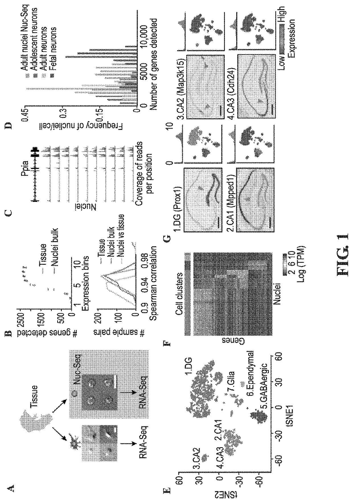
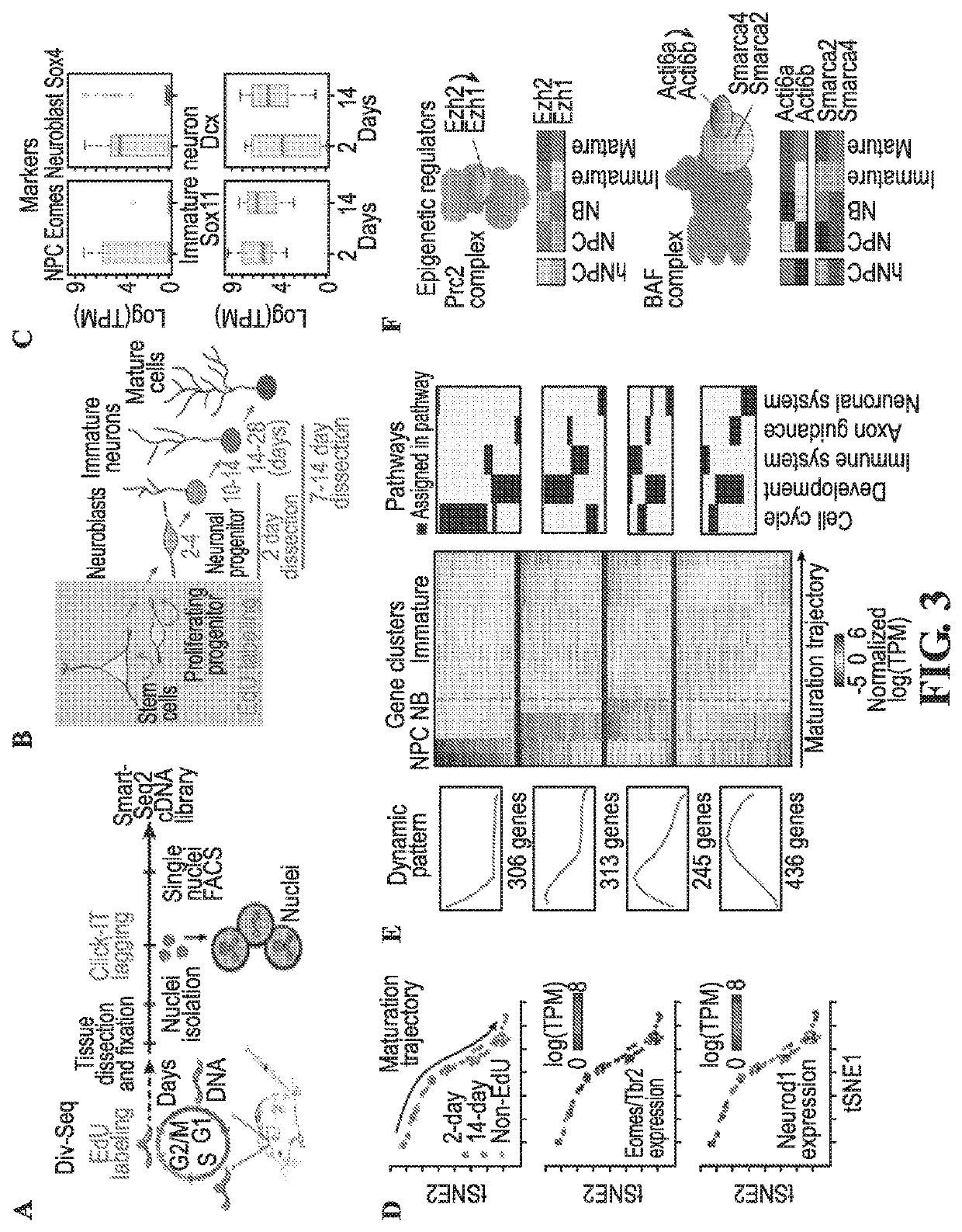
[0861]Applicants have developed methods of performing a high-throughput single-nucleus isolation and RNA-Seq method compatible with fresh, frozen, or fixed tissue (Nuc-seq). The uniform shape and fixation of the isolated nuclei (FIG. 1A) combined with nuclei labeling (FIG. 5) enables enrichment of rare cell populations by fluorescent-activated cell sorting (FACS). The method was further developed for temporal analysis of dividing cells by addition of unbiased labeling with 5-ethynyl-2′-deoxyuridine (EdU), which is incorporated into the DNA of dividing cells (8), and using Click-IT to fluorescently tag the isolated EdU labeled nuclei, which can be readily captured by FACS (FIG. 5) (Div-seq).

[0862]Earlier studies have shown the feasibility of single neuronal nuclei RNA-seq (9-11), however, it was previously unclear whether the type and complexity of nuclear mRNA could be effectively used for sensitive classification of cell types and states in the CNS on a large scale. Furthermore, gi...

example 2

[0864]The present invention also provides for novel methods to analyze the Nuc-Seq data and generate high resolution maps (see materials and methods). Nuc-Seq analysis sensitively identified both major cell types and refined sub-types. Cluster analysis of Nuc-Seq data revealed seven major clusters of cells with distinct gene expression patterns (FIG. 1E-G, FIG. 8 and FIG. 9) that clearly correspond to known cell types and major anatomical distinctions in the hippocampus. Cluster identities were consistent with Applicants' microdissection scheme, and their gene expression patterns globally agreed with Allen Brain Atlas ISH data (Allen ISH(13), FIG. 9). Iterative re-clustering of the glia nuclei (cluster 7 in FIG. 1E. FIG. 10) recovered five known glial cell sub-types (14), and averaged expressions across each sub-cluster well-correlated with published population RNA-Seq data (14) (FIG. 10).

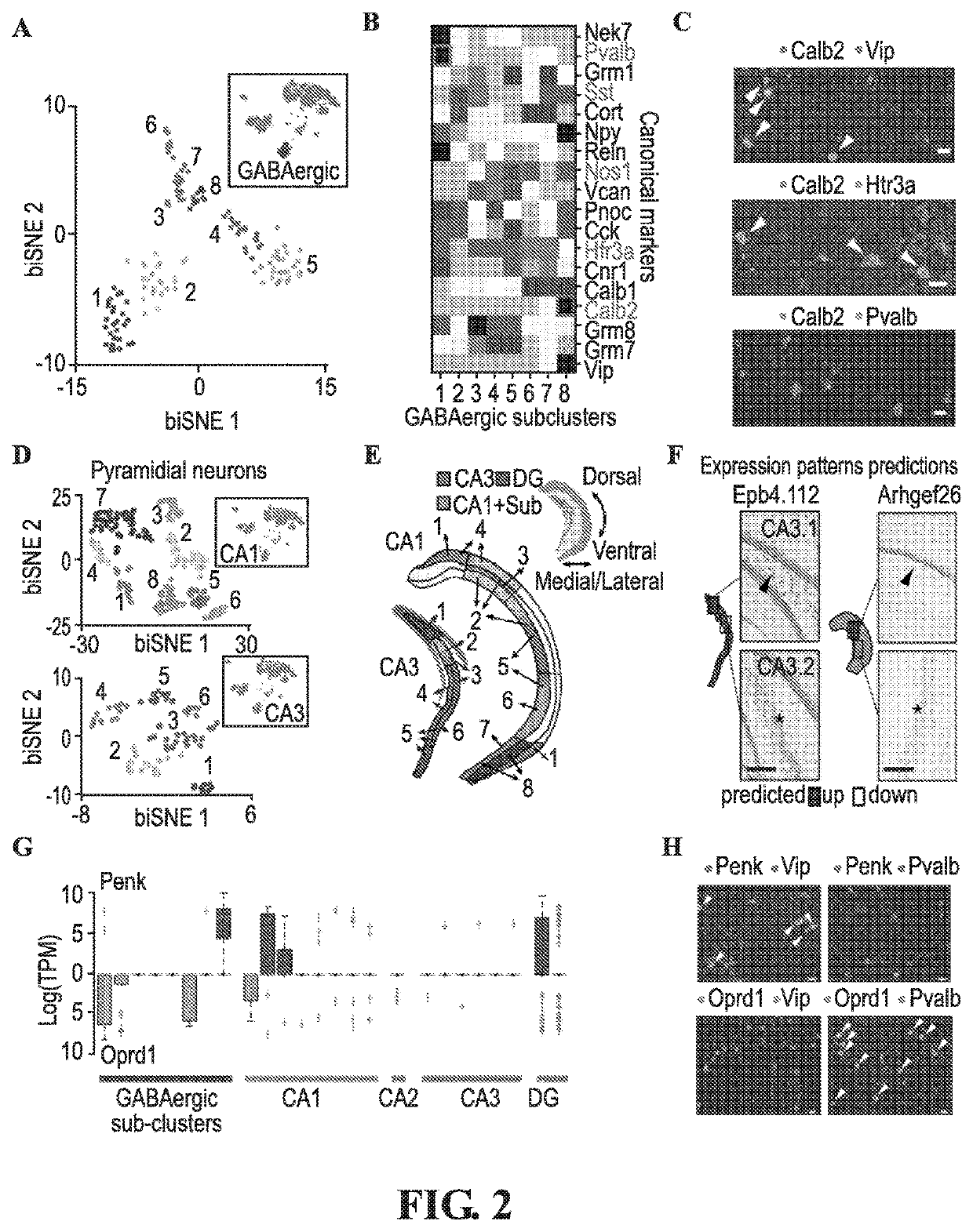
[0865]Applicants captured finer distinctions between closely related cell types using a new clu...

example 3

[0868]Applicants next combined Nuc-Seq with EdU labeling of dividing cells, in a method Applicants call Div-Seq (FIG. 3A). In contrast to commonly used genetic labeling techniques (3, 19, 20), which might be limited to specific cell types and requires cell types or developmental stage marker genes (3, 19, 20), EdU tags newly synthesized DNA in dividing cells at a given time window, allowing for unbiased isolation of nuclei of neural stem cells and their progeny with high temporal resolution. To study transcriptional dynamics during adult neurogenesis in the DG, one of the canonical neurogenic sites in the mammalian CNS (7), Applicants used Div-Seq to isolate nuclei at 2 and 14 days after cell division, representing neural precursor cells (NPC), neuroblasts, and immature neuronal stages of adult neurogenesis, respectively (7) (FIG. 3B, FIG. 22). Div-Seq enriched for a broad range of newborn cells (FIG. 20). Expression of stage-specific marker genes confirmed that Div-Seq captured cel...


PUM

PropertyMeasurementUnit
Temperatureaaaaaaaaaa
Fractionaaaaaaaaaa
Fractionaaaaaaaaaa

Abstract

Provided herein are methods of recovering single nuclei from a tissue sample comprising chopping or dounce homogenizing the tissue sample in a nuclear extraction buffer at 4° C. to produce a tissue homogenate; centrifuging the tissue homogenate to produce a nuclear pellet; resuspending the nuclear pellet in a nuclear resuspension buffer comprising bovine serum albumin, RNase inhibitor, and salts to produce a resuspension; and filtering the resuspension through a strainer, wherein the single nuclei are present in a supernatant passed through the strainer. The invention also provides a method of single cell sequencing comprising extracting nuclei from a population of cells under conditions that preserve a portion of the outer nuclear envelope and rough endoplasmic reticulum; sorting single nuclei into separate reaction vessels; extracting RNA from the single nuclei; generating a cDNA library, whereby gene expression data from single cells are obtained. The tissue sample may be fresh or frozen.

Description

CROSS-REFERENCE TO RELATED APPLICATIONS[0001]This application claims the benefit of U.S. Provisional Application No. 62 / 841,408, filed May 1, 2019 and U.S. Provisional Application No. 62 / 865,829, filed Jun. 24, 2019. The entire contents of the above-identified applications are hereby fully incorporated herein by reference.FIELD OF THE INVENTION[0002]The present invention generally relates to methods of determining cell type, subtype, cell state, spatial location and developmental stages of single cells obtained from a sample, preferably a tissue sample. The present invention also relates to a combination of molecular barcoding and emulsion-based microfluidics to isolate, lyse, barcode, and prepare nucleic acids from individual nuclei in a high-throughput manner.BACKGROUND OF THE INVENTION[0003]Single cell RNA-Seq has greatly extended the understanding of heterogeneous tissues, including the Central Nervous System, and is reshaping the concept of cell type and state. However, some ke...

Claims


Application Information

Patent Timeline
IPC IPC(8): C12Q1/6869G01N1/34C40B50/18C12Q1/6844C12Q1/6886
CPCG01N1/34C12Q1/6869C40B50/18C12Q1/6844C12Q1/6886C12Q2563/143C12N15/1017C12N15/1096
Inventor ROZENBLATT-ROSEN, ORIT
Owner HOWARD HUGHES MEDICAL INST
Why Eureka
Try Eureka