Devices, systems, and methods for treating urinary and fecal incontinence

a technology for fecal incontinence and urinary tract, applied in the field of devices, systems and methods for treating urinary tract and fecal incontinence, can solve problems such as difficult implementation of therapies, and achieve the effects of improving or stabilizing the condition of the subject, and reducing the likelihood of urinary

Pending Publication Date: 2021-03-11
RENOVIA INC
View PDF0 Cites 2 Cited by
  • Summary
  • Abstract
  • Description
  • Claims
  • Application Information

AI Technical Summary

Benefits of technology

[0035]As used herein, the term “treating” refers to providing electrical stimulation to the sacral nerve of a subject in need thereof for therapeutic purposes (e.g., to treat or reduce the likelihood of urinary or fecal incontinence, or urge associated therewith), in particular in conjunction with the use of a device (e.g., an intravaginal device), system, or method described herein. “Treating” may also refer to the use of an intravaginal device to assess whether a subject has, or is in need of, electrical stimulation to the sacral nerve of a subject (e.g., to treat or reduce the likelihood of urinary or fecal incontinence, or urge associated therewith). To “treat disease” or use for “therapeutic treatment” includes administering treatment to a subject already suffering from a disease to improve or stabilize the subject's condition. To “prevent” or “reduce likelihood of developing” disease refers to prophylactic treatment of a subject who is not yet ill or symptomatic, but who is susceptible to, or otherwise at risk of, a particular

Problems solved by technology

These therapies can be challenging to implement due to difficulties associated with positioning of the neur

Method used


Image

  • Devices, systems, and methods for treating urinary and fecal incontinence
  • Devices, systems, and methods for treating urinary and fecal incontinence
  • Devices, systems, and methods for treating urinary and fecal incontinence

Examples


example 1

Data Output in Live Mode and Use with a Smartphone Application

[0204]An intravaginal device described herein is connected to a transmitter box that wirelessly (via Bluetooth) sends the positional data gathered from the device sensors to a smartphone or computer for display on a graphical user interface, e.g., through a smartphone application. The shape of the vagina (data from the MEMS sensors in the device) reflects the position of the subject's pelvic floor in her body. The data is captured as a score based on the angles of the sensors. The score is a measure of the strength and / or position of the subject's pelvic floor muscles. The data created by the device is transmitted to a centralized database creating a personal health record for the subject, providing care and measurable results.

[0205]These data can provide predictive information that notifies a subject, or their health care professional, about the potential need for various treatment options or the status of treatment (e.g...

example 2

Metrics of Sensor Readout as Patient Improves Pelvic Muscle Strength

[0207]A patient with symptoms of urinary incontinence uses an intravaginal device for ˜2.5 minutes twice daily. After performing pelvic floor exercises, the application computes an average weekly score, which increases from a baseline (screen) of 9 to a range of 44-52, reflecting the vaginal angle changes during the lifting of the pelvic floor during exercises. An increase in score correlates with an increase in pelvic muscle strength. The application can also track endurance, which calculates the duration of time holding a lift during an exercise. After a 3 week exercise regimen, the incontinence issue is assessed and, in many cases, would be resolved. If not resolved, the intravaginal device could be used to assess the need for, and possibly to determine the timing of electrical stimulation during, SNM therapy.

example 3

ng Optimal Sensor Positioning

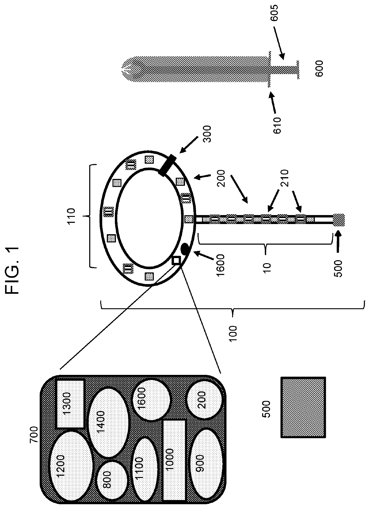
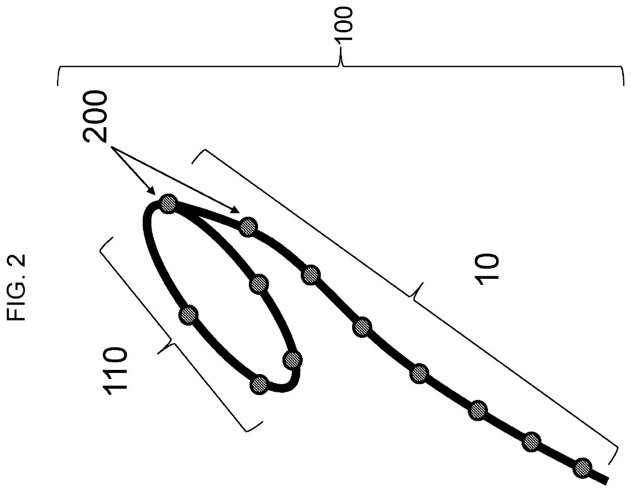
[0208]An intravaginal device (see FIG. 2) was used to characterize specific sensor placement within the intravaginal device (e.g., within main body 110 or tether 10) in ten subjects. The sensors in the main body were within the anterior fornix, lateral fornices and posterior fornix. The sensors in the tether 10 were within the posterior fornix (shared by the main body) and along the vaginal canal caudal to the fornices. The main body contained 5 sensors, while the tether contained 8 sensors (including the one shared with the main body). The subjects had a range of vaginal lengths; these subjects were studied to determine if a subset of the original 12 sensors consistently correlated with pelvic floor movement (e.g., PFL) when the subjects performed a variety of maneuvers and demonstrated superior signal-to-noise characteristics. The sensors in the fornix exhibited lower signal and provided less robust visualization of pelvic floor movement. Of the sensor...


PUM


Abstract

Featured are intravaginal devices and electrical stimulation devices, systems, thereof, and methods of using the devices and systems thereof to observe pelvic floor movements in order to diagnose, treat, or prevent urinary and fecal incontinence disorders (e.g., urge incontinence) and their accompanying symptoms or to diagnose and/or improve the efficacy of neuromodulation therapy.

Description

BACKGROUND[0001]Urinary and fecal incontinence are a group of conditions that occur predominantly in women and that are associated with weakened (e.g., hypotonic) or tense (e.g., hypertonic) pelvic floor (PF) muscles. Many common factors contribute to the weakening or tightening of the pelvic floor muscles in women, such as, for example, pregnancy, vaginal childbirth, pelvic surgery, aging, genetic predisposition, neurological disease, and weight gain. In the United States, pelvic floor disorders (PFDs) occur in 24% of women, with 16% of women experiencing urinary incontinence (UI), 3% experiencing pelvic organ prolapse (POP), and 9% experiencing anal or fecal incontinence (FI). Current methods of treatment include electrical stimulation therapy and neuromodulation to nerves that innervate the bladder and muscles that manipulate the pelvic floor. These therapies can be challenging to implement due to difficulties associated with positioning of the neuromodulation devices, as well as...

Claims


Application Information

Patent Timeline
IPC IPC(8): A61N1/36A61N1/372
CPCA61N1/36135A61N1/36007A61B5/202A61N1/37247A61N1/3606A61N1/0558A61N1/37217A61N1/36139A61B5/0002A61B5/1104A61B5/1122A61B5/11A61B5/20A61B5/227A61B5/4318A61B5/746A61B5/6847A61B2562/0219A61B2562/028
Inventor BEER, MARC D.BOHORQUEZ, JOSEPULLIAM, SAMANTHA J.MCKINNEY, JESSICA L.
Owner RENOVIA INC
Why Eureka
Try Eureka