Method of in situ gene sequencing

a gene and in situ technology, applied in the field of in situ gene sequencing, can solve the problems of low efficiency, inability to apply current sequencing methods to 3d volumes of intact tissue, and the potential value of such in-tissue sequencing could be enormous

Pending Publication Date: 2021-06-03
THE BOARD OF TRUSTEES OF THE LELAND STANFORD JUNIOR UNIV
View PDF0 Cites 22 Cited by
  • Summary
  • Abstract
  • Description
  • Claims
  • Application Information

AI Technical Summary

Benefits of technology

[0006]Also provided is a device used according to the methods described herein. In some embodiments, provided is a fluidics system for automation of the methods described herein, allowing for continual operation. In some embodiments, the system includes a fluidics device, and a processor configured to perform the methods described herein.

Problems solved by technology

Current in situ sequencing approaches face the challenge of implementing enzymatic reactions in the dense, complex tissue environment and currently suffer from low efficiency, but the potential value of such in-tissue sequencing could be enormous; in comparison to hybridization-based multiplexing / readout which utilizes multiple polynucleotide probes to encode gene identity, sequencing operates with single-nucleotide resolution, and thus inherently provides vastly greater information.
However, current sequencing methods are not yet applicable to 3D volumes of intact tissue, due to fundamental limitations in requisite sensitivity, fidelity, and scalability for throughput in tissues such as the mammalian brain.
Retrieving high-content gene-expression information while retaining 3D positional anatomy at cellular resolution has been difficult, limiting integrative understanding of brain structure and function.

Method used


Image

  • Method of in situ gene sequencing
  • Method of in situ gene sequencing
  • Method of in situ gene sequencing

Examples


example 1

STARmap

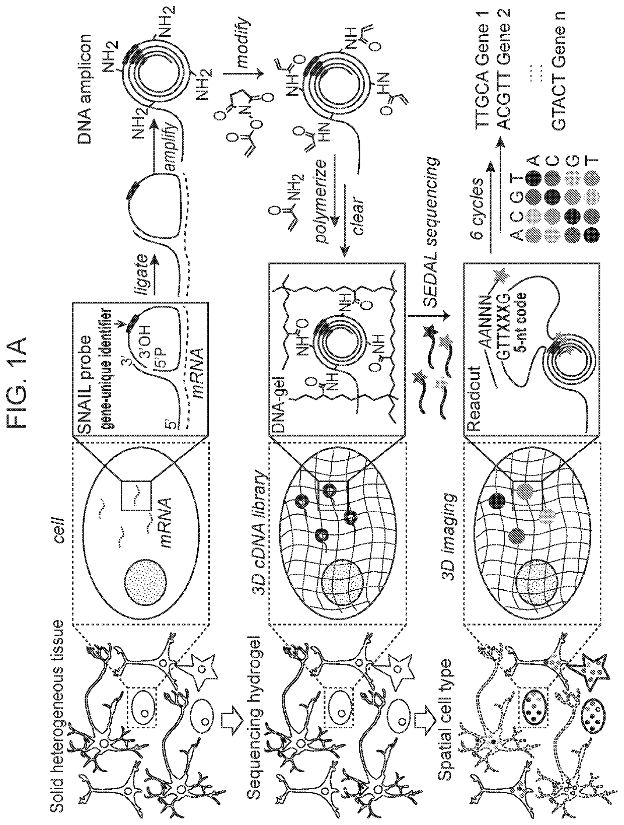
[0299]The methods disclosed herein including the in situ sequencing technology enabled highly-multiplexed gene detection (up to 1000 genes) in intact biological tissue. The methods began with raw biological tissue (fresh or preserved, as little as one-cell or as large as millimeter(s) in size) and resulted in output image-based gene quantification at subcellular and cellular resolution and with high efficiency, low error rate, and fast processing time.

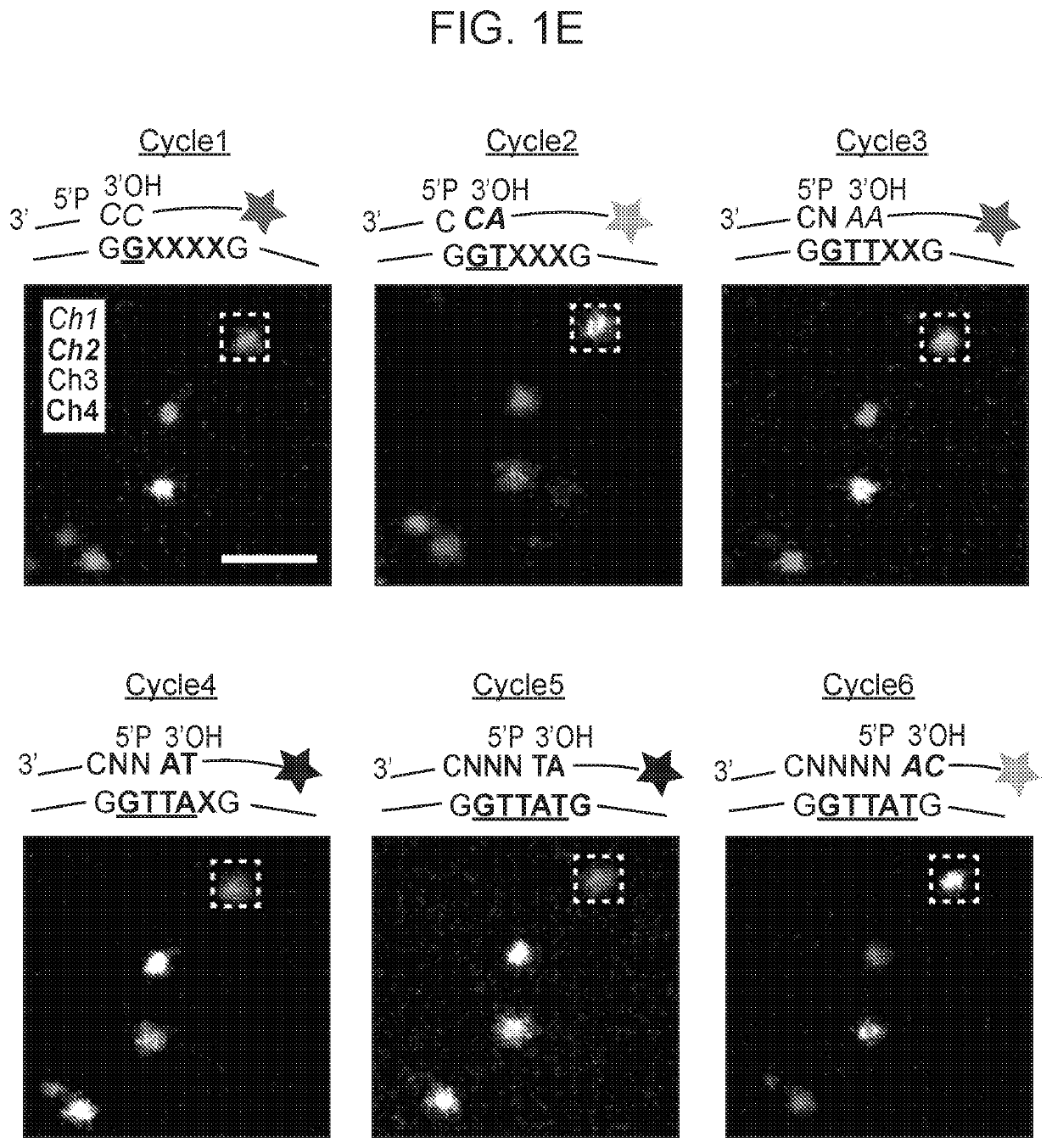
[0300]A schematic diagram of STARmap as described herein was depicted in FIG. 1A. After the brain tissue was prepared (see Methods for mouse brain protocols), the custom SNAIL probes (FIG. 2A) that encountered with and hybridized to intracellular mRNAs (dashed lines) within the intact tissue were enzymatically replicated as cDNA amplicons. The amplicons were constructed in situ with an acrylic acid N-hydroxysuccinimide moiety modification and then copolymerized with acrylamide to embed within a hydrogel network (wavy lines), fol...

example 2

SNAIL

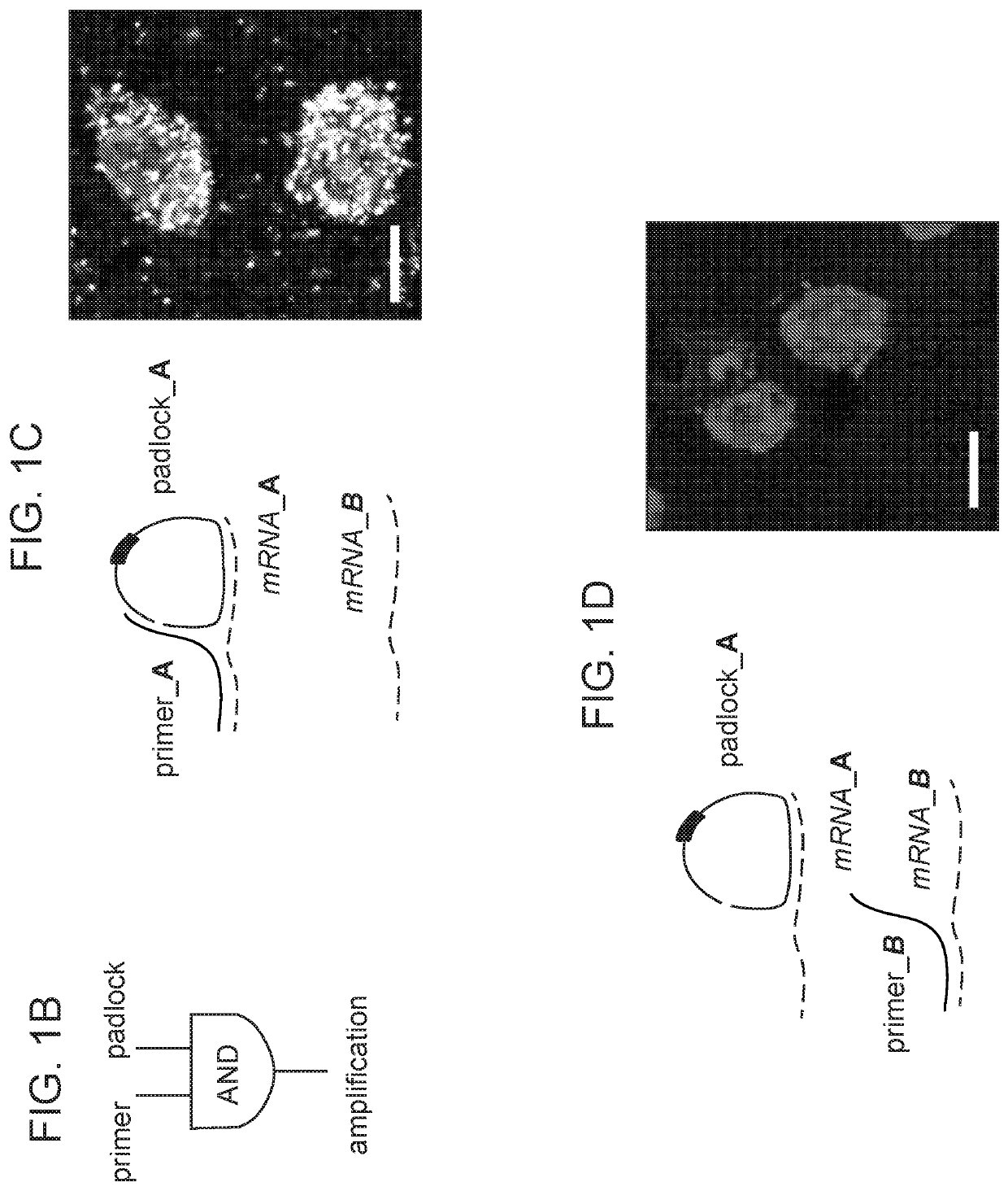
[0302]Reverse transcription may be the major efficiency-limiting step for in situ sequencing, and SNAIL bypassed this step with a pair of primer and padlock probes (FIG. 2A) designed such that only when both probes hybridize to the same RNA molecule. Design of SNAIL probes (one component of STARmap) included the following: each primer or padlock probe (19-25 nucleotides; nt, blue) had a designed Tm (melting temperature of nucleic acids) of 60° C. or higher to hybridize with target RNA, while the complementary sequence between primer and padlock was only 6 nt on each arm with Tm below room temperature, so that primer-padlock DNA-DNA hybridization was negligible during DNA-RNA hybridization at 40° C., but allowed DNA ligation by T4 DNA ligase in the following step.

[0303]The padlock probe was circularized and rolling-circle amplified to generate a cDNA nanoball (amplicon) containing multiple copies of the cDNA (FIG. 1A-1D). This mechanism ensured target-specific signal amplificati...

example 3

cDNA Amplicon Embedding in the Tissue-Hydrogel Setting

[0304]To enable cDNA amplicon embedding in the tissue-hydrogel setting, amine-modified nucleotides were spiked into the rolling circle amplification reaction, functionalized with an acrylamide moiety using acrylic acid N-hydroxysuccinimide esters, and copolymerized with acrylamide monomers to form a hydrogel (FIG. 1A, FIG. 3A). A schematic diagram of hydrogel-tissue chemistry for STARmap in thin tissue slices was depicted in FIG. 3A. DNA amplicons were synthesized in the presence of minor levels of 5-(3-aminoallyl)-dUTP, which replaced T at a low rate and allowed further functionalization with the polymerizable acrylamide moiety using acrylic acid N-hydroxysuccinimide ester (AA-NHS), so that the DNA amplicons were covalently anchored within the polyacrylamide network at multiple sites (FIG. 3A, right). The resulting tissue-hydrogel was then subjected to protein digestion and lipid removal to enhance transparency (FIG. 3B-3E). Flu...


PUM

PropertyMeasurementUnit
thicknessaaaaaaaaaa
thicknessaaaaaaaaaa
temperatureaaaaaaaaaa

Abstract

Provided herein are devices, methods, and systems for in situ gene sequencing of a target nucleic acid in a cell in an intact tissue. Methods of screening a candidate agent to determine whether the candidate agent modulates gene expression of a nucleic acid in a cell in an intact tissue are also provided herein.

Description

CROSS-REFERENCE[0001]This application claims the benefit of U.S. Provisional Patent Application Ser. No. 62 / 655,052 filed on Apr. 9, 2018, U.S. Provisional Patent Application Ser. No. 62 / 687,490 filed on Jun. 20, 2018, and U.S. Provisional Patent Application Ser. No. 62 / 808,159 filed on Feb. 20, 2019, which applications are incorporated herein by reference in their entirety.INTRODUCTION[0002]In biological tissues, the diversity of function arises from the diversity of form, in part via the complexity of cell-specific gene expression, which defines the unique three-dimensional molecular anatomy and cellular properties of each tissue. In situ transcriptomic tools for the spatial mapping of gene expression with subcellular resolution have emerged that may be applicable to probing these tissue structure-function relationships, including both multiplexed in situ RNA hybridization and in situ RNA sequencing. Current in situ sequencing approaches face the challenge of implementing enzymati...

Claims


Application Information

Patent Timeline
Patent Type & Authority Applications(United States)
IPC IPC(8): C12Q1/6874C12Q1/6876
CPCC12Q1/6874C12Q2600/158C12Q2600/136C12Q1/6876C12Q1/6869C12Q1/682C12Q1/6841C12Q2521/501C12Q2525/307C12Q2531/125C12Q2563/159C12Q2563/185C12Q2535/122C12Q2525/10C12Q2563/107
Inventor WANG, XIAOALLEN, WILLIAM E.DEISSEROTH, KARL A.
Owner THE BOARD OF TRUSTEES OF THE LELAND STANFORD JUNIOR UNIV
Why Eureka
Try Eureka