System and method for determining cardiac output

a cardiac output and system technology, applied in the field of system and method for determining cardiac output, can solve the problems of severe risks involved in prolonged intravascular blood flow measurement in the aorta, and inability to accurately determine co in extracorporeal ultrasound. to achieve the effect of reducing the problem

Inactive Publication Date: 2021-09-30
KONINKLJIJKE PHILIPS NV
View PDF0 Cites 2 Cited by
  • Summary
  • Abstract
  • Description
  • Claims
  • Application Information

AI Technical Summary

Benefits of technology

[0007]It is an object of the present invention to provide an improved system, method and software product for determining a representation of cardiac output in a patient that mitigates the problems of the known systems and approaches.

Problems solved by technology

Extracorporeal ultrasound cannot determine CO accurately enough.
Intravascular measurements are called for, but there are severe risks involved with prolonged intravascular blood flow measurements in the aorta.
However, its use has been associated with various complications during and after the measurement procedure.
However, these methods all have their drawbacks, either in practicality (limited frequency of measuring, hands-on operation, time needed for a measurement and / or patient discomfort) or accuracy in a large range of hemodynamic conditions.
However, hemodynamic instability (e.g. during surgery, or in ICU patients) may cause these ratios to change rapidly over time.
This means that measuring the flow in the Superior Vena Cava alone cannot serve as a surrogate for CO.

Method used


Image

  • System and method for determining cardiac output
  • System and method for determining cardiac output

Examples


Embodiment Construction

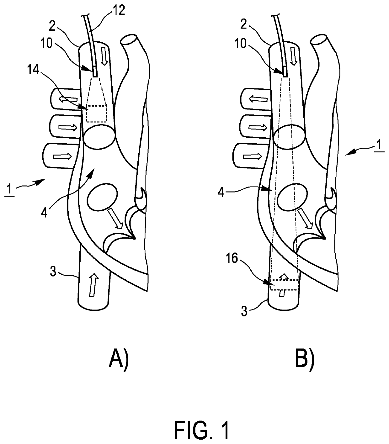
[0047]FIG. 1 shows a schematic illustration of a setup in accordance with an embodiment of the invention. FIG. 1 schematically illustrates a part of a heart 1, including the Superior Vena Cava 2 and the Inferior Vena Cava 3, both leading to the Right Atrium 4.

[0048]The illustrated setup according to the present invention provides a method and system to monitor cardiac output using a single device inserted into a Central Venous Catheter (CVC) or Peripheral Inserted Central Catheters (PICC) line. In the present preferred embodiment, the device contains an elongated body that fits the lumen of a Central Venous Catheter or Peripheral Inserted Central Catheters line and has an ultrasound transducer at the tip. By operating the device such that the velocity in both the Superior Vena Cava and Inferior Vena Cava can be sampled continuously the CO is monitored.

[0049]In an exemplary embodiment, the main elements include an elongated body 10 with an ultrasound transducer at the tip that fits t...


PUM


Abstract

The invention relates to determining a representation of cardiac output in a patient. Provided are a method and a system for monitoring a cardiac output using a single device inserted into a Central Venous Catheter (CVC) or Peripheral Inserted Central Catheters (PICC) line. In an embodiment, the device contains an elongated body that fits the lumen of a Central Venous Cava or Peripheral Inserted Central Catheters line and has an ultrasound transducer at the tip. By operating the device such that the velocity in both the Superior Vena Cava and Inferior Vena Cava can be sampled continuously the CO is monitored.

Description

FIELD OF THE INVENTION[0001]The invention relates to a system, method and software product for determining a representation of cardiac output in a patient.BACKGROUND OF THE INVENTION[0002]Cardiac output (CO) measurement is an important tool in monitoring the hemodynamic status of patients during surgery and during their stay on the intensive care unit (ICU). Cardiac output is the volume of blood pumped by the heart in a given time. As the blood circulation is a closed system, this can mean either the volume of blood pumped into the aorta by the left ventricle, but equally the volume of blood pumped into the pulmonary artery by the right ventricle, or the volume of blood flowing into the right atrium (venous return) or left atrium.[0003]Extracorporeal ultrasound cannot determine CO accurately enough. Intravascular measurements are called for, but there are severe risks involved with prolonged intravascular blood flow measurements in the aorta. The Pulmonary Artery Catheter (PAC) or S...

Claims


Application Information

Patent Timeline
Patent Type & Authority Applications(United States)
IPC IPC(8): A61B8/06A61B8/00A61B8/12A61B8/08
CPCA61B8/065A61B8/4483A61B2562/0247A61B8/12A61B8/5215A61B8/54A61B8/04A61B8/0883A61B8/445A61B8/4477A61B8/488G16H50/20
Inventor VAN DER HORST, ARJENSIO, CHARLES FREDERIKKUENEN, MAARTEN PETRUS JOSEPH
Owner KONINKLJIJKE PHILIPS NV
Why Eureka
Try Eureka