
Helical Tissue Anchor Device and Delivery System

Active Publication Date: 2021-12-30
BINMOELLER KENNETH F +3
View PDF0 Cites 1 Cited by
  • Summary
  • Abstract
  • Description
  • Claims
  • Application Information

AI Technical Summary

Benefits of technology

The patent text describes a delivery system for closing tissue openings that has three or four individual lumens. The system includes a sheath with separate lumens for the helical devices, the suture or strap assembly, and a locking mechanism. The use of independent lumens in the sheath reduces the potential for twisting around the delivery elements and ensures better placement of the suture or strap. The system allows for easy handling and placement of the suture or strap, with a minimal invasive procedure.

Problems solved by technology

Clipping devices are limited by a fixed distance and relationship between the prongs.
The fixed distance between the prongs limits the operator's ability to close defects that exceed this distance, which limits applicability to small defects.
The fixed relationship between the prongs limits the operator's ability to position the clip appropriately in relation to the area in need of treatment.
For instance, the clips may not be able to close a defect that is approached tangentially at a curve or angle.
Further, because the proximal ends of the legs are joined, the operator may not be able to adjust the positioning of one clip prong without affecting the positioning of the second.
Positioning also may be limited because the clip may not be properly oriented when it is deployed or the clip may slip out of alignment during application.
If unequal pressure is applied to the legs during anchoring, closure may be sub-therapeutic and scissoring of the legs may occur that can result in tissue damage.
Presently-used clips are only capable of capturing the mucosa and do not penetrate into the deeper wall layers (submucosa and muscular propria layers).
This device is capable of full-thickness (entire bowel wall) tissue capture and the closure of larger defects, however, the device cannot be delivered through the working (operative) channel of the endoscope and must be pre-mounted on the end of the endoscope.
It also involves multiple time consuming, technically demanding maneuvers and manipulations.
The size of the defect that can be closed is small, limited by the diameter of the cap attachment.

Method used


Image

  • Helical Tissue Anchor Device and Delivery System
  • Helical Tissue Anchor Device and Delivery System
  • Helical Tissue Anchor Device and Delivery System

Examples


Embodiment Construction

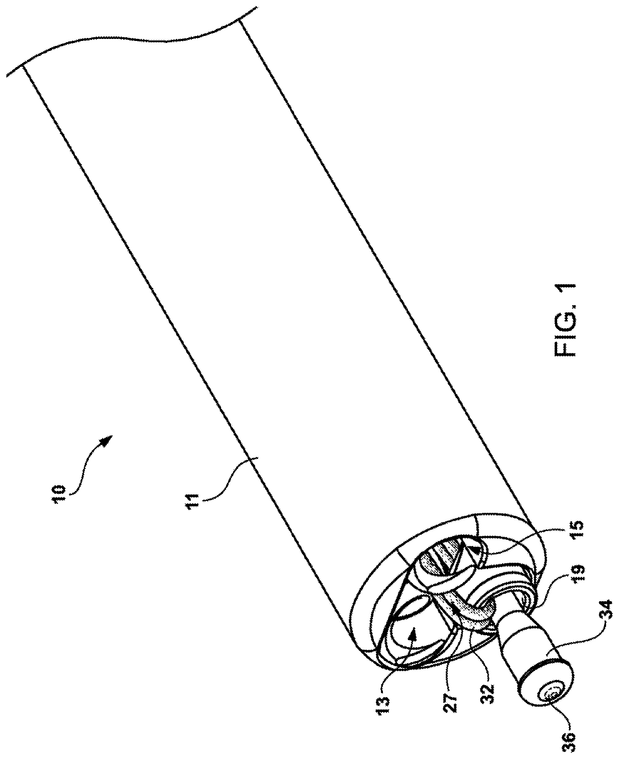
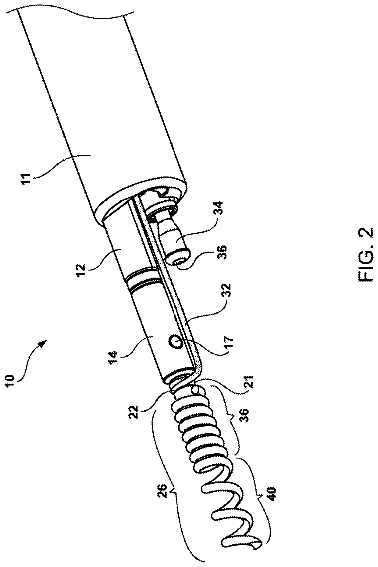
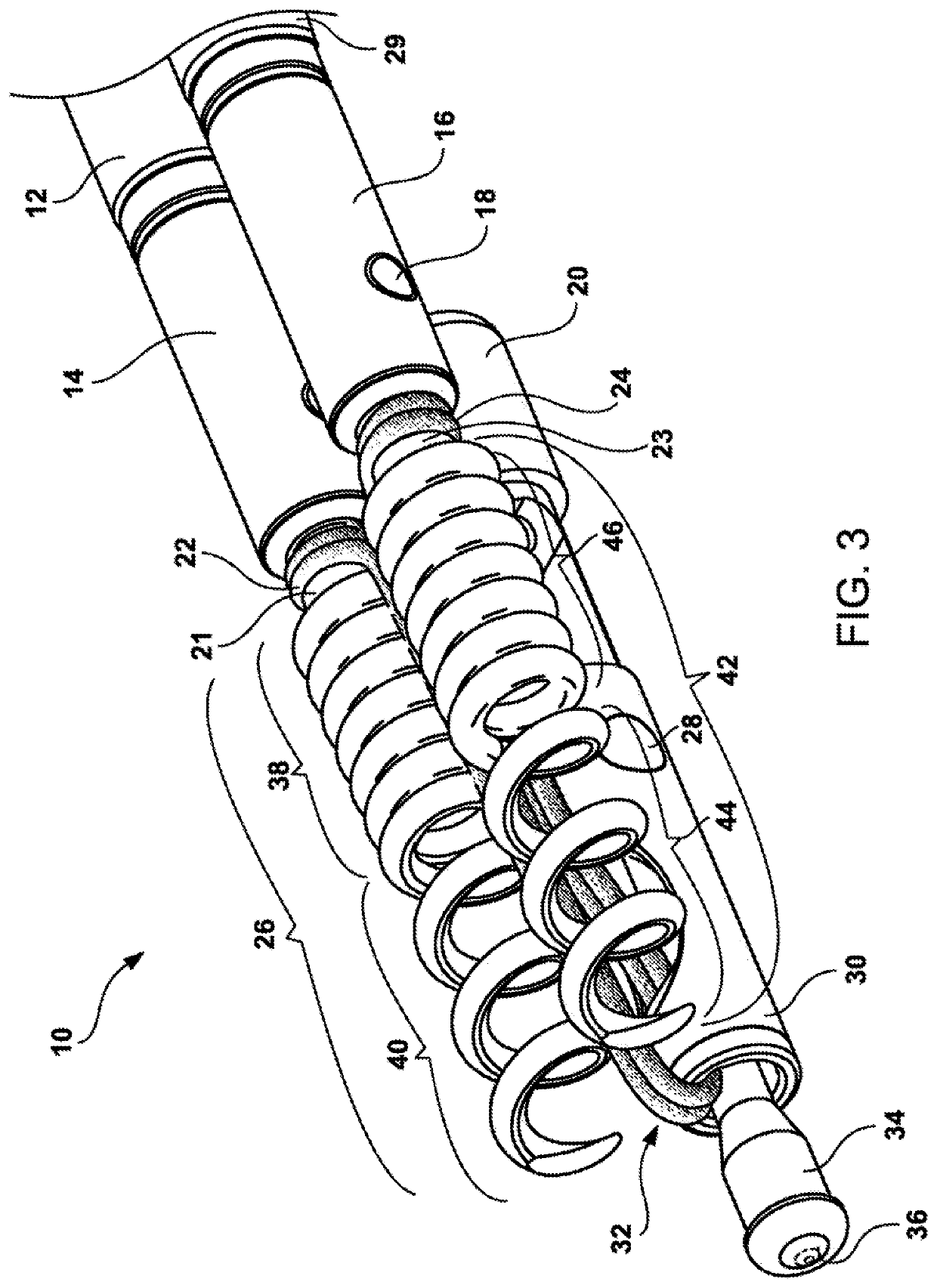
[0033]Referring now to the drawings and particularly to FIG. 1 which is a perspective view of the present invention 10 which includes a distal end of the catheter system showing the multi-lumen catheter 11, a first inner tubular lumen 13 and second inner tubular lumen 15 which correspond to the first and second helical tissue anchors and associated shaft members and a third and fourth inner tubular lumen 19 and 27 with the suture strap and cinching mechanism engaged to the proximal ends of elongated shaft that travels throughout the length of the multi-lumen catheter and terminates in a connection to the handle mechanism 70.

[0034]The multi-lumen catheter can be fabricated from a number of polymeric materials, such as polytetrafluoroethylene (PTFE), FEP, ETFE, polyvinyl chloride (PVC), polyethylene, polypropylene, PEEK, polybutylene, acryaontirile-butadiene-styrene (ABS), rubber modified styrene, polyacetal, polyethylene, graphite or nylon, or a combination of metal coil or braid enc...


PUM


Abstract

A delivery system for delivering a plurality of helical tissue anchors to repair a wall defect. Coaxially contained within the outer sheath are inner tubular members. The first inner tubular member designed to deploy a first helical tissue anchor, a second inner tubular member designed to deploy a second helical tissue anchor, and a centered inner tubular member contains a cinching mechanism. The two helical tissue anchors are connected to a suture or strap that pull the two helical tissue anchors together to close a tissue defect. A cinching mechanism holds the anchors and tissue defect together and cut the suture or strap.

Description

RELATED APPLICATIONS[0001]This patent application is a non-provisional of Provisional Application No. 62 / 367,592 filed on Jul. 27, 2016. This Provisional Application is incorporated by reference in its entirety.FIELD OF THE INVENTION[0002]This present invention relates to an apparatus and method for approximating or closing tissue defects with anchors that can be controlled independently of each other. More specifically, the present invention relates to a delivery system that deploys two or more anchoring elements through an endoscope. The device and methods also may be used for plicating or otherwise reconfiguring tissue and for fixation of tissue or other material to tissue.BACKGROUND OF THE INVENTION[0003]During a gastrointestinal endoscopic procedure, the operator often desires to repair or reconstruct a tear or defect or otherwise approximate or fixate tissue or other material by suturing.[0004]Metallic clipping devices were first introduced for the primary purpose of achieving...

Claims


Application Information

Patent Timeline
IPC IPC(8): A61B17/064A61B17/04A61B1/018A61B17/072
CPCA61B17/064A61B17/0467A61B17/0487A61B17/0401A61B2017/0034A61B17/07207A61B2017/0441A61B2017/0464A61B2017/0488A61B1/018A61B2017/0409A61B2017/00243A61B2017/0649
Inventor BINMOELLER, KENNETH F.YUREK, MATTHEW THOMASHARTSFIELD, MICHAEL P.GREELIS, JOHN
Owner BINMOELLER KENNETH F
Features
  • Generate Ideas
  • Intellectual Property
  • Life Sciences
  • Materials
  • Tech Scout
  • Unparalleled Data Quality
  • Higher Quality Content
  • 60% Fewer Hallucinations
Learn More