

Implanting grafts to valve leaflets for cardiac procedures
a technology for implanting grafts and valve leaflets, which is applied in the field of implanting grafts to valve leaflets for cardiac procedures, can solve the problems of prolapse and regurgitation of mitral valves, affect the proper functioning of one or more of the valves of the heart, and affect the function of the valves in the heart, so as to reduce the regurgitation of the mitral valv
- Summary
- Abstract
- Description
- Claims
- Application Information
AI Technical Summary
Benefits of technology
Problems solved by technology
Method used
Image

Examples
Embodiment Construction
[0043]The headings provided herein, if any, are for convenience only and do not necessarily affect the scope or meaning of the disclosed subject matter.
Overview
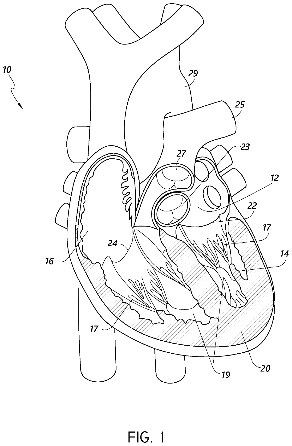
[0044]During conventional, on-pump cardiac operations, the heart is stopped, and the doctor has vision of and direct access to the internal structures of the heart. In conventional operations, doctors perform a wide range of surgical procedures on a defective valve. In degenerative mitral valve repair procedures, techniques include, for example and without limitation, various forms of resectional repair, chordal implantation, and edge-to-edge repairs. Clefts or perforations in a leaflet can be closed and occasionally the commissures of the valve sutured to minimize or eliminate MR. Although devices have been developed to replicate conventional mitral valve procedures on a beating heart (see, e.g., International Patent Application No. PCT / US2012 / 043761, published as WO 2013 / 003228 A1, and referred to herein as “the '761 PCT Ap...
PUM
 Abstract
Description
Claims
Application Information