Surgical navigation system, computer for performing surgical navigation method, and storage medium

a surgical navigation system and navigation system technology, applied in the field of surgical navigation system, computer for performing surgical navigation method, storage medium, surgical navigation system, etc., to achieve the effects of reducing the risk of operation, improving the accuracy of operation, and simplifying the operation process

Pending Publication Date: 2022-11-10
SHENZHEN XINJUNTE SMART MEDICAL EQUIP CO LTD
View PDF0 Cites 0 Cited by
  • Summary
  • Abstract
  • Description
  • Claims
  • Application Information

AI Technical Summary

Benefits of technology

[0032]Compared with prior arts, the present invention has the following advantages:
[0033]the technical solution of the present invention is based on the digital navigation technology of visual positioning, the binocular camera cooperates with the tracer on the bone and the spatial positioning marker component on the surgical instrument to acquire real-time information of the tracer and the spatial positioning marker component; the spatial position information of the bone to be operated on and the surgical instrument is calculated using the binocular vision positioning algorithm, and is fused with the three-dimensional models in real time to obtain the dynamic images of the real-time position relationship between the bone and the surgical instrument to realize the surgical navigation, which can simplify the operation process, improve the accuracy of the operation, reduce the risk of the operation, and realize the personalized treatment needs.
[0034]Further, the tracer adopts 3D printing technology to produce a surgical guide that fits the bone surface, because the 3D-printed surgical guide is customized according to the patient's bone, and a fitting surface thereof can perfectly fit the patient's bone with minimal error and being combined with a complex bone stably; the navigation tracking surface and the bone are fixed in a unique relative spatial position, and without registration or intraoperative image support, can be directly used for tracking the operation, which can reduce intraoperative imaging process and simplify the operation process, solve the problem of navigating and positioning patient's anatomy and surgical instrument during orthopedic surgery, optimize the operation process and reduce the use of intraoperative images. The visual navigation can shorten the operation time, reduce radiation to the medical staff and patients during the operation, and reduce the risk of surgery. It is applicable for small and medium-sized hospitals; in addition, the surgical guide generally fits well with the area to be operated on, which enables minimally invasive surgery without additional position fixation.

Problems solved by technology

However, this technical solution requires a substantial transformation of surgical instrument, and the positioning device is also very complicated.
Both the device itself and the working principle are quite complicated, and registration is difficult.
The tracer is pre-designed, is fixed near the bone of the surgical site, and needs an additional space, which results an enlarged open area of the surgery, and increases the patient's pain and is difficult in the operation.
Further, the predesigned tracer cannot work well with different bones, it is difficult to fix, which may cause a decrease in navigation accuracy and secondary injury during the operation.
With this technology, fluoroscopy image registration is required during the operation, which increases the operation time, and the medical staff and patients are also exposed to more radiation during the operation.
The fluoroscopy process requires a C-arm X-ray machine, which takes up a large space and is not applicable for small hospitals.
The navigation tracer of the current orthopedic surgical navigation system has the disadvantages of inconvenient fixation, instability, and difficulty for minimally invasive surgery, or a problem of the complicated positioning of surgical instruments.
During the operation, the fluoroscopy registration is generally required, which occupies a greater space, the registration process also prolongs the operation time, and medical staff and patients are exposed to additional radiation.

Method used


Image

  • Surgical navigation system, computer for performing surgical navigation method, and storage medium
  • Surgical navigation system, computer for performing surgical navigation method, and storage medium
  • Surgical navigation system, computer for performing surgical navigation method, and storage medium

Examples


first embodiment

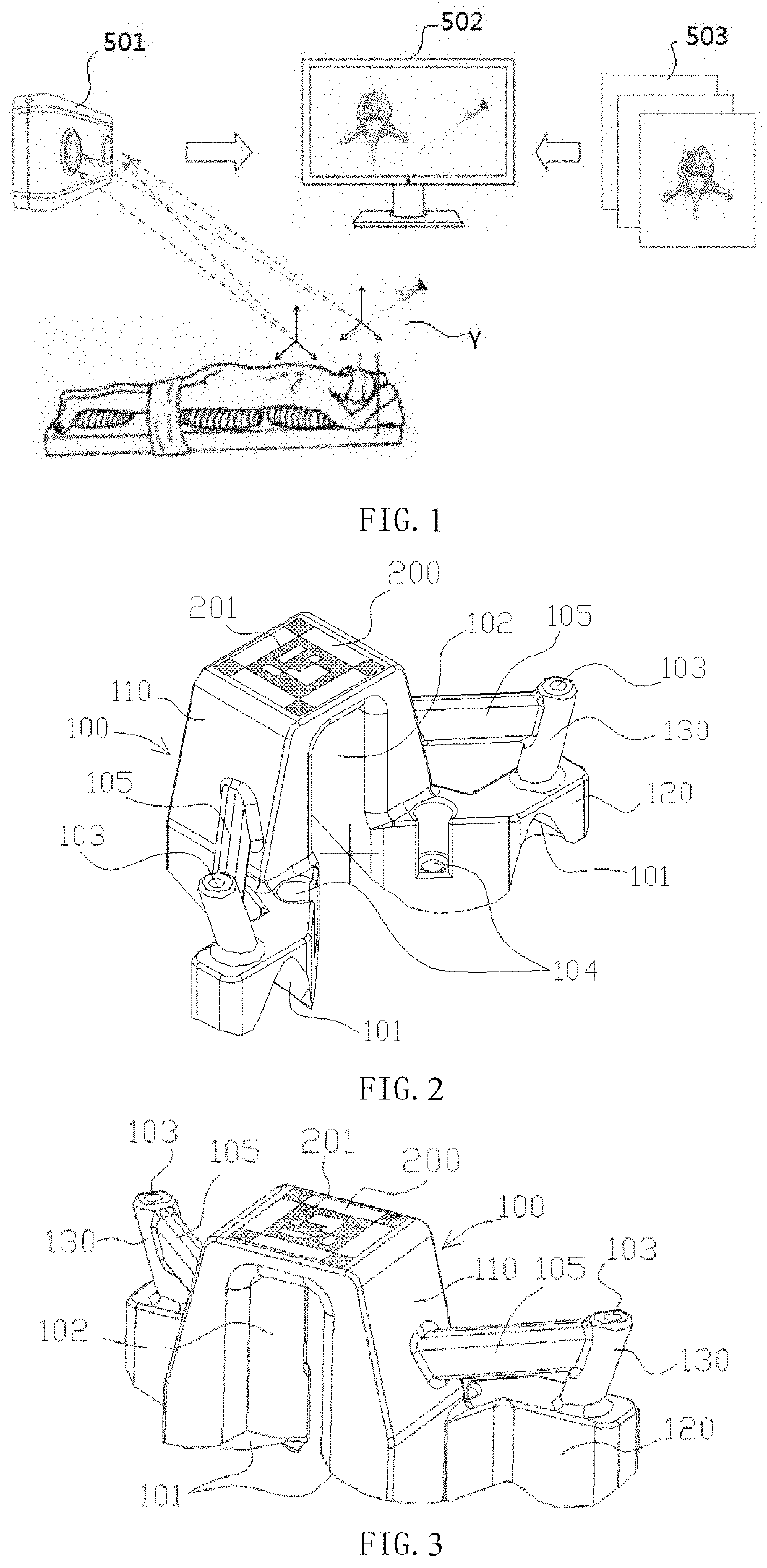
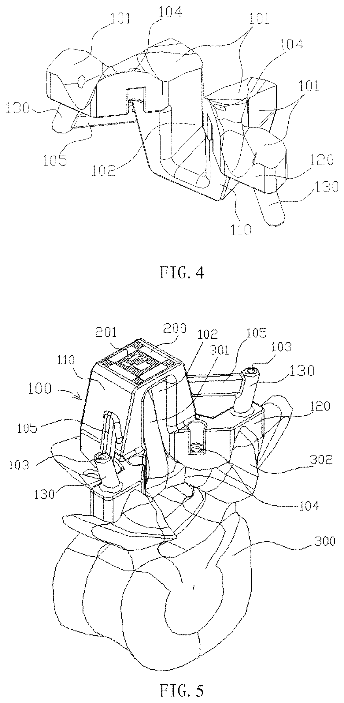
[0052]Please refer to FIGS. 2-7, the tracer is made by 3D printing. The tracer in the first embodiment includes a guide body 100. A bone fitting surface 101 is provided under the guide body. The surgical guide is made based on 3D reconstruction of preoperative bone images. The bone fitting surface 101 completely fits with a bone surface 302 of the vertebra 300 to be operated on. A navigation tracking surface 200 is also provided on the surgical guide, and the navigation tracking surface 200 is directly set on the surface of the guide body 100.

[0053]In this embodiment, the tracer defines a needle guide hole 103 and a fixing hole 104. The fixed hole 104 is used for engaging with screws or other fixing nails to strengthen fixation of the tracer on the vertebra 300 and make the fixation more stable. The needle guide hole 103 is used to guide surgical needles or other instruments. Specifically, the guide body 100 in this embodiment includes a main body 110 and a base 120, the fixing hole...

second embodiment

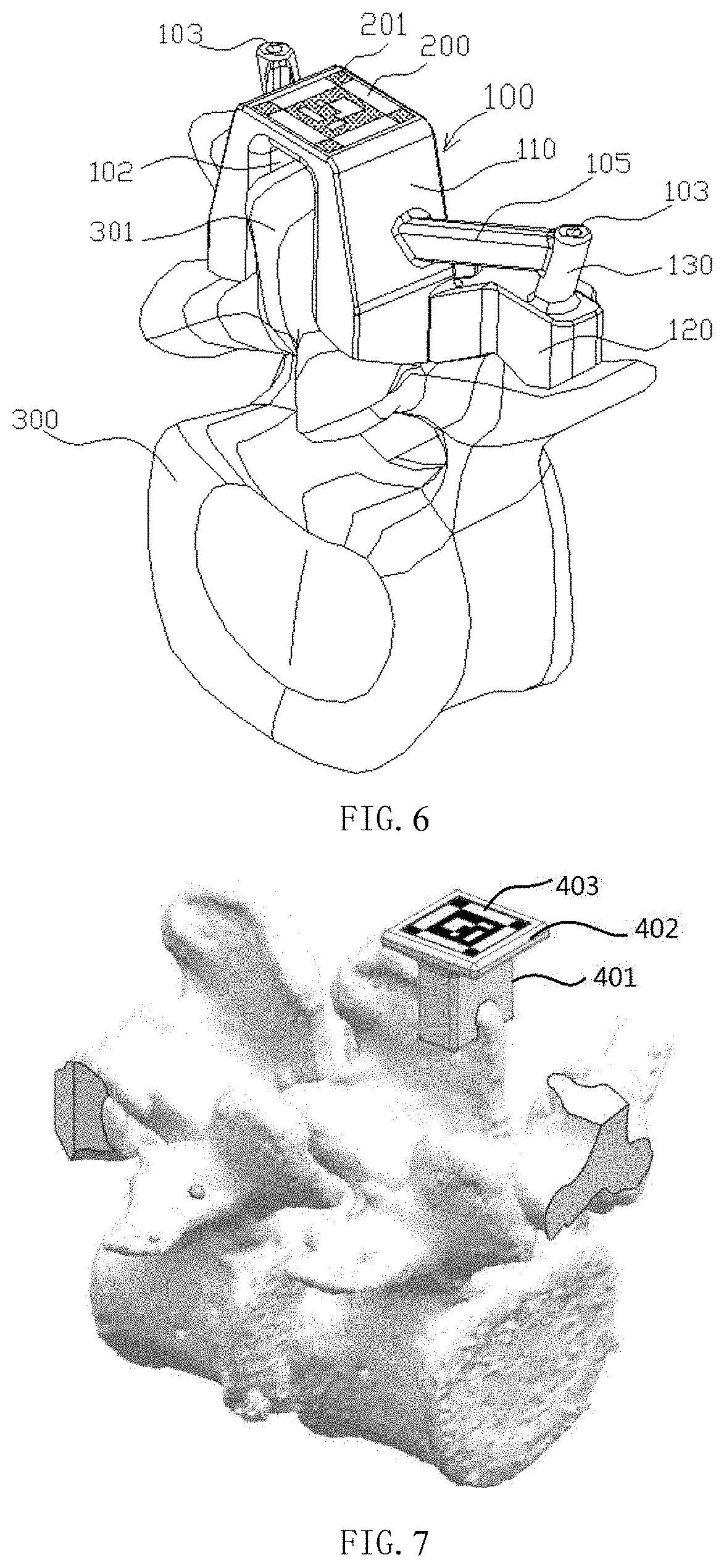
[0057]As shown in FIG. 7, in the second embodiment, the tracer includes a guide body 401 and a navigation tracking surface thereon. The guide body 401 is directly fixed on the bone, is attached to the bone with a complete fit between the bone fitting surface of the guide body and the bone surface, so that the tracer can be clamped at the spinous process of the vertebra. The guide body 401 is provided with a platform 402 as a navigation tracking carrier, and the platform 402 is provided with a tracking pattern 403 of visual recognition by visible light.

[0058]The bone fitting surface is in perfect fit with the bone to be operated on, and the error is small. After the tracer is fixed to the bone, the relative spatial position is unique, and the tracer acts as an extension of the bone. The spatial position of the navigation tracking surface on the tracer is known, therefore, there is no need for fluoroscopic image and registration during the operation, and tracer can be directly used fo...


PUM


Abstract

A surgical navigation system comprising a spatial positioning marker component (600) on a surgical instrument, a tracer on a bone structure, a binocular camera (501), and a computer (502). The tracer is provided with a navigation tracer surface (200). The spatial positioning marker component (600) comprises a navigation surface (601). The binocular camera (501) is connected to the computer (502), and transmits acquired information of the tracer and the spatial positioning marker component (600) to the computer (502). The computer (502) and a computer readable storage medium each separately store a computer program for performing a surgical navigation method. The method comprises; generating three-dimensional models of a bone and the surgical instrument, and obtaining tracer registration information and calibration information of the spatial positioning marker component (600); receiving real-time information of the tracer and the spatial positioning marker component (600) acquired by the binocular camera (501); and calculating spatial position information of the bone and the surgical instrument, and fusing, in real-time, the information with the three-dimensional models to obtain a dynamic image of a real-time positional relationship therebetween. The invention eliminates intraoperative registration used for surgical navigation, simplifies surgical processes, and improves surgical precision.

Description

FIELD OF THE INVENTION[0001]The present invention relates to the field of medical device, specifically relates to a surgical navigation system that can be used in orthopedic surgery, and particularly to a navigation system for orthopedic surgery based on a 3D-printed tracer and optical positioning technology, a method for performing the navigation system, a computer, and a storage media.BACKGROUND OF THE INVENTION[0002]With the development of modern medicine and computer technology, medical imaging technology and computer image processing technology are gradually being applied to the medical field. Computer-assisted surgery technology has become a major development direction in surgery. This technology extends the limited vision of surgeons, breaks through the boundaries of traditional surgery, and redefines the surgery and surgical instruments. It is of great significance for improving the accuracy of surgical positioning, reducing surgical trauma, shortening operation duration, an...

Claims


Application Information

Patent Timeline
Patent Type & Authority Applications(United States)
IPC IPC(8): A61B34/10A61B34/20
CPCA61B34/10A61B34/20A61B2034/105A61B2034/107A61B2034/2055A61B2034/2068B33Y80/00B33Y40/00A61B2034/2065A61B2090/3983
Inventor LIU, YANZHISUN, DONGHUIHUANG, WEIZHU, SHENGXIAO
Owner SHENZHEN XINJUNTE SMART MEDICAL EQUIP CO LTD
Why Eureka
Try Eureka