Lipid nanoparticle lyophilized composition

a technology of lipid nanoparticles and compositions, applied in the direction of non-active genetic ingredients, oil/fat/waxes non-active ingredients, non-active genetic ingredients, etc., to achieve the effect of increasing the encapsulation rate of nucleic acids and preparing lipid nanoparticles

Pending Publication Date: 2022-11-10
NOF CORP +1
View PDF0 Cites 0 Cited by
  • Summary
  • Abstract
  • Description
  • Claims
  • Application Information

AI Technical Summary

Benefits of technology

[0079]The present invention relates to a lyophilized composition of lipid nanoparticles not containing a nucleic acid, and a preparation method of lipid nanoparticles by using same.
[0080]The lyophilized composition of the present invention can prepare lipid nanoparticles containing any nucleic acid highly efficiently and easily by rehydration with an aqueous solution containing a nucleic acid.
[0081]The preparation method of lipid nanoparticles of the present invention can further increase the encapsulation rate of nucleic acid by incubating the lyophilized composition of the present invention by adding an aqueous solution of any nucleic acid, and further, alcohol.
[0082]When gene transfer is performed using lipid nanoparticles prepared by the method of the present invention, the gene can be uniformly introduced into cells. Further, the lipid nanoparticles prepared by the method of the present invention have a particle size of 50-200 nm and a narrow particle size distribution, which is advantageous for gene transfer in vivo.

Problems solved by technology

However, it is difficult to control the particle size of lipoplex produced by such method, and cytotoxicity derived from positively-charged cationic lipids becomes a problem.
However, since nucleic acids are generally unstable compounds, there are still problems with their stability as pharmaceutical preparations.
Although these methods are useful as a method for enhancing the storage stability of lipid nanoparticles encapsulating a specific nucleic acid, there is a problem as a method for more easily encapsulating any nucleic acid in lipid nanoparticles.
Thus, the method of preparing a lyophilized composition not containing a nucleic acid and then rehydrating same with an aqueous solution of nucleic acid, disclosed in Patent Literatures 1 and 2, cannot prepare nucleic acid-encapsulating lipid nanoparticles.
As described above, no means were available for easily encapsulating any nucleic acid in lipid nanoparticles with a high nucleic acid encapsulation rate.

Method used


Image

  • Lipid nanoparticle lyophilized composition
  • Lipid nanoparticle lyophilized composition
  • Lipid nanoparticle lyophilized composition

Examples


experimental example 1

Evaluation of Expression Intensity and Expression Efficiency Per Cell by EGFP-mRNA-Encapsulating LNP

[0307]An LNP solution encapsulating mRNA expressing EGFP was prepared by the method described in Examples.

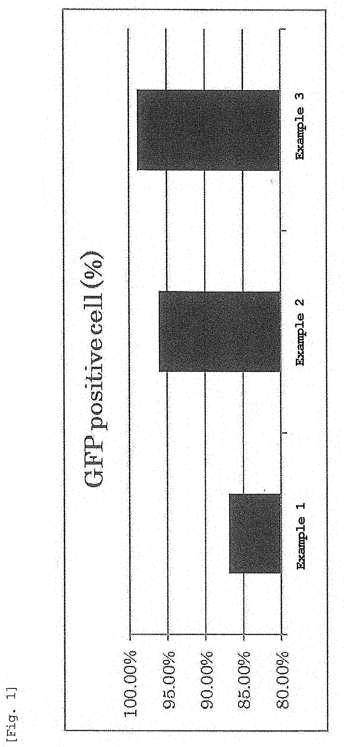
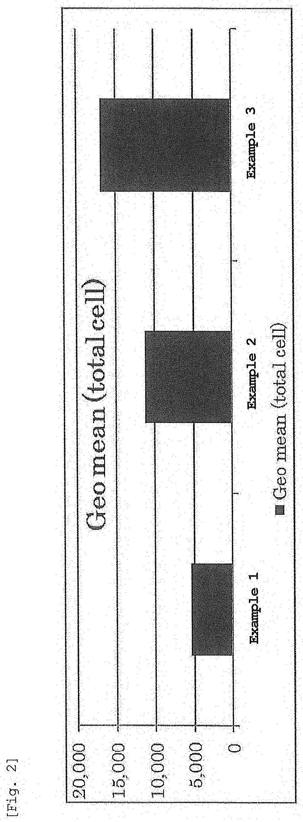
[0308]Jurkat cells which are human leukemia T cells were seeded in a 1.2 cm well at 5.0×10⁴ cells / 1 mL / well 24 hr before transfection. After 24 hr, the prepared LNP solution was added to 1.2 cm well such that 0.1 μg of mRNA was contained, and the cells were cultured for 24 hr in an incubator. The culture medium was exchanged with FACS buffer (PBS containing 0.5% bovine serum albumin (BSA), 0.1% NaN3), measurement was performed by a flow cytometer (NovoCyte; manufactured by ACEA Biosciences), and transgenic cells were analyzed. The proportion of cells expressing EGFP is shown in FIG. 1, and the expression intensity of EGFP is shown in FIG. 2.

[0309]As shown in FIGS. 1 and 2, nucleic acid-encapsulating lipid nanoparticles prepared using the lyophilized composition of the present inve...

experimental example 2

Evaluation of Gene Transfer Efficiency Into Cells Using Luc-mRNA Encapsulating Lipid Nanoparticles

[0310]An LNP solution encapsulating mRNA expressing luciferase was prepared by the method described in Examples.

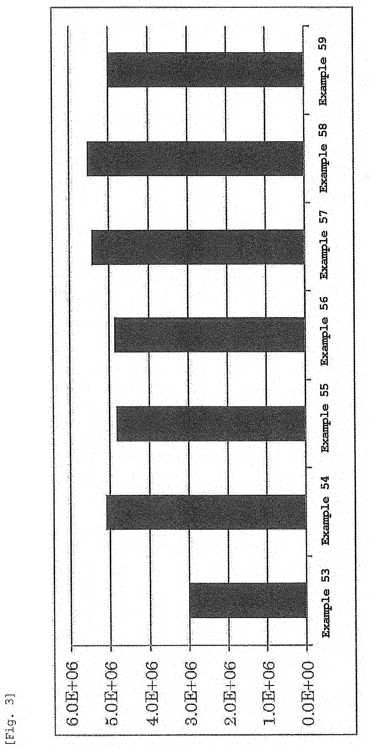
[0311]Jurkat cells which are human leukemia T cells were seeded in a 3.5 cm dish at 2.0×10⁵ cells / 1.9 mL / Dish 24 hr before transfection. After 24 hr, 100 μL of a D-luciferin-containing medium (RPMI1640) was added to each dish at a final concentration of 0.1 mM. The prepared LNP solution was added thereto such that 0.4 μg of mRNA was contained, and the mixture was set in an incubator luminometer KronosDio. The luminescence intensity of luciferase was measured for 2 min every 3 hr. The cumulative luminescence intensity for 24 or 48 hr was calculated from the obtained time change of expression. The results are shown in FIGS. 3-6.

[0312]As shown in FIGS. 3-6, genes could be efficiently introduced into cells by using any of DOPC, POPC, DOPE, and POPE as a phospholipid, and the gene ...

experimental example 3

[0313]Evaluation of In Vivo mRNA Expression Efficiency in Mouse

[0314]An LNP solution encapsulating mRNA expressing erythropoietin was prepared by the method described in Examples.

[0315]The prepared LNP solution was diluted with PBS such that the concentration of mRNA was 5 μg / mL. The diluted mRNA-encapsulated LNP was administered into the tail vein of 6-week-old female Balb / c mice at 10 μL per 1 g body weight (0.05 mg / kg as dose of mRNA). The blood (15 μL) was collected from the tail vein of the mice 1, 3, 6, 9, 24 hr after the administration. The collected blood was immediately mixed with 0.3 μL of heparin solution (5000 U / 5 mL). Each blood sample was centrifuged under centrifugation conditions (25° C., 2000 g, 20 min), and the supernatant was recovered. The concentration of erythropoietin in the supernatant was measured using Mouse Erythropoietin Quantikine ELISA Kit (manufactured by R&D Systems) and by the method described in the protocol of the Kit. The results are shown in FIG....


PUM

PropertyMeasurementUnit
weight ratioaaaaaaaaaa
concentrationaaaaaaaaaa

Abstract

A lyophilized composition capable of encapsulating any nucleic acid with high efficiency and easily is provided. A lyophilized composition of lipid nanoparticles not containing a nucleic acid but containing an ionic lipid, a sterol, a PEG lipid, an acidic buffer component that shows a buffering action at pH 1-6, and a cryoprotectant, wherein a weight ratio of the cryoprotectant and a total lipid is 10:1-1000:1.

Description

TECHNICAL FIELD[0001]The present invention relates to a lyophilized composition of lipid nanoparticles not containing a nucleic acid and a method for producing nucleic acid-encapsulating lipid nanoparticles by using same.BACKGROUND ART[0002]For practicalization of nucleic acid therapy using oligonucleic acids such as siRNA, and gene therapy using mRNA, pDNA, and the like, an effective and safe nucleic acid delivery carrier is demanded. While virus vectors are nucleic acid delivery carriers with good expression efficiency, the development of non-viral nucleic acid delivery carriers that can be used more safely is ongoing.[0003]Since cationic liposomes using cationic lipids with quaternary amine are positively charged, they can form a complex (lipoplex) by electrostatic interaction with negatively-charged nucleic acids, and can deliver nucleic acids into cells. Utilizing the electrostatic interaction of quaternary amine with nucleic acid, it is also possible to prepare a lyophilized c...

Claims


Application Information

Patent Timeline
Patent Type & Authority Applications(United States)
IPC IPC(8): A61K9/19A61K47/10A61K47/24A61K47/28A61K47/44A61K47/69
CPCA61K9/19A61K47/10A61K47/24A61K47/28A61K47/44A61K47/69A61K31/7088A61K47/26A61K48/0008C12N15/87C12N15/88A61K48/005A61K48/0041A61K9/5123A61K9/0019A61K47/6929A61K48/0025
Inventor TANGE, KOTANAKAI, YUTATAMAGAWA, SHINYATANEICHI, SAKURAAKITA, HIDETAKATANAKA, HIROKISHIRANE, DAIKIHAGIWARA, SHINYA
Owner NOF CORP
Why Eureka
Try Eureka