Acrylic microchannels and their use in electrophoretic applications

a technology of electrophoretic applications and microchannels, applied in the field of electrophoretic applications, can solve the problems of uniformity of the eof, treatment failure to completely mask the negatively charged silica surface, and the modification layer on the surface resulting from such treatment may not be entirely stabl

Inactive Publication Date: 2000-04-25
MONOGRAM BIOSCIENCES
View PDF26 Cites 464 Cited by
  • Summary
  • Abstract
  • Description
  • Claims
  • Application Information

AI Technical Summary

Problems solved by technology

Furthermore, sample components such as proteins and other analytes can adsorb to the negatively charged surface of the fused silica and thereby unpredictably disrupt the uniformity of the EOF, thereby contributing to the irreproducibility of the results obtained in the electrophoretic application.
Although under ideal circumstances these treatments can reduce or even eliminate the problems associated with EOF and solute adsorption, in practice these treatments can fail to completely mask the negatively charged silica surface.
Furthermore, the modified layer on the surface resulting from such treatments may not be entirely stable under the conditions of electrophoresis.
Finally, surface modified fused silica microvessels such as capillaries are difficult to manufacture, as the manufacturing process can be laborious and time consuming.

Method used


Image

  • Acrylic microchannels and their use in electrophoretic applications
  • Acrylic microchannels and their use in electrophoretic applications
  • Acrylic microchannels and their use in electrophoretic applications

Examples


example 1

CE of dsDNA in Polymethylmethacrylate Capillaries

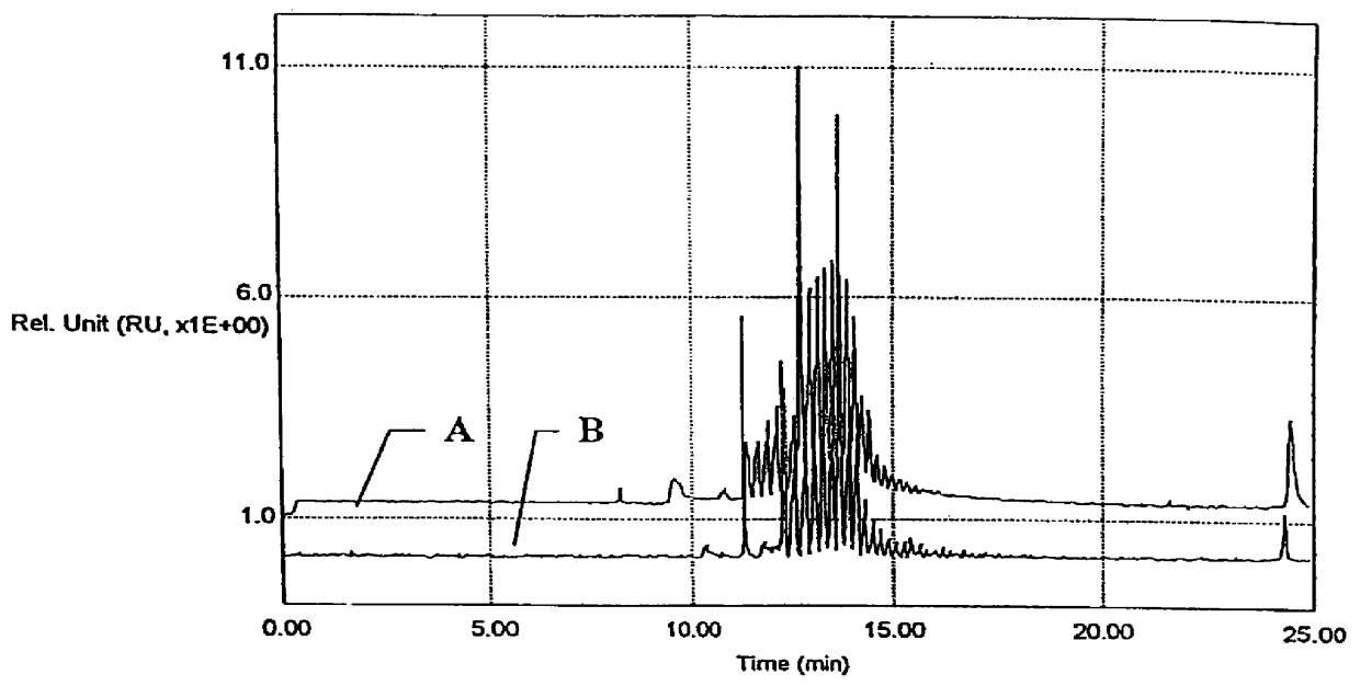
A 34 cm polymethylmethacrylate (PMMA) capillary (Biogeneral, Inc.) having an inner diameter of 75 .mu.m and an outer diameter of 375 .mu.m was pressure-loaded with a separation media prepared from 0.4 g of hydroxyethylcellulose (HEC) (MW 90,000-105,000) and 1.5 g of hydroxypropylcellulose (HPC) (MW 300,000) dissolved in 98.1 g of 0.5.times. TBE at 400 psi. A 10 base pair DNA ladder consisting of about 30 10-bp repeats (GibcoBRL) was loaded electrokinetically for five seconds at 5 kV. Electrophoresis was performed at 5 kV using a prototype electrophoresis instrument with a confocal fluorescence detector having Spindler & Hoyer (Medford, Mass.) optical components and an Omnichrome Argon Ion Laser operating at about 12 mW and 488 nm.

The results are provided in FIG. 1. Trace A is the separation of the 10 base pair ladder in a polymethylmethacrylate capillary, while trace B is the separation of an identical sample in a coated fused silica ...

example 2

CE of Single Stranded DNA Ladder in Polymethylmathacrylate Capillary

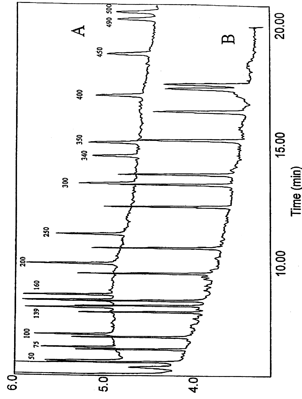
A 10 cm length of polymethylmethacrylate (PMMA) capillary (Biogeneral, Inc.) having an inner diameter of 75 .mu.m was loaded with sieving buffer containing 2% (by wt) hydroxyethylcellulose (HEC, MW 90,000-105,000), 6M urea, 10% formamide, and 1.times. TBE. The capillary was placed into a horizontal capillary holder in a capillary electrophoresis unit. DNA was injected eletrokinetically at 150 V / cm for 15 seconds. For the DNA sample, TAMRA(N,N,N',N'-tetramethyl-6-carboxyrhodamine) labelled GeneScan 500 ladder (Perkin Elmer) was used. GeneScan 500 ladder consists of 16 fragments of 500, 490, 450, 400, 350, 340, 300, 250, 200, 160, 150, 139, 100, 75, 50 and 35 bases. Electrophoresis was performed at 150 V / cm for 20 minutes, with sieving buffer in the anodic reservoir. Detection was performed using confocal fluorescence detection. Separated fragments of DNA were detected by excitation of TAMRA with a xenon lamp (440-490...

example 3

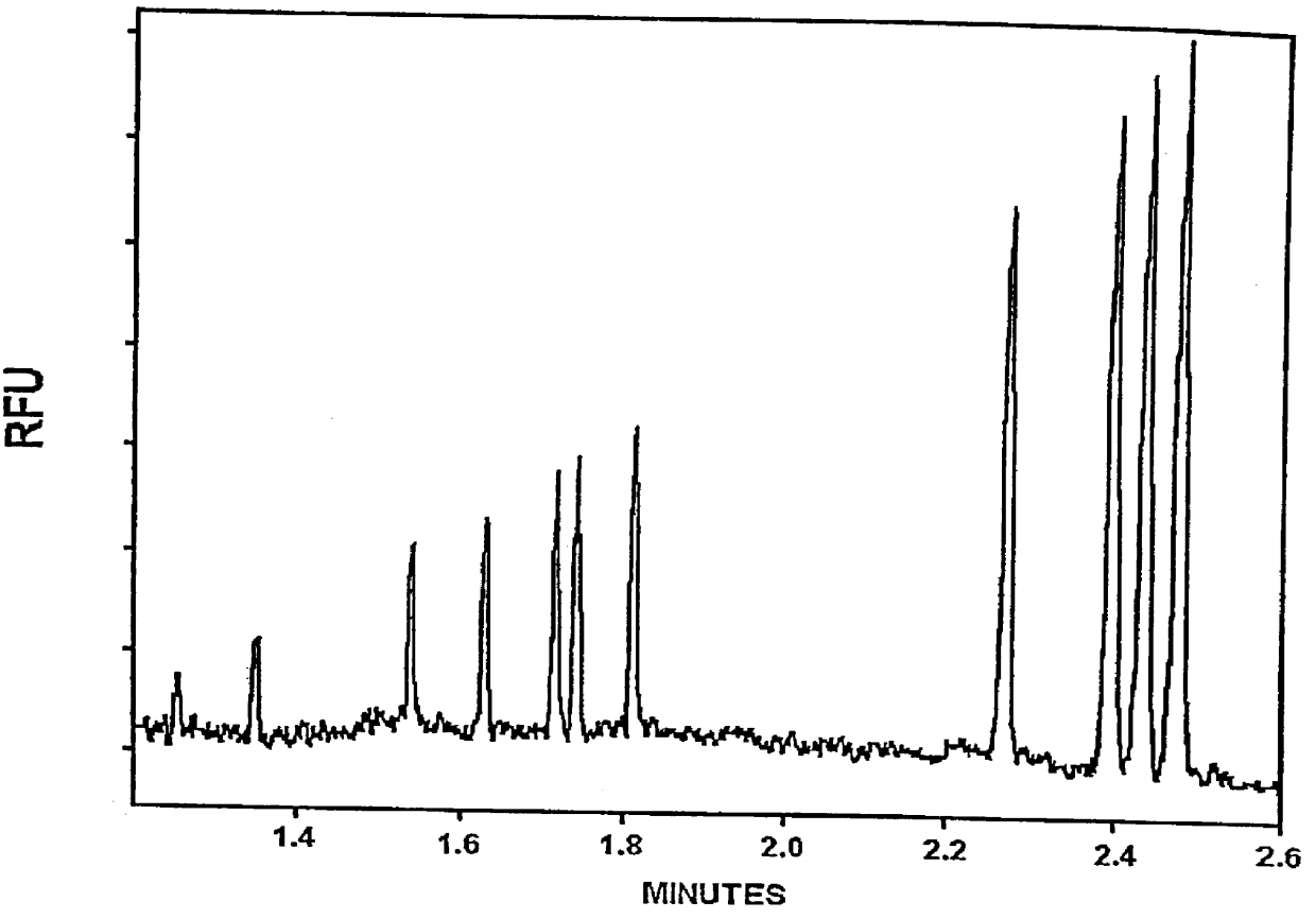
CE of the Fragments in the HAE III Digest of .PHI.X174 RF DNA in a Sealed Plastic PMMA / Mylar.TM. Microchannel Structure

Photolithographic, electroforming and injection molding techniques were used to prepare an acrylic polymer (AtoHaas, Plexiglas.TM.V825NA-100) microchannel base plate. The microchannel structure corresponds to two crossed linear channels of dimensions 2 cm and 5.5 cm in length respectively. The channels cross-section is trapezoidal, at widest it measures 120 .mu.m and at narrowest 30 .mu.m, with an average depth of 40 .mu.m. At the termini of the channels, holes of 3 mm in diameter were drilled as buffer reservoirs. The channels were covered by thermal lamination of a 2 mil thick sheet of Mylar.TM. coated with a thermally-activated adhesive (MonoKote.TM., made by Top Flight Co.) at 105.degree. C. for 5 minutes. Electrodes of 76 micron diameter platinum wire were routed to each of the four reservoirs and terminated at one edge of the chip with a 4-prong 2.54 mm pitch ...


PUM

PropertyMeasurementUnit
thickaaaaaaaaaa
diameteraaaaaaaaaa
diameteraaaaaaaaaa

Abstract

Microchannels having at least an acrylic inner surface and methods of their use in electrophoretic applications are provided. The subject microchannels may be in the form of a variety of configurations suitable for holding an electrophoretic medium. The subject microchannels give rise to substantially reduced EOF and/or adsorption as compared to fused silica under conditions of electrophoresis and find use in a variety of electrophoretic applications in which charged entities are moved through a medium under the influence of the an applied electric field.

Description

1. Field of the InventionThe field of this invention is electrophoresis.2. Background of the InventionElectrophoresis has become an indispensable tool of the biotechnology industry, as it is used extensively in the separation, identification and preparation of pure samples of nucleic acids, proteins and carbohydrates. For example, electrophoresis is critical to DNA sequencing applications such as the Maxam-Gilbert and Sanger dideoxy methods, where a major step of these methods is the electrophoretic separation of labeled DNA fragments.Of increasing interest in the field of electrophoresis is the use of devices having micro cross-sectional dimensions, such as microbore capillaries and microchannels. Use of devices in which the electrophoretic medium is housed in a container having micro cross-sectional dimensions can provide a number of different advantages over conventional slab gel electrophoretic configurations. For example, in capillary electrophoresis (CE) where electrophoresis ...

Claims


Application Information

Patent Timeline
Patent Type & Authority Patents(United States)
IPC IPC(8): B01D57/02B01L3/00B01J19/00B29C65/02C07K1/00C07K1/26G01N27/447G03F7/00B01F13/00B29C65/14
CPCB01D57/02B01F13/0076B01J19/0093B01L3/502753B29C65/02B29C66/54B82Y10/00B82Y40/00C07K1/26G01N27/44752G01N27/44773G01N27/44791G03F7/0002B29C66/542B29C65/483B29C65/4865B01L2200/0689B01L2200/12B01L2300/0816B01L2400/0415B29C65/1425B29C65/524B29L2031/756B29L2031/757B01F13/0059B29C65/4845B29C65/4835B29C65/00B29C66/71B01F33/3031B01F33/30B29K2033/12
Inventor SOANE, DAVID S.SOANE, ZOYA M.HOOPER, HERBERT H.AMIGO, M. GORETTY ALONSO
Owner MONOGRAM BIOSCIENCES
Why Eureka
Try Eureka