Method and apparatus for motion estimation within biological tissue

a biological tissue and motion estimation technology, applied in applications, ultrasonic/sonic/infrasonic image/data processing, ultrasonic/sonic/infrasonic diagnostics, etc., can solve the problem of large decorrelation errors persisting, biological tissue deformation is typically very complex, and temporal stretching fails to remove significant decorrelation errors

Inactive Publication Date: 2001-08-21
UNIV KANSAS MEDICAL CENT
View PDF3 Cites 147 Cited by
  • Summary
  • Abstract
  • Description
  • Claims
  • Application Information

AI Technical Summary

Problems solved by technology

Unfortunately, the deformation of biological tissue is typically very complex and large decorrelation errors persist.
Temporal stretching, however, fails to remove significant decorrelation errors because when a nearly incompressible material such as a biological tissue is compressed in one direction, for example, along the axis of the ultrasonic beam, it necessarily bulges in directions not axial to the beam.
Accordingly, five main problems exist with the current acoustic motion estimation and strain imaging techniques: 1) Noise; 2) Contrast; 3) Out of Plane Displacement; 4) Shear Motion; and 5) Rotational Motion.
Noise increases with the amount of applied compression.
As noted above, however, increased compression also increases noise, which distorts the strain image.
Limiting compression also limits contrast making abnormal tissue more difficult to detect.
Consequently, a target that may have moved in the elevational direction ...

Method used


Image

  • Method and apparatus for motion estimation within biological tissue
  • Method and apparatus for motion estimation within biological tissue
  • Method and apparatus for motion estimation within biological tissue

Examples


Embodiment Construction

While the present invention may be embodied in many different forms there is shown in the drawings and discussed herein one specific embodiment and specific fabrication method with the understanding that the present disclosure is to be considered only as an exemplification of the principles of the invention and is not intended to limit the invention to the embodiments illustrated.

Warping

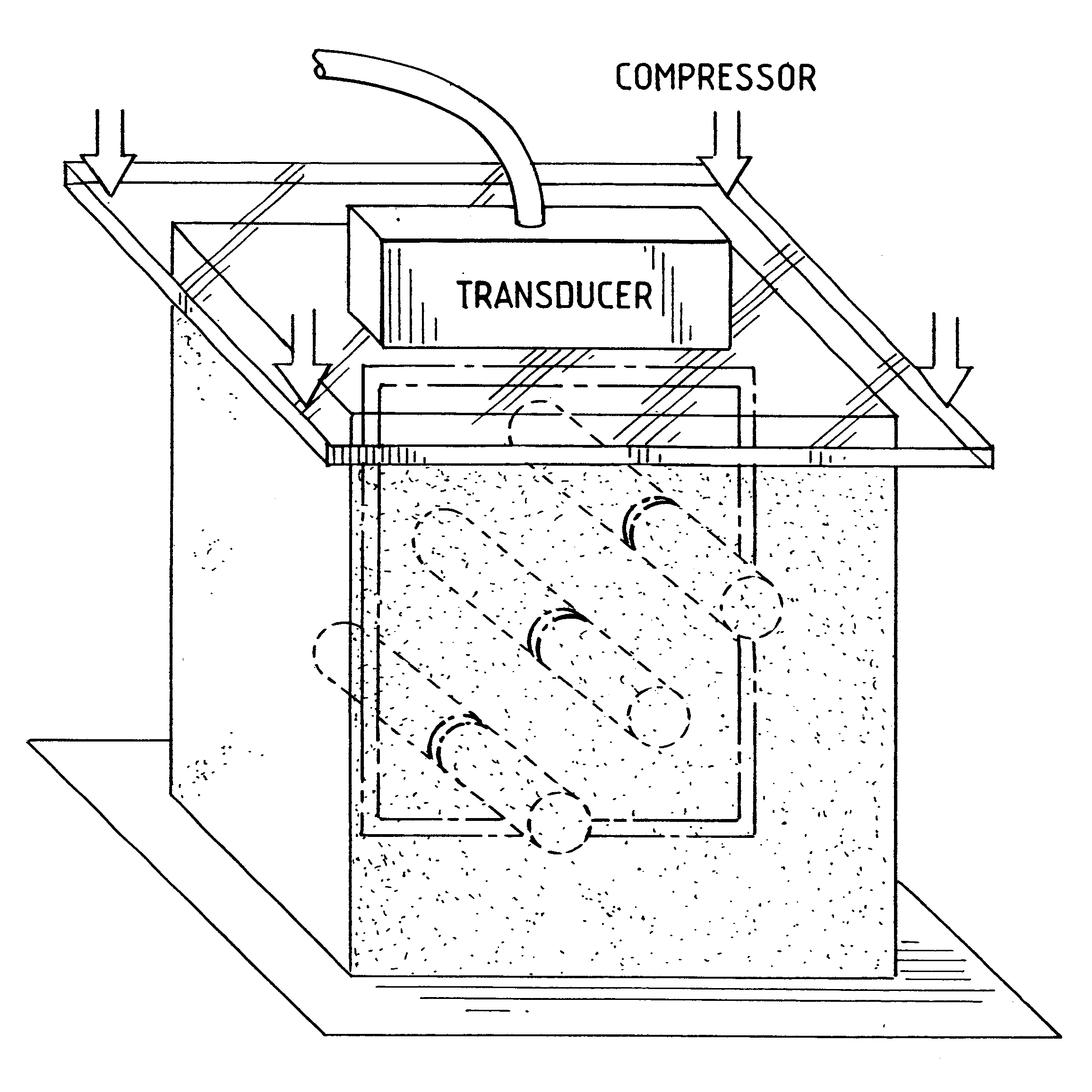
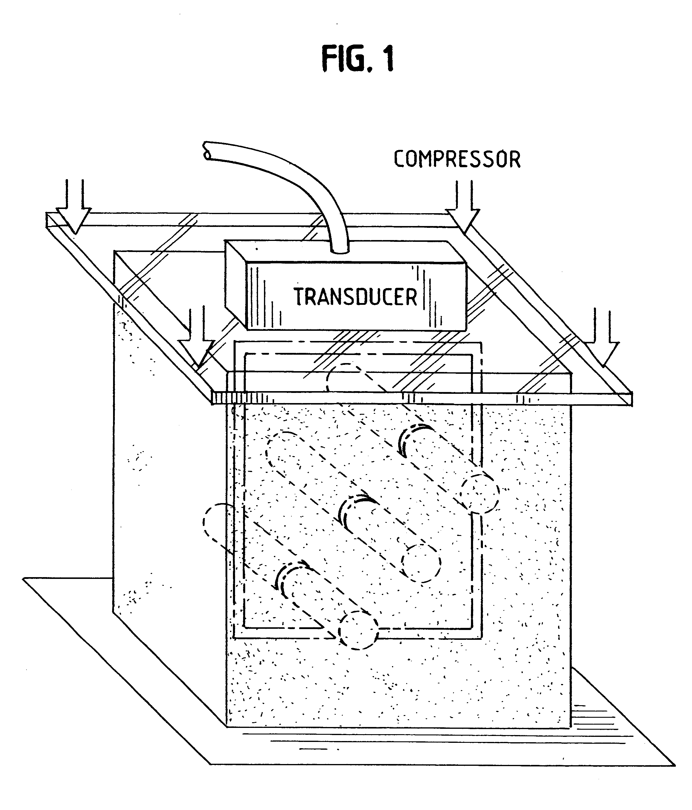
As shown in FIG. 1, a transducer having a rectangular grid of sensors is flush mounted to a planar compressor surface. While a two dimensional planar array transducer is preferred, a transducer having sensors in concentric circles or a linear array transducer may be used. If only a one dimensional detector array is available, a plurality of pre-compression images or post-compression images, or both may be taken to obtain the three dimensional data set, if required, around the target object. All the data from the multiple scans is then combined to form the data sets.

In the preferred embodiment, the pl...


PUM


Abstract

A method for motion estimation within biological tissue is disclosed. The method involves acoustically coupling a transducer to a target biological tissue, which transducer emits an ultrasonic signal and collects the energy back-scattered by the target issue. A first set of energy data is collected and stored, then target tissue is axially compressed and a second set of ultrasonic energy data is collected and stored. One of the first and second data sets is warped to account for the anticipated compression forming a warped data set. This warped data set is cross-correlated with the unwarped one of the first and said second data sets to obtain a fine scale displacement of said target biological tissue from the displacement estimated by the warping. This fine scale displacement is summed with the warped data set to obtain a total axial displacement. A gradient of the total axial displacement is taken and used to form a strain image. An apparatus for practicing this method is also disclosed.

Description

1. Field of the InventionThe present invention relates generally to a method and apparatus for improving motion estimation within a biological tissue and specifically to local Warping for improving contrast and reducing noise in strain imaging.2. Background ArtUltrasonicsIt is generally known to use ultrasonics for non-invasive imaging of soft biological tissue. An acoustic transducer is used as both a transmitter and receiver of sound pulses in the 1-50 MHz frequency range. The transducer is used in a pulse-echo system where the transducer converts between electrical and acoustic energy to transmit and receive sound pulse to and from the target tissue. Received signals are translated by a computer to produce an image.ElastographyA technique called elastography was developed in concert with ultrasonics in order to detect inhomogeneities or stiff regions within soft biological tissues. Elastography is a remote sensing technique for imaging elastic properties of biological tissues. El...

Claims


Application Information

Patent Timeline
IPC IPC(8): A61B8/08
CPCA61B5/0048A61B8/485A61B8/0833A61B5/0053
Inventor CHATURVEDI, PAWANINSANA, MICHAEL F.HALL, TIMOTHY J.ZHU, YANNING
Owner UNIV KANSAS MEDICAL CENT
Why Eureka
Try Eureka