
System and method for intraluminal imaging

a technology of intraluminal imaging and catheter system, applied in the field of medical devices and methods, can solve the problems of reduced catheter wall strength, restricted blood flow, and serious risk to the health of peopl

Inactive Publication Date: 2002-04-02
BOSTON SCI LTD +1
View PDF27 Cites 96 Cited by
  • Summary
  • Abstract
  • Description
  • Claims
  • Application Information

AI Technical Summary

Benefits of technology

In another aspect of the present invention, an improved catheter system includes (a) a tubular catheter body having a proximal portion, a distal portion, and a lumen therethrough, and (b) a drive cable rotatably received in the lumen. In a first aspect of the improved catheter system, the improvement comprises forming the flexible tubular body at least in part from a material capable of withstanding high hoop stresses. The material may be a polymer or plastic material, such as polyetheretherketone (PEEK), having increased hoop strength and bending stiffness.
In an alternate preferred method a further step of exchanging the distal portion of the catheter body is provided. For example, a distal portion, having a short guidewire access lumen may not provide adequate stiffness for accessing a specific lesion in a patient's vascular system. Thus, the distal portion may be exchanged with a second distal portion, having a long guidewire access lumen which provides increased stiffness to the distal portion of the catheter body.

Problems solved by technology

Blood flow is restricted and the person's health is at serious risk.
As the catheter body size is minimized, the strength of the catheter wall is decreased and the danger that the wall will rupture or collapse may become significant.
A reduction in strength can lead to slight closure or collapse of the tubular structure under normal operating pressures incurred during a normal procedure.
This type of failure causes an increase in friction on the drive cable, commonly referred to as NURD.
The NURD effect can cause the motor powering the drive cable to be overworked causing a failure of the catheter.
While the danger of catheter wall failure is present in any catheter system, it may be particularly troublesome in a common lumen catheter of the type described in U.S. Pat. No. 5,314,408.
When the rotating work element encounters a kink in the catheter body, it will impact the wall at the kink and in some cases may cause failure of the catheter body wall.
While a significant improvement, use of the improved tracking tip can sometimes result in users confusing the solder ball, located at the distal end of the tracking tip, for the working element, when the procedure is viewed via x-ray.
This leads to a mistaken determination by the user that the target location has been reached with the work element.
Another possible disadvantage of using the tracking tip is its effect on the shape of the work element carrier.
Since ultrasound does not travel through air bubbles, this phenomenon generally causes a blurred or smeared image to appear on viewing monitors.
This buckling or distortion creates additional friction forces which quickly creates enough drag to cause the imaging core assembly to fail in a safe manner.
Moreover, a user having difficulties crossing a lesion, does not have the option to exchange the short guide wire engagement sheath (low stiffness) for the smaller profile longer guide wire engagement common lumen design (high stiffness).
Thus, creating waste and increased costs.
For example, a distal portion, having a short guidewire access lumen may not provide adequate stiffness for accessing a specific lesion in a patient's vascular system.

Method used


Image

  • System and method for intraluminal imaging
  • System and method for intraluminal imaging
  • System and method for intraluminal imaging

Examples


Embodiment Construction

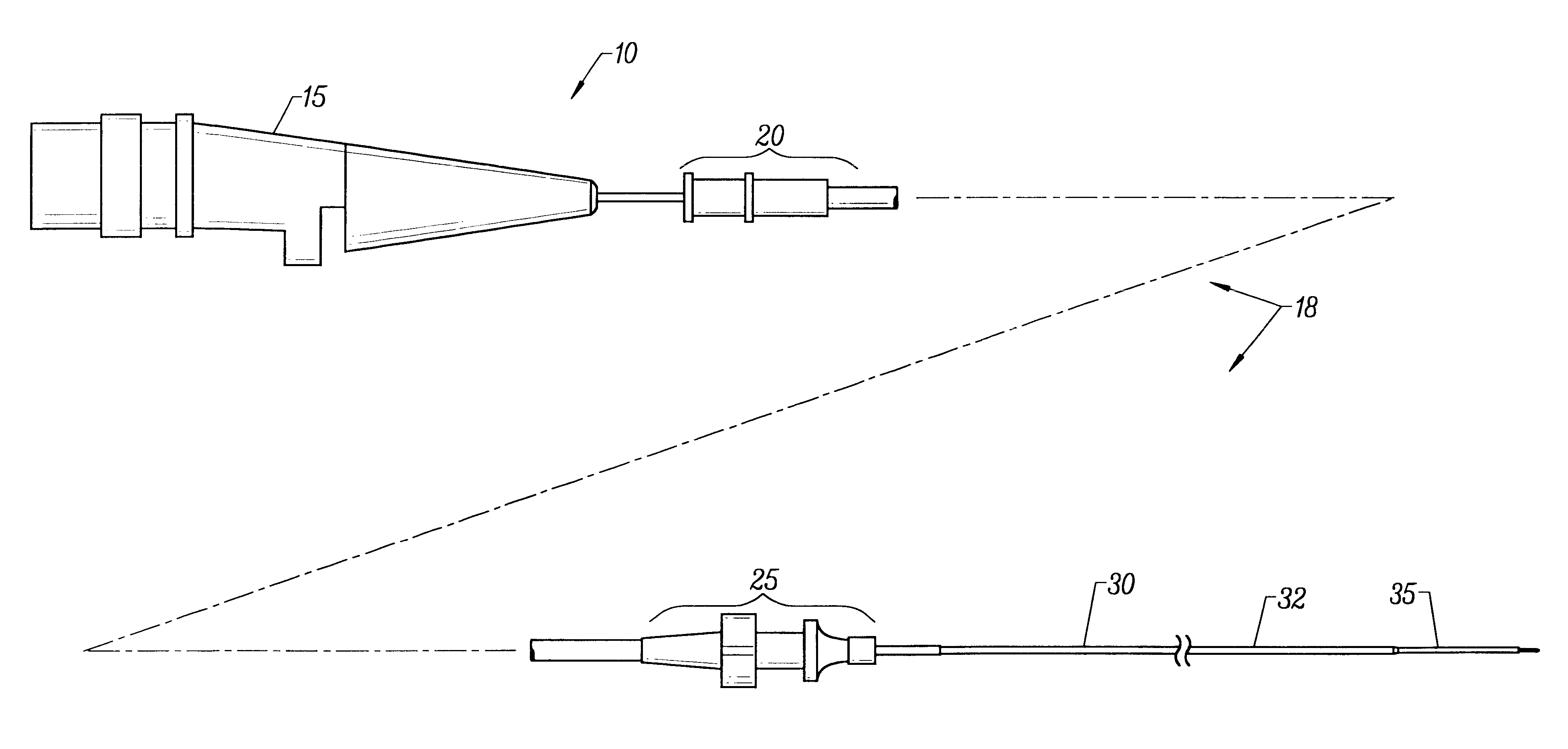
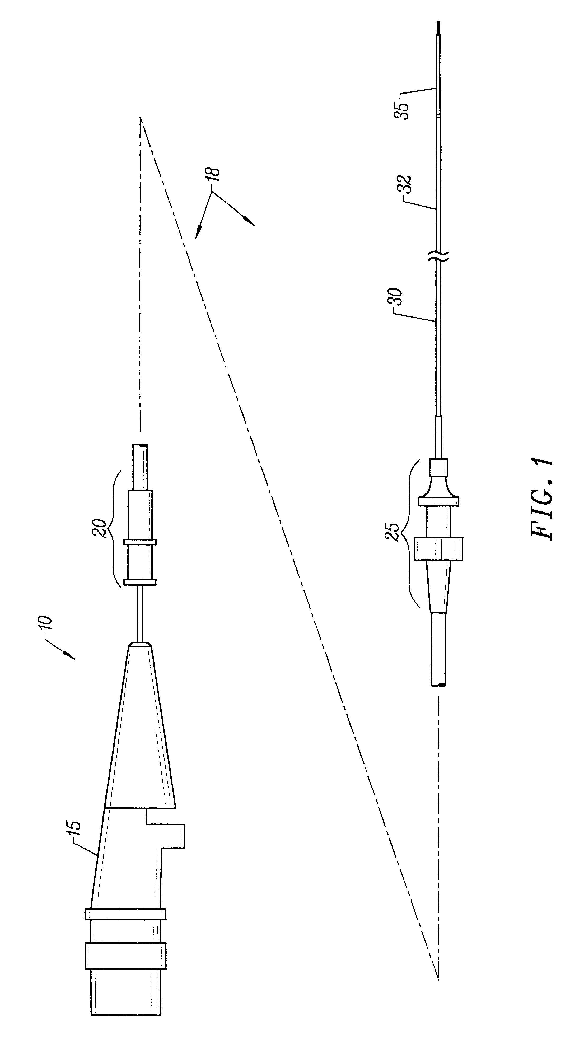
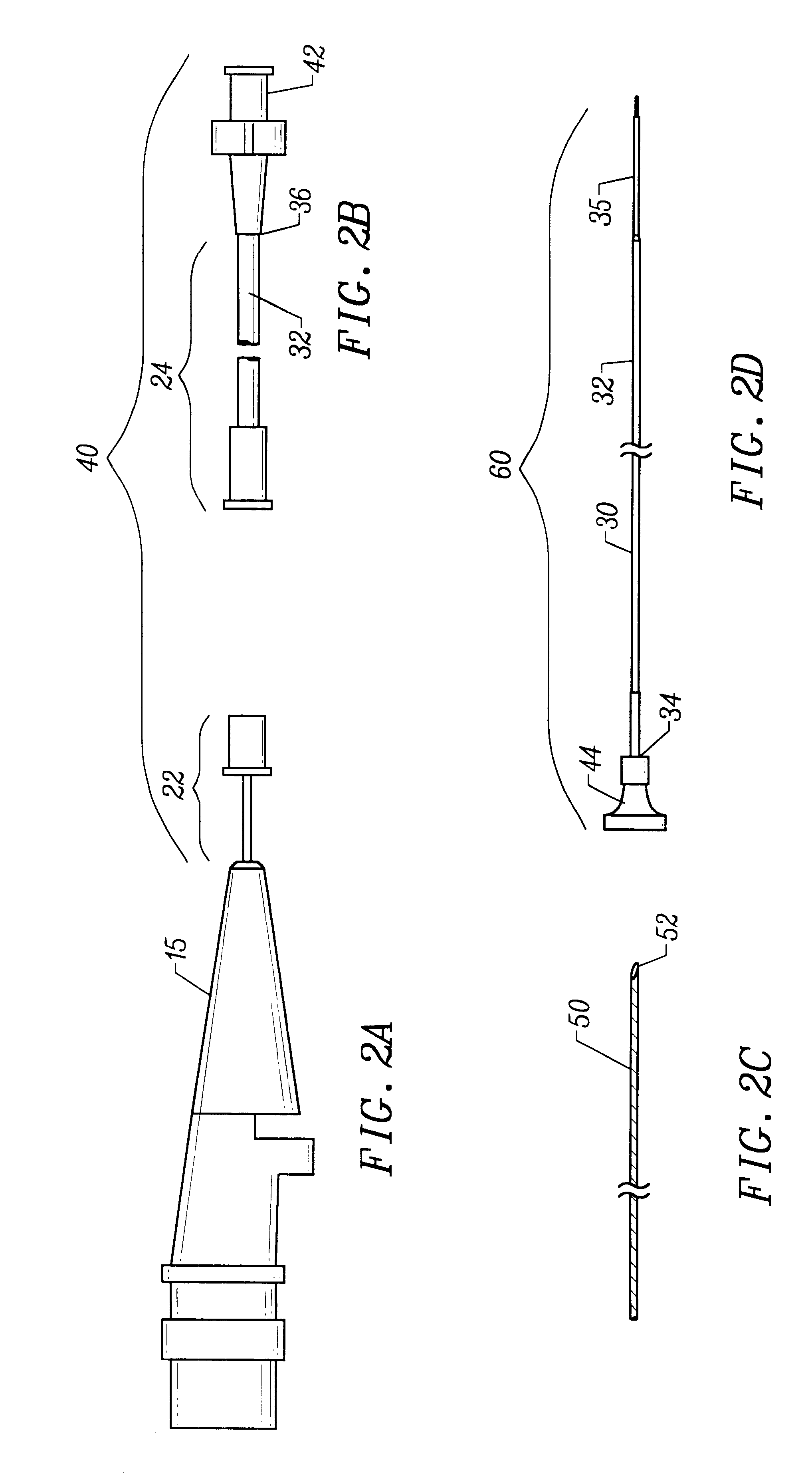
The present invention provides an improved vascular catheter system comprising a catheter body having a reduced profile distal portion to facilitate entry into coronary blood vessels and / or tight stenotic regions. The catheter body will comprise two regions, a proximal portion and a distal portion, each having a single common lumen therebetween. The distal portion will extend from the distal end of the catheter body to a point of adhesion to and including a connector. The proximal portion will extend from a point of adhesion to and including a second connector to the distal end of the tuning hub.

The proximal portion will have a somewhat larger cross-sectional area to accommodate a distal tube and a proximal tube, joined together in a telescopic engagement and used to increase the working length of the drive cable.

Preferably, the catheter body will be made of a polymer or plastic material, which increases the bending stiffness and has improved hoop strength, i.e. accommodates large h...


PUM


Abstract

A vascular catheter system comprises a catheter body having a proximal and distal portion and a single common lumen therebetween. The catheter body includes a first connector secured to the distal end of the proximal portion and a second connector secured to the proximal end of the distal portion. The connectors can be selectively connected to each other to join the lumens of the proximal and distal portions together in a continuous, axially fixed relationship. Disposed within the lumens, when the proximal and distal portions are joined together, is a drive cable. The drive cable may be movably, rotatable about its own longitudinal axis and carries at its distal end, a work element, which is typically an ultrasonic imaging transducer or interventional device. The lumen carrying the cable will be sufficiently large along a proximal portion to permit preferential collapse of the cable should rotation of the distal end become impeded. The catheter body may be made of a polymer or plastic material, such as polyetheretherketone (PEEK), which provides adequate bonding stiffness and resistance to kinking from large hoop stresses.

Description

1. Field of the InventionThe present invention relates generally to medical devices and method and more particularly to a catheter system having a work element coupled to a drive cable disposed within a lumen of a flexible tubular catheter body.2. Description of the Background ArtArteriosclerosis, also known as atherosclerosis, is a common human ailment arising from the deposition of fatty-like substances, referred to as atheromas or plaque, on the walls of blood vessels. Such deposits occur in both the peripheral blood vessels, which feed the limbs of the body, and the coronary vessels, which feed the heart. When deposits accumulate in localized regions of a blood vessel, stenosis, or narrowing of the vascular channel, occurs. Blood flow is restricted and the person's health is at serious risk.Numerous approaches for reducing and removing such vascular deposits are known, including balloon angioplasty, in which a balloon-tipped catheter is used to dilate a region of atheroma; ather...

Claims


Application Information

Patent Timeline
Patent Type & Authority Patents(United States)
IPC IPC(8): A61B8/12A61M25/00A61M39/10A61M39/00
CPCA61B8/12A61M25/00A61B8/4461A61M39/10
Inventor WHITE, DAVID A.WILLIAMS, ALFRED E.
Owner BOSTON SCI LTD
Learn More