Inhibitors of prion formation

a prion and inhibitor technology, applied in the field of prion formation inhibitors, can solve the problems of compound inability to cross the blood-brain barrier, compound inability to efficiently infect one species of animal (e.g., a human) and cannot efficiently infect another, so as to prevent prion disease or symptoms, and determine the infectivity of prions in a sample quickly

Inactive Publication Date: 2002-04-02
RGT UNIV OF CALIFORNIA
View PDF7 Cites 3 Cited by
  • Summary
  • Abstract
  • Description
  • Claims
  • Application Information

AI Technical Summary

Benefits of technology

An advantage of the present invention is that infectivity of prions in a sample can be determined rapidly.
is that infectivity of prions in a sample can be determined rapidly.
In another aspect of the invention, the pharmacophore is any one of a collection of molecules that repress prion infectivity and or progression of prion-mediated disease. Any pharmacophore of the invention may inhibit initial infectivity, conversion of PrP.sup.C to PrP.sup.Sc and / or progression of neurodegeneration by any number of mechanisms, including but not limited to binding a molecule involved in prion complexing, e.g. PrP.sup.C or PPMF or inhibiting .beta.-sheet formation or elongation.
Yet another aspect of the invention features a method of repressing conversion of PrP.sup.C to PrP.sup.Sc, comprising administering an inhibitor that meets the criteria specified by the pharmacophore model. This may be administered prophylactically to a subject at risk of developing a prion-mediated disorder, e.g. a mammal exposed to infectious prions, or to treat a subject that is exhibiting signs of prion-mediated neurodegeneration.
A feature of the invention is that the inhibitors can be used to treat subjects suffering from prion-mediated disorders.
Yet another aspect of the invention features an assay to identify a PrP pharmacophore, a geometric and chemical specification of a collection of small molecules that could inhibit PrP.sup.Sc formation. The assay utilizes the steps of determining functional residues of the PrP protein involved in prion complex interactions, developing three dimensional structures based on these functional residues, comparing the three dimensional structures with a series of compounds having known or calculated tertiary structures, and identifying compounds having a spatial orientation consistent with binding to components of the PrP.sup.Sc replication complex (PrP.sup.C, PrP.sup.Sc, PPMF) at these functional residues.

Problems solved by technology

Further, a prion which infects one species of animal (e.g., a human) will not efficiently infect another (e.g., a mouse).
No systematic drug discovery efforts have been attempted owing to difficulties in developing a robust screening assay.
However, these compounds are unable to cross the blood-brain barrier, and therefore have no therapeutic benefit after the infection has reached the central nervous system (Caughey, et al.

Method used


Image

  • Inhibitors of prion formation
  • Inhibitors of prion formation
  • Inhibitors of prion formation

Examples


example 1

Binding Site in PrP.sup.C for PPMF

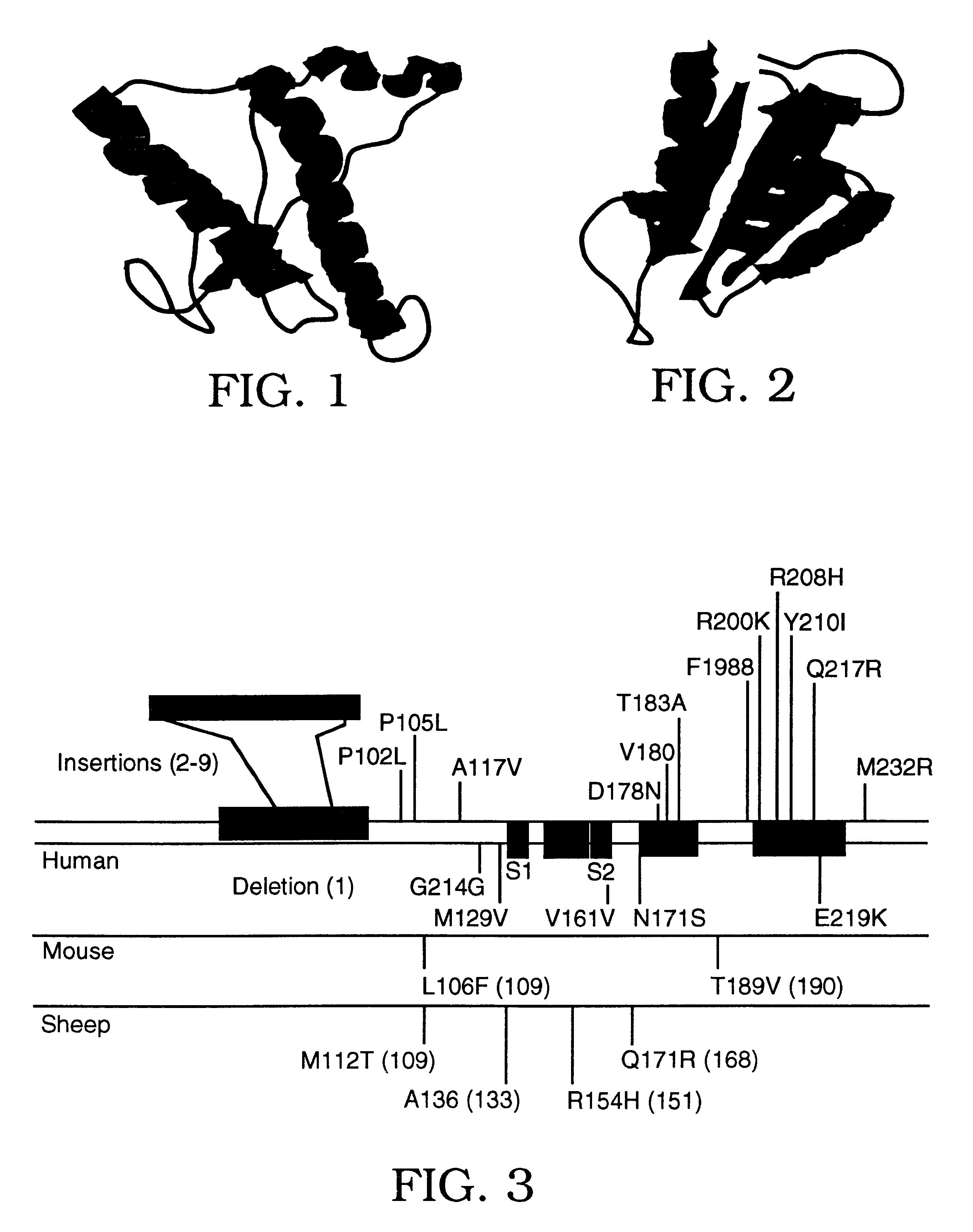
An examination of the amino acids which distinguish Hu PrP from Mo PrP shows only seven residues at the C-terminus (168-231) that are different. Four ofthese residues are close to the glycophosphatidylinosytol (GPI) anchor attached to Ser231 while the remaining three residues were within or near the C-terminus of a postulated -helix which has been conformed by NMR structural studies. To identify the critical binding site within PrP.sup.C for PPMF the seven residues were divided into two groups: those at the C-terminal end of the last -helix (HuA) and those at the extreme C-terminus (Hub). The Mo residues were replaced with Hu counterparts in positions that were critical for binding of PrP.sup.C to Mo PPMF to determine the effect of such on inhibiting the formation of recombinant PrP.sup.Sc. Recombinant PrP.sup.Sc was distinguished from endogenous wild-type (wt) Mo PrP.sup.Sc by using the SHa / Mo chimeric PrP designated MHM2 that contains a binding si...

example 2

Identification of Inhibitory Pharmacophores

Alanine scanning mutagenesis (Wells, 1991, Methods Enzymol. 202:390-411) has been used to estimate experimentally the binding contribution of single residues to a protein-protein interaction. For prion replication, residues 168, 172, 215 and 219 on the surface of the human PrP.sup.C molecule contribute the most to the integrity of the PrP-PPMF interface. Indeed, substitution for basic residues at this site increases the affinity of PrP for PPMF sufficiently to block the replication cycle. Thus, those side chain sites define a plausible 3D pharmacophore target for mimetic design. In particular, the sidechain coordinates of residues Q168, Q172, T215 and Q219 from the PrP(90-231) NMR structure as well as the coordinates of residue Q168 when helix B is extended to residue 166.

Since basic residues at Q168, Q172 and Q219 and acidic or hydrophobic residues at T215 increase the affinity of PPMF for PrPC, Arg, Lys, His, Asp, Glu and Trp were modeled...

example 3

Targeting the Inoculum to the Caveolar Space of Neuronal Cells

From a variety of studies, it has become clear that PrP.sup.Sc formation is likely to take place in the caveolar space. However, there is no specific reason why the refolded .beta.-rich peptides should preferentially concentrate in this region. PrP targeted to clatharin coated pits is not converted into PrP.sup.Sc. Since PrP is GPI anchored, lipidating the C-terminus of a polypeptide pharmacophore should improve cell localization. Accordingly, the Mo 89-143, P101L peptide was myristylated and applied to cells. The lipidated peptide can then localize to the cell surface and be internalized into endocytic vesicles.


PUM

PropertyMeasurementUnit
incubation timeaaaaaaaaaa

Abstract

Molecules are disclosed that interact with the cellular components involved in conversion of PrPC to PrPSc. The molecules disclosed can be small molecules, peptides or protein analogs, e.g. analogs of PrPC. In one embodiment, these molecules interfere with prion formation and / or replication, e.g. by preventing interactions of proteins involved in a prion complex or by interfering with beta-sheet formation. In another embodiment, the molecules of the invention promote PrPC conversion to PrPSc, e.g. by binding to PrPC and facilitating a conformational change from PrPC to PrPSc.

Description

This invention relates generally to proteins, functionally equivalent pharmacophores and methods of creating and / or detecting inhibitors of prion formation. Specifically, the invention relates to small molecules, peptides and peptide analogs with the ability to either inhibit prion formation or replication and methods of treating a neuropathology such as a prion-mediated neuropathology.Prions are infectious pathogens that cause central nervous system spongiform encephalopathies in humans and animals. Prions are distinct from bacteria, viruses and viroids. The predominant hypothesis at present is that no nucleic acid component is necessary for infectivity of prion protein. Further, a prion which infects one species of animal (e.g., a human) will not efficiently infect another (e.g., a mouse).From a clinical perspective, the prion diseases represent a variety of neurodegenerative states characterized at the neuropathologic level by the presence of spongiform degeneration and astrocyti...

Claims


Application Information

Patent Timeline
Patent Type & Authority Patents(United States)
IPC IPC(8): A61K31/417A61K31/4178A61K31/4164C07K1/04C07H21/00C07K1/00C07K14/435C07K14/47A61K38/00A01K67/027A61K31/4172A61K31/662A61K31/7064A61K31/7076A61K31/7084A61K45/00A61P25/00A61P43/00C07K16/18C12N5/10C12N15/09C12Q1/02G01N33/53
CPCA61K31/4178A61K31/7048A61K31/7076A61K31/417C07K1/045C07K1/047C07K14/47C07H21/00A61K38/00A61P25/00A61P43/00Y02P20/582
Inventor PRUSINER, STANLEY B.COHEN, FRED E.JAMES, THOMAS L.KANEKO, KIYOTOSHI
Owner RGT UNIV OF CALIFORNIA
Why Eureka
Try Eureka