Computer radiographic oncology portal imaging

a portal imaging and computer radiographic technology, applied in the direction of instruments, radiation beam directing means, conversion screens, etc., can solve the problems of normal tissues being overexposed, healthy tissues being overexposed, adversely affecting image quality,

Inactive Publication Date: 2002-08-06
CARESTREAM HEALTH INC
View PDF8 Cites 15 Cited by
  • Summary
  • Abstract
  • Description
  • Claims
  • Application Information

AI Technical Summary

Problems solved by technology

Usually, this causes some healthy tissues to be overexposed.
Usually this leads to some normal tissues being overexposed.
However, this adversely affects image quality as pointed out in several publications [for example, Wilenzink et al., Med. Phys., 14(3), 1987, pp.
However, the weight of such image storage assemblies is consid

Method used


Image

  • Computer radiographic oncology portal imaging
  • Computer radiographic oncology portal imaging

Examples


example 2

Scanning of Image Storage Assemblies

The imaging assemblies described in Example 1 were "erased" using the conventional "CR400 smart erase" feature. The original images in the phosphor layers were erased completely. A 0.25 mR exposure was then directed to each imaging assembly, followed by scanning using a conventional KODAK CR 400 reader. The results of scanned showed the 0.25 MR signal, but there was no evidence of the original images.

example 3

Portal Imaging

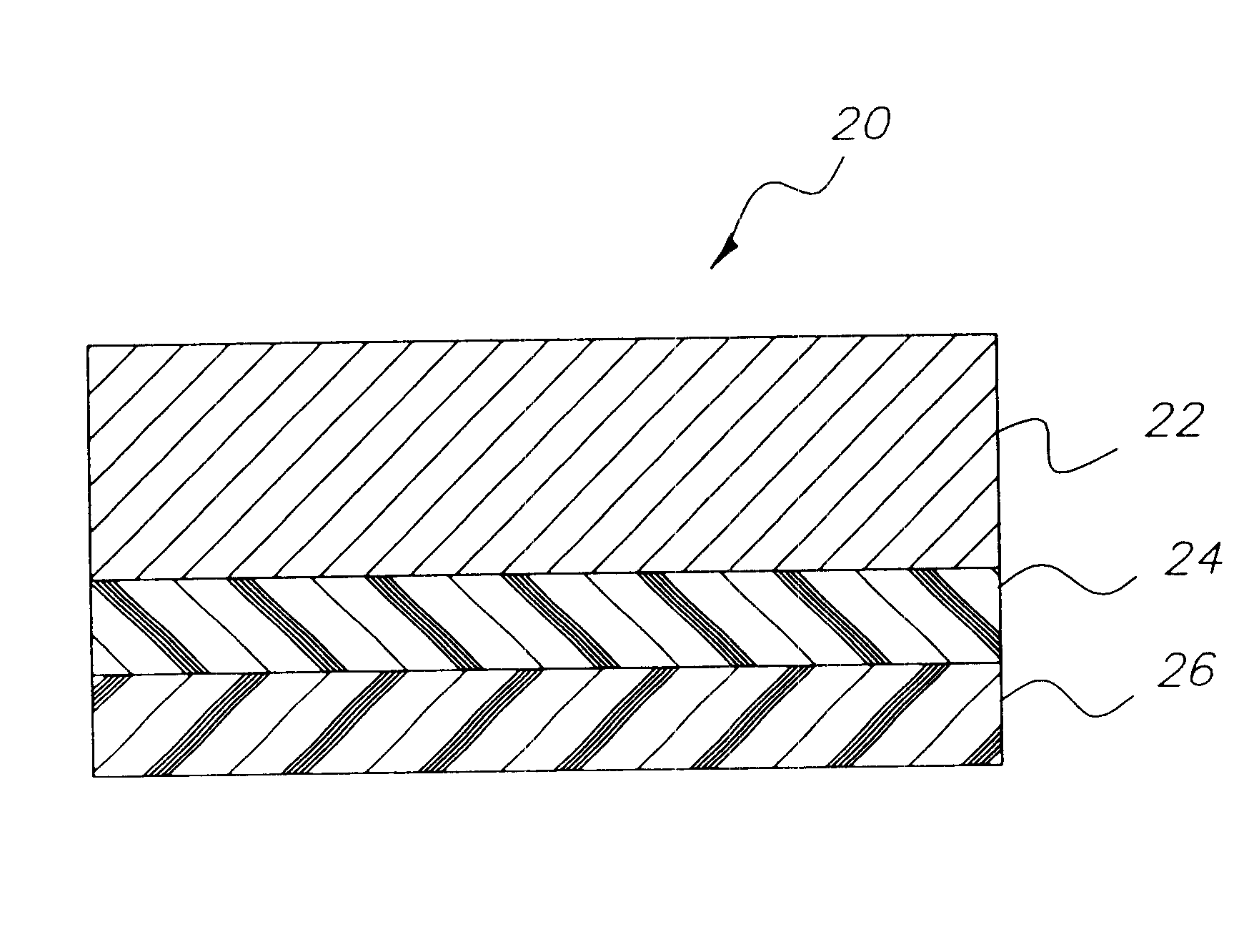
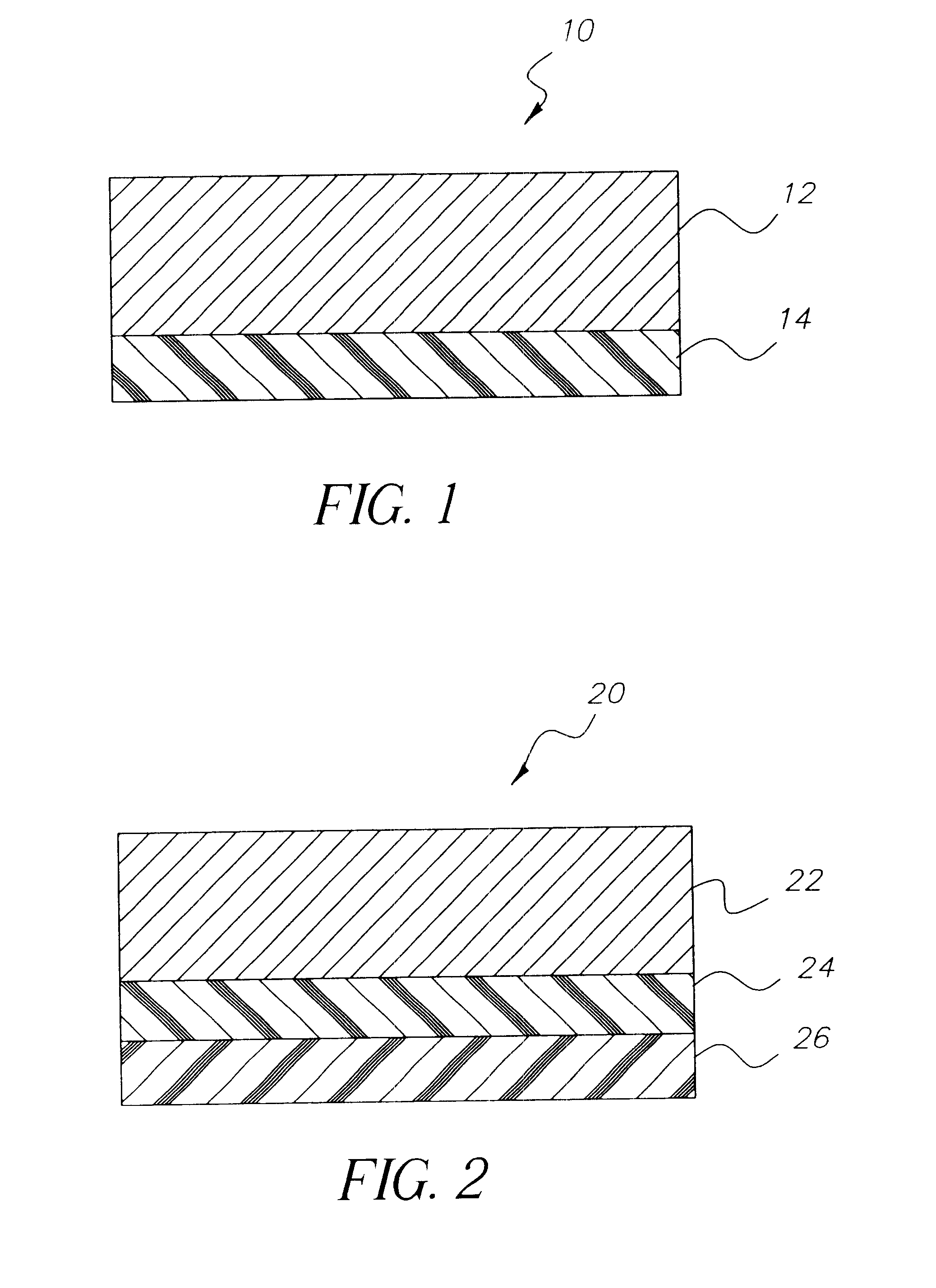
Using a conventional Varian, Inc. radiation therapy treatment machine, a 1.5-cm plastic patient tissue phantom was exposed through the "back" of each imaging assembly described in Example 1. A 10-cm plastic patient tissue phantom was on the front side of each imaging assembly. This provided calibrated exposures to each imaging assembly. Each of the imaging assemblies was exposed to 1 Rad, 3 Rads, 7 Rads, and 10 Rads in 10 cm square fields at the comers of the storage phosphor layers using 6 Mvp. We were surprised to observe that the 1-Rad signals in the storage phosphor layers behind each of the different lead screens were the same, .+-.3%. Surprisingly, the signal in the phosphor layer behind the copper screen (Control assembly) was 10% smaller.

The signal results in the imaging assemblies were the same for the other exposure levels. The imaging assemblies with the lead screens gave similar signals but the signal from the Control (copper) assembly was smaller by about ...

example 4

Scanning of Image Storage Assemblies

The imaging assemblies exposed and evaluated in Example 3 were erased completely as described in Example 2. A 0.25 mR exposure was directed to each of the storage phosphors in the assemblies, and the resulting images were scanned using a conventional KODAK CR 400 reader. The scanned 0.25 mR images were seen but not the initial higher exposure.


PUM


Abstract

Image storage assemblies comprise thin metal screens adjacent phosphor storage screens. The thin metal screen is from about 0.01 to about 0.75 mm in thickness when composed of copper and from about 0.05 to about 0.4 mm when composed of lead. These image storage assemblies can be used in portal imaging whereby imaging radiation is directed through the thin metal screens before the phosphor storage screens. Stimulating radiation can then be directed to the phosphor storage screen before it reaches the thin metal screen.

Description

This invention is directed to radiography and in particular to image storage assemblies that are useful for oncology portal imaging. It also relates to radiation image recording and reproducing method. Thus, this invention is useful in portal radiography.BACKGROUND OF TIE INVENTIONIn conventional medical diagnostic imaging the object is to obtain an image of a patients internal anatomy with as little X-radiation exposure as possible. The fastest imaging speeds are realized by mounting a dual-coated radiographic element between a pair of fluorescent intensifying screens for imagewise exposure. About 5% or less of the exposing X-radiation passing through the patient is adsorbed directly by the latent image forming silver halide emulsion layers within the dual-coated radiographic element. Most of the X-radiation that participates in image formation is absorbed by phosphor particles within the fluorescent screens. This stimulates light emission that is more readily absorbed by the silve...

Claims


Application Information

Patent Timeline
IPC IPC(8): A61B6/08G01N23/02G01N23/04G03C5/17G21K4/00
CPCG03C5/17G21K4/00A61B6/583
Inventor STEKLENSKI, DAVID J.MOORE, WILLIAM E.BYNG, JEFFREY W.
Owner CARESTREAM HEALTH INC
Why Eureka
Try Eureka