Endoscope

a technology of endoscope and endoscope body, which is applied in the field of medical instruments, can solve the problems of insufficient macroscopic magnification of the previously known endoscope, insufficient examination of the organ tissue in sufficient detail to determine, and unnecessary harm and even loss of organ function to the patient, so as to prevent unnecessary biopsies and/or organ removal, the effect of enhancing the imaging of microscopic images

Inactive Publication Date: 2002-09-17
INNER VISION IMAGING
View PDF37 Cites 39 Cited by
  • Summary
  • Abstract
  • Description
  • Claims
  • Application Information

AI Technical Summary

Benefits of technology

The ability to provide microscopic imaging of internal organs permits in vivo pathological examination of internal organs without the necessity of a biopsy. Furthermore, to enhance the imaging of the microscopic images, preferably infrared radiation is provided directly through the lens tube and stage window to the tissue. Any conventional source of infrared illumination can be utilized, but preferably a diode laser is utilized.
In the preferred embodiment of the invention, the optical images through the endoscope are digitized and coupled as an input signal to a computer system. The computer system, in turn, communicates the digitized images via a network and / or telephone lines to a pathologist remote from the patient. Consequently, the pathologist is capable of viewing the images through the endoscope on a real-time basis. Since the endoscope of the present invention enables real-time pathological examination of suspect tissue, unnecessary biopsies and / or organ removal is prevented.

Problems solved by technology

Perhaps the most significant disadvantage of these previously known endoscopes is that, since the optical lenses are fixed within the endoscope, the angular magnification for the endoscope remains constant.
For example, in certain situations where cancerous growths within body organs are suspected, the macroscopic magnification provided by the previously known endoscopes is insufficient to examine the organ tissue in sufficient detail to determine whether the tissue abnormality is cancerous or benign.
First, in the event that the organ abnormality is benign, the biopsy and possible removal of the entire organ from the body results in unnecessary harm and even loss of organ function to the patient.
Second, since the subsequent pathological examination of the body tissue oftentimes occurs long after the end of the operation, in the event that the pathological examination reveals a cancerous growth within the body tissue, it is oftentimes necessary for the surgeon to re-enter the body cavity and remove additional body tissue in an attempt to completely eradicate the cancer.
This disadvantageously, however, subjects the patient to a second operation.
These prior devices, however, have used complex systems for moving the lenses which disadvantageously increase the overall diameter of the endoscope.

Method used


Image

  • Endoscope
  • Endoscope
  • Endoscope

Examples


Embodiment Construction

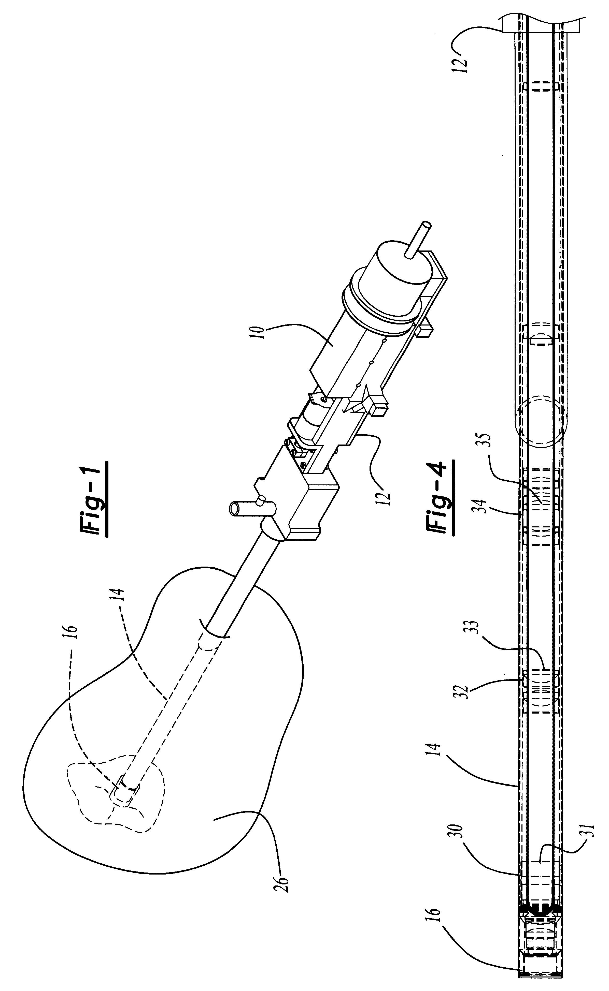
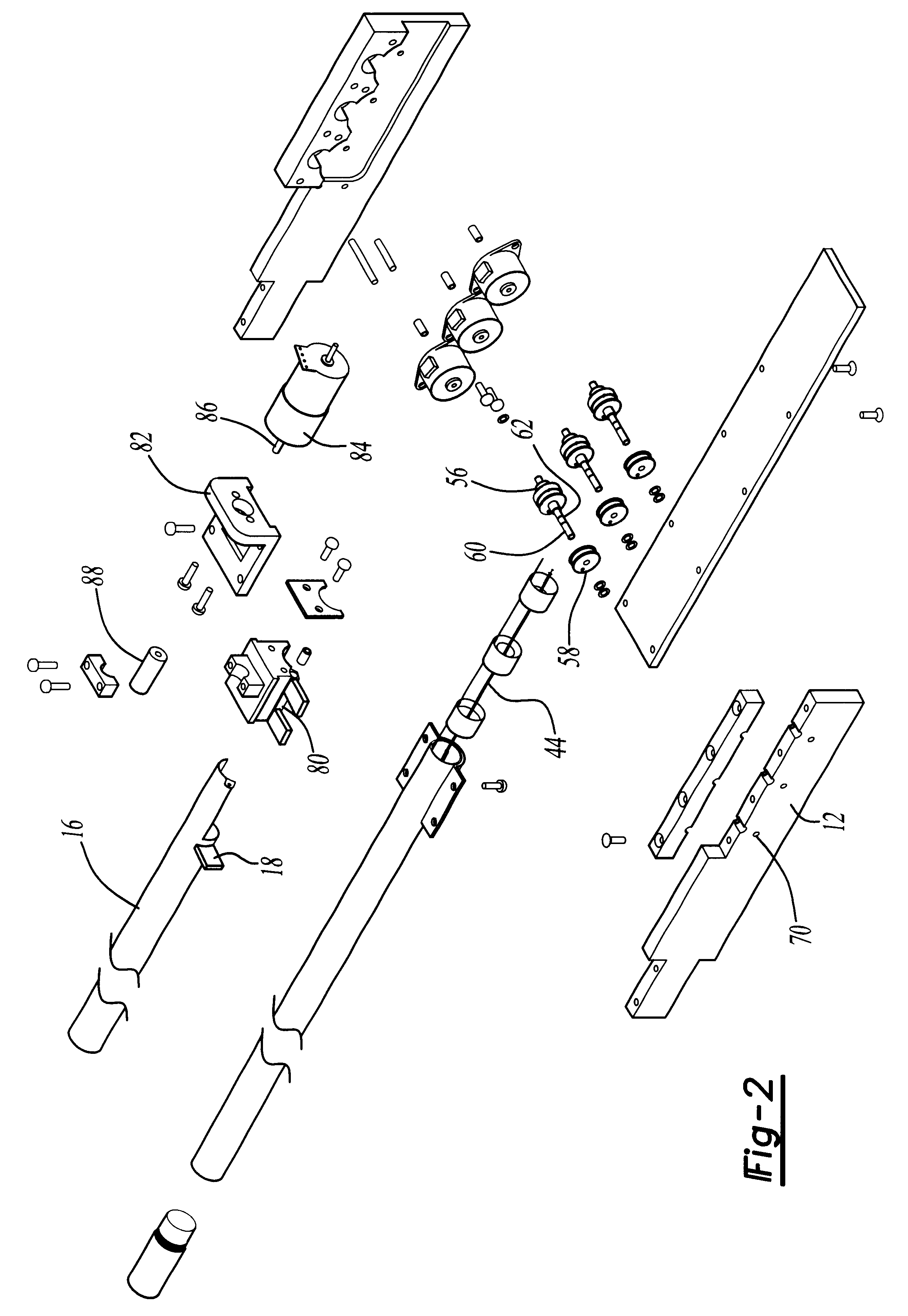
With reference first to FIGS. 1 and 3, a first preferred embodiment of the endoscope 10 of the present invention is there shown and comprises a housing 12 dimensioned to be hand held by medical personnel. An elongated lens tube 14 is secured to and extends outwardly from the housing 12. An elongated tubular stage 16 is open at one end and has a transparent window 20 (FIG. 6) disposed across its opposite end. The stage 16, furthermore, is dimensioned to be slidably received over the free end 15 of the lens tube 14 and detachably secured to the housing 12 by a bayonet coupling 18 (FIG. 2). Furthermore, with the stage 16 secured to the housing 12, the stage and lens tube are adapted for insertion into a body cavity 26.

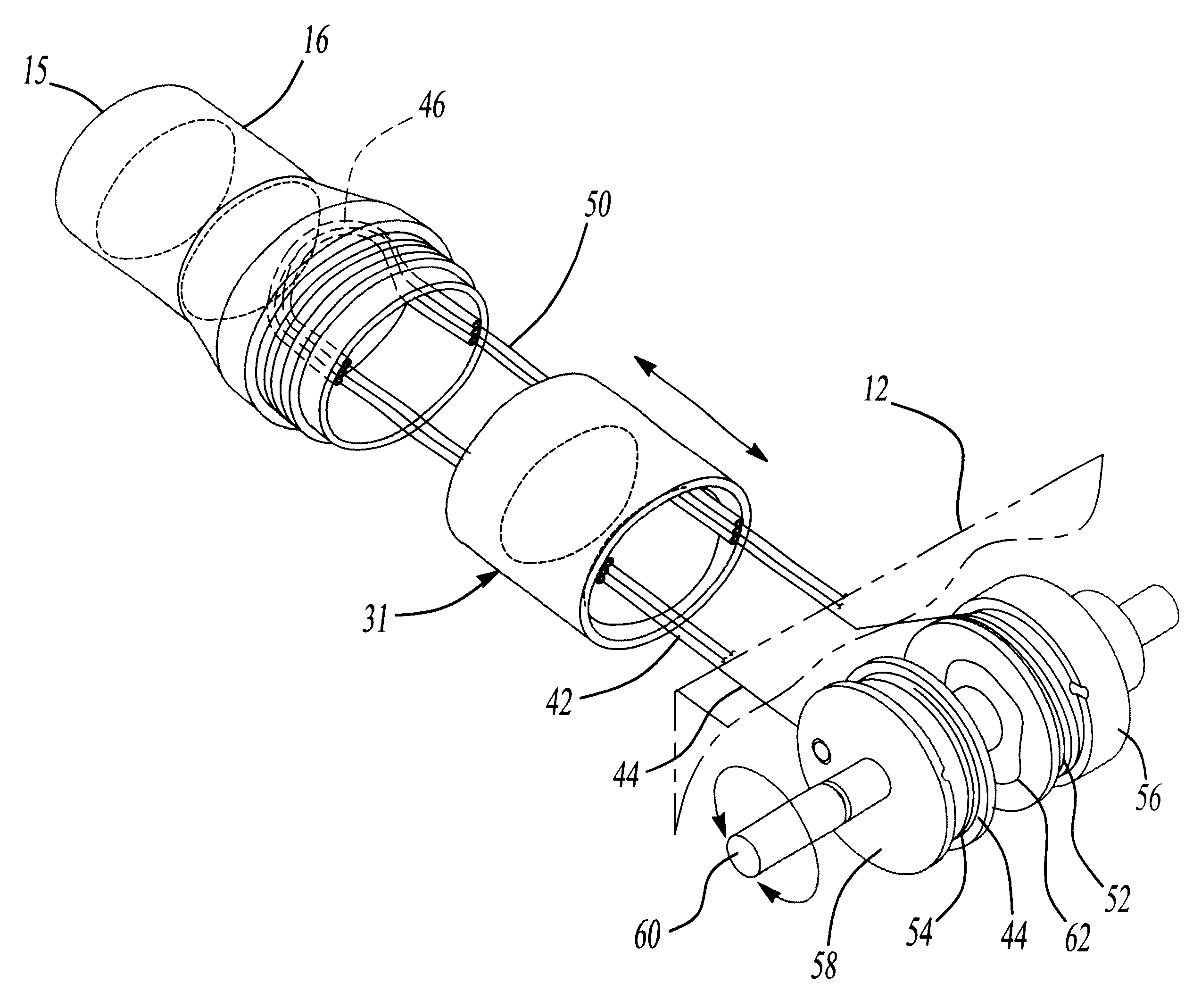
With reference now to FIGS. 3, 4 and 8, the endoscope 10 further comprises at least one, and preferably three moveable lens groups 30, 32 and 34. Each lens group 30, 32 and 34 is secured within its own support tube 31, 33 and 35, respectively, and these lens support tubes...


PUM


Abstract

An endoscope assembly is disclosed having a housing adapted to be manipulated by medical personnel, such as a surgeon. An elongated lens tube has one end secured to the housing while the free end of the lens tube is adapted for insertion into a cavity of a body. At least one, and preferably several, lens assemblies are longitudinally slidably mounted within the lens tube and movable between longitudinally spaced positions within the lens tube. A drive mechanism is mounted to the housing and mechanically coupled to each lens assembly to move the lens assemblies longitudinally within the lens tube independently of one another. In doing so, the lens assemblies provide varying degrees of magnification between low or macroscopic magnification and microscopic magnification at the free end of the lens tube. A stage is also detachably secured over the lens tube and has a transparent window across its end adjacent the free end of the lens tube. In use, the stage window is positioned against target tissue within a human cavity while the lens assemblies are longitudinally displaced within the lens tube to obtain the desired magnification. Strands extending through and/or connected to the lens groups are used to longitudinally displace the lens groups.

Description

I. Field of the InventionThe present invention relates generally to medical instruments and, more particularly, to an endoscope.II. Discussion of Related ArtLaparoscopic surgery has enjoyed increasing acceptance, particularly for surgery involving the abdominal cavity. In such surgery, one or more incisions are made through the patient's skin. Thereafter, various medical instruments, including endoscopes, are inserted through the incisions and into a body cavity, such as the abdominal cavity.In order for the surgeon to see into the abdominal cavity, the surgeon typically uses an endoscope which is inserted through a cannula and into the abdominal cavity. The previously known endoscopes typically comprise an elongated tube having one or more fixed lenses. These lenses provide an optical view of the interior of the body cavity to an eyepiece or other display means accessible to the surgeon outside the body. Illumination for the endoscope is typically provided by optical fibers which e...

Claims


Application Information

Patent Timeline
Patent Type & Authority Patents(United States)
IPC IPC(8): A61B1/00G02B23/24A61B5/00H04N5/225
CPCA61B1/00135A61B1/00188A61B5/0068G02B23/2438G02B23/2446A61B5/0002A61B5/0084A61B2562/146A61B1/00
Inventor FARKAS, RICHARDFISHER, RICHARDHENKE, STEVEN
Owner INNER VISION IMAGING
Why Eureka
Try Eureka