Diffraction enhanced x-ray imaging of articular cartilage

a technology of articular cartilage and x-ray imaging, which is applied in the field of contrast mechanism, can solve the problems of insufficient pain and early warning signs, failure to achieve successful conservative treatments that could lead to tissue regeneration, and deformation of the entire join

Inactive Publication Date: 2003-06-10
ILLINOIS INSTITUTE OF TECHNOLOGY +2
View PDF11 Cites 33 Cited by
  • Summary
  • Abstract
  • Description
  • Claims
  • Application Information

AI Technical Summary

Problems solved by technology

The degeneration of articular cartilage is a component of pathological processes that result in the destruction of the tissue and leads to the deformation of the entire joint.
Use-related joint pain is one of the first signs of disease; however, pain is not always an early warning sign.
By the time pain becomes a symptom, successful conservative treatments that could lead to regeneration of the tissue are too late.
Conventional procedures for repairing of transplanting articular cartilage do not restore a normal articular surface.
Therefore cartilage cannot be easily seen in conventional radiograph.
However, condensed collagen fibrils may also cause such effects.
Phase contrast images are limited to either thin objects or high x-ray imaging energies.

Method used


Image

  • Diffraction enhanced x-ray imaging of articular cartilage
  • Diffraction enhanced x-ray imaging of articular cartilage
  • Diffraction enhanced x-ray imaging of articular cartilage

Examples


example

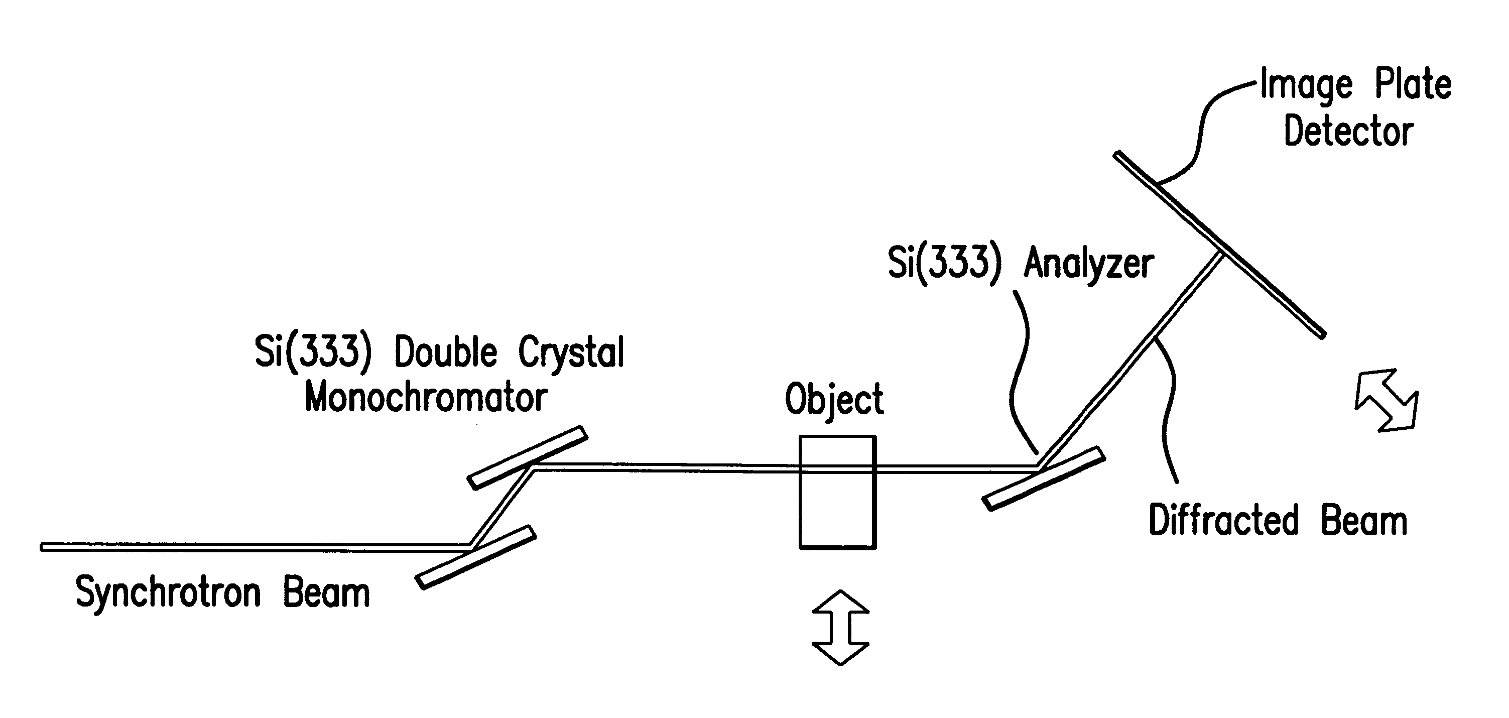
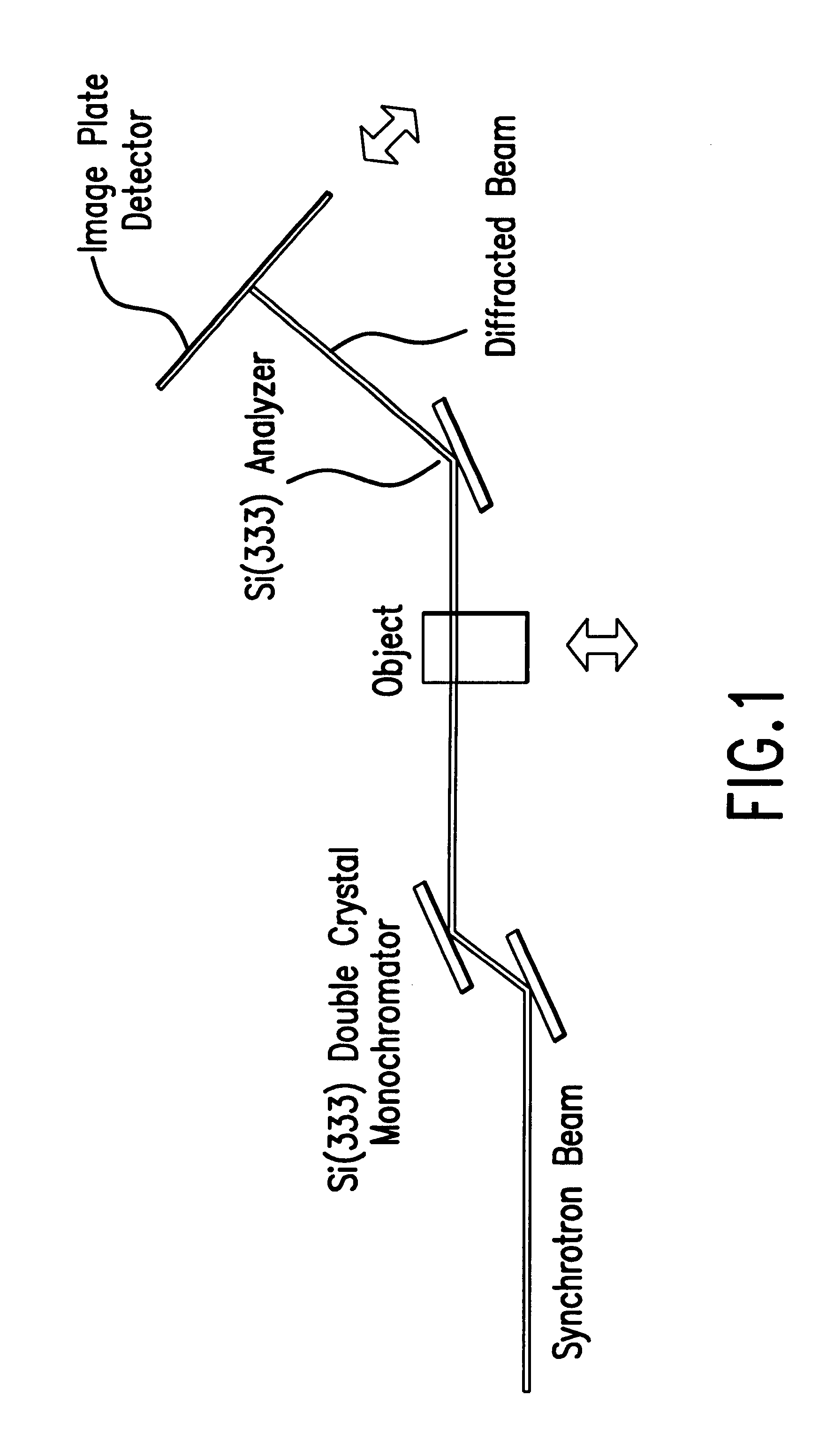
Experiments using the method and the system of this invention were performed at the National Synchrotron Light Source 15A Imaging PRT Beamline, at Brookhaven National Laboratory, Upton, N.Y. Preliminary results have rendered an interpretation of refraction and a qualitative description of extinction. One experimental setup is shown in FIG. 1. A white synchrotron beam was made nearly monochromatic by a silicon double-crystal monochromator. One usable energy range of this system, as used, was 15 keV -40 keV. For the measurements described here the beam energy was 18 keV with a bandwidth of 1.5 eV, but could also be 16 keV to 100 keV with a bandwidth of 1.5 eV and 2.6 eV, respectively. An in-hutch monochromator was used to monochromate the beam to provide the imaging beam. The monochromator had silicon (3,3,3)-lattice planes. The type of lattice planes can be selected to determine the refraction and scatter rejection sensitivity. All of the crystals in the system were in the parallel g...


PUM

PropertyMeasurementUnit
energy levelaaaaaaaaaa
photon energyaaaaaaaaaa

Abstract

A method and system for detecting an image of an object, particularly a soft tissue material. A generated x-ray beam is transmitted through the soft tissue material. A transmitted beam is directed at an angle of incidence upon a crystal analyzer. An image of the object is detected from a beam diffracted from the crystal analyzer either at or near a peak of a rocking curve of the crystal analyzer. The method and system of this invention is particularly useful for analyzing images of cartilage.

Description

1. Field of the InventionThis invention relates to a contrast mechanism referred to as extinction contrast which can be used for a variety of x-ray imaging applications. The method and system of this invention relates to an x-ray imaging modality that uses an analyzer crystal after the object. The imaging contrast of this invention is based on attenuation and refraction.2. Description of Related ArtOne type of diffraction enhanced imaging (DEI) is described in Chapman et al., U.S. Pat. No. 5,987,095.Articular cartilage covers the ends of bones in synovial joints and provides elasticity, distribution of load, resistance to compressive forces, smooth articulation and cushioning of the subchondral bone during joint movements. Tissue is composed of collagen, primarily type II collagen, entrapping compressed proteoglycan aggregates.The degeneration of articular cartilage is a component of pathological processes that result in the destruction of the tissue and leads to the deformation of ...

Claims


Application Information

Patent Timeline
Patent Type & Authority Patents(United States)
IPC IPC(8): A61B6/00G01N23/225G01N23/22G01N23/20
CPCA61B6/50A61B6/502G01N23/20G01N23/2258A61B6/4092A61B6/505A61B5/4514A61B5/4528G21K2207/005A61B6/583
Inventor CHAPMAN, LEROY DEANHASNAH, MOUMEN O.OLTULU, ORALZHONG, ZHONGMOLLENHAUER, JUERGENMUEHLEMAN, CAROLKUETTNER, KLAUSAURICH, MATTHIASPISANO, ETTA D.JOHNSTON, R. EUGENETHOMLINSON, WILLIAM CRAIGSAYERS, DALE
Owner ILLINOIS INSTITUTE OF TECHNOLOGY
Why Eureka
Try Eureka