Non-viable keratinocyte cell composition or lysate for promoting wound healing

a keratinocyte and non-viable technology, applied in the field of non-viable keratinocyte cell composition or lysate for promoting wound healing, can solve the problems of insufficient amount to cover the entire wound surface, risk of transferring pathogens such as human immunodeficiency virus, hepatitis b virus, cytomegalovirus, from the skin donor to the acceptor patient,

Inactive Publication Date: 2003-07-01
CELLTRAN LTD
View PDF17 Cites 27 Cited by
  • Summary
  • Abstract
  • Description
  • Claims
  • Application Information

AI Technical Summary

Problems solved by technology

Even then, the amount may not be sufficient to cover the entire wound surface.
However, this time period may be critical for the patient, especially in the case of extensive third degree burns.
A main disadvantage of cultured allografts is the risk of transferring pathogens such as human immunodeficiency virus, hepatitis B virus, and cytomegalovirus, from the skin donor to the acceptor patient.
The availability of these sheets in terms of time and quantity is a major drawback of fresh keratinocyte grafts.
However, it is impossible to predict when a patient will enter the hospital and how many keratinocyte grafts will then be needed.
Another drawback of using fresh (autologous) keratinocyte sheets is that a certain period of time is still required before a sufficient amount of keratinocyte sheets can be grown and then applied.
Thawed cryopreserved keratinocyte grafts have to be handled carefully since the sheets are extremely fragile.
In the case of keratinocyte sheets, the wound is covered with a sheet and it is assumed to be useless to put another sheet on top.
Fresh keratinocyte grafts cannot be tested and, in the case of cryopreserved keratinocyte sheets, the preparation of the material requires additional work.
This is obviously impossible when fresh keratinocyte sheets are used.
Moreover, fresh sheets must be used at an optimal time and may not be stored at all.
If there are no patients to treat at that optimal moment of culture, the cells become worthless for this purpose.
Moreover, these keratinocyte grafts may detach from the supporting gauze, and start to float or curl up, hampering the exact method of application, i.e., basal keratinocytes facing downwards.
The drawback is that this process results in low yields of keratinocyte sheets.
It is not clear from this article whether confluency is reached, which makes the cultured cells inappropriate for wound healing.
The drawbacks are the necessity of high seeding density and the slow growth of the cell cultures.
Moreover, epidermal growth factor exposure resulted in the rapid appearance of a cell type which lacked the characteristic keratinocyte morphology and proliferated rapidly.
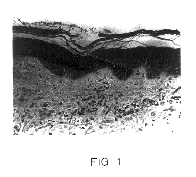
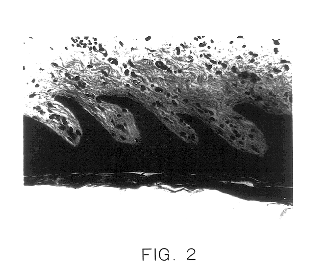
Collagen bundles and blood capillaries cannot be evaluated on this picture because of the low magnification.

Method used


Image

  • Non-viable keratinocyte cell composition or lysate for promoting wound healing
  • Non-viable keratinocyte cell composition or lysate for promoting wound healing
  • Non-viable keratinocyte cell composition or lysate for promoting wound healing

Examples


example 1

Skin Source and Isolation of Keratinocytes

Human keratinocytes were isolated from surgical skin specimens (foreskin, mammoplasties, abdominal skin, and other locations on the body), and from cadaver skin.

Skin was removed with a dermatome at a thickness up to 1 mm. Thicker pieces of skin, sometimes containing hypodermis, were used as well. The skin was immediately placed in phosphate buffered saline (PBS): 8 g NaCl, 0.2 g KCl, 3.1 g Na.sub.2 HPO.sub.4.12H.sub.2 O, 0.2 g KH.sub.2 PO.sub.4 in MILLI-Q.RTM. water to 1 L; pH 7.4) and kept at 4.degree. C. Specimens were then transferred to the culture lab within 4 h. The skin was rinsed extensively with PBS containing 100 U / ml penicillin, and 100 mg / ml streptomycin. If necessary, skin samples were stored at 4.degree. C. in solution A, which is an isotonic buffered solution containing 30 mM Hepes, 10 mM glucose, 3 mM KCl, 130 mM NaCl, 1 mM Na.sub.2 HPO.sub.4.12H.sub.2 O and 3.3 nM phenol red. Thin pieces of skin may be stored for about 16 h,...

example ii

Role of EGF, Cholera Toxin, Hydrocortisone, and Insulin in Feeder Layer-free Keratinocyte Grown In Vitro

Summary

Epidermal growth factor (EGF) is necessary to induce rapid proliferation of human keratinocytes. However, a low yield of attached keratinocytes is seen when EGF alone is present in the medium. Cholera toxin (CT) allows a high yield of attached and spread keratinocytes. On the other hand, growth arrest is seen when CT alone is present in the culture medium.

Hydrocortisone induces keratinocyte proliferation but is less potent than EGF. Insulin has a beneficial effect on keratinocytes, although its effect is more important when the medium contains EGF and cholera toxin, or EGF and hydrocortisone, than when EGF, cholera toxin, and hydrocortisone are present, particularly at the preferred concentrations.

The optimal concentrations of EGF, hydrocortisone, and cholera toxin in the culture medium are respectively for instance 10 ng, 0.13 .mu.g and 9 ng per ml. Insulin may be added, p...


PUM

PropertyMeasurementUnit
temperatureaaaaaaaaaa

Abstract

Cultures of keratinocyte cells are provided which are free from nonautologous fibroblasts and organ extracts, and which have a high speed of cell amplification for a minimum seeding density. The cultures can be cryopreserved in a buffered isotonic medium containing serum and a cryoprotectant. The cultures are produced by a process that does not involve the use of a feeder layer and organ extracts. A culture medium which can be used contains Medium 199, serum, epidermal growth factor, cholera toxin and/or hydrocortisone, and optionally insulin. A substance for wound healing and for cosmetic applications is derived from cultured human keratinocytes. A non-viable total keratinocyte lysate for use in promoting wound healing is produced by growing keratinocyte cells on a support, detaching the cells from the support, and lysing the detached cells to obtain the lysate which may be frozen and lyophilized. The cells may be grown without using a support to produce the lysate, or to produce a non-viable keratinocyte cell culture lyophilisate or spray dried non-viable keratinocyte cell composition for use in healing wounds.

Description

The invention relates to new cultures of keratinocytes.The invention also relates to a process for preparing the same.The invention also relates to the use of new cultures of keratinocytes as wound healing substances.The invention also relates to pharmaceutical compositions containing as active substances, said new cultures of keratinocytes.The invention also relates to cosmetic compositions, containing as active substances, new cultures of keratinocytes.DESCRIPTION OF THE PRIOR ARTSkin is presumably the organ most subject to injury. Skin repair is a complex process that can be divided in 4 phases usually described as inflammation, granulation tissue formation, epithelialization and remodeling of the connective tissue matrix. Each of these phases is complex in itself, and it is clear that for good wound healing, the processes must occur successively and in coordination. Good wound healing can be defined as restoration of the skin, including the dermal and epidermal part, in such a w...

Claims


Application Information

Patent Timeline
Patent Type & Authority Patents(United States)
IPC IPC(8): A61K35/36C12N5/06A61K35/12C12N5/071
CPCA61K35/36C12N5/0629A61K35/12C12N2501/01C12N2501/11C12N2501/33C12N2501/39Y02A50/30
Inventor VAN BOSSUYT, HANS
Owner CELLTRAN LTD
Why Eureka
Try Eureka