Ultrasound probe for ultrasound examination system

a technology of ultrasound and probe, which is applied in the direction of ultrasonic/sonic/infrasonic diagnostics, tomography, catheters, etc., can solve the problems of difficult to reduce the diameter of the probe to a size, and difficult to insert the probe into a narrow guide channel or endoscope passag

Inactive Publication Date: 2004-01-06
FUJI PHOTO OPTICAL CO LTD
View PDF10 Cites 4 Cited by
  • Summary
  • Abstract
  • Description
  • Claims
  • Application Information

AI Technical Summary

Benefits of technology

It is another object of the present invention to make it possible to insert into and pass through a narrow guide passage a connector portion which is provided at the proximal end of a flexible cord of an electronic scan type ultrasound probe and which usually incorporates a large number of terminals for connection to an ultrasound signal processor of an ultrasound image observation terminal.
When coiled into the shape of a helical roll, the flexible wiring film should be easily passable through a narrow guide passage. For this purpose, the flexible wiring film is rolled into a cylindrical shape gradually from the root end portion which is connected to the flexible cord of the probe. In the case of a flexible wiring film with a broader surface area, in order to hold the flexible wiring film stably in the coiled shape, that is, in the cylindrical shape, it is desirable to extend the flexible wiring film obliquely in the rearward direction relative to the longitudinal axis of the flexible cord of the probe. By so doing, the flexible wiring film strip can be easily coiled into a partially overlapped helical roll. To prevent uncoiling or unrolling of the rolled flexible wiring film, it is necessary to stop the free end of the film. For this purpose, a rubber band or an adhesive tape can be used. Alternatively, a flexible sheath member may be fitting on the free end of the film. Further, the flexible wiring film may be imparted with a convolving traits beforehand in order to facilitate the job of coiling same into a partially overlapped helical roll.

Problems solved by technology

Since the ultrasound transducer on an electronic scan type probe contains a large number of transducer elements, it is difficult to reduce its diameter down to a size which can easily pass through a biopsy channel of an endoscope.
However, in the case of an electronic scan type ultrasound probe which requires a large number of contact points, it becomes necessary to provide a large connector at the proximal end of the flexible cord.
Therefore, even if the flexible cord of an electronic scan type probe is thin and narrow enough for passage through an endoscopic biopsy channel, it has been often found difficult to insert the probe into a narrow guide channel or passage of an endoscope because of bulkiness of the ultrasound scanner head and the connector which are provided at the head and tail ends of the flexible cord, despite various advantages of electronic scan type probe in ultrasound examinations.
However, the ultrasound is not introduced into a body cavity by itself.

Method used


Image

  • Ultrasound probe for ultrasound examination system
  • Ultrasound probe for ultrasound examination system
  • Ultrasound probe for ultrasound examination system

Examples


Embodiment Construction

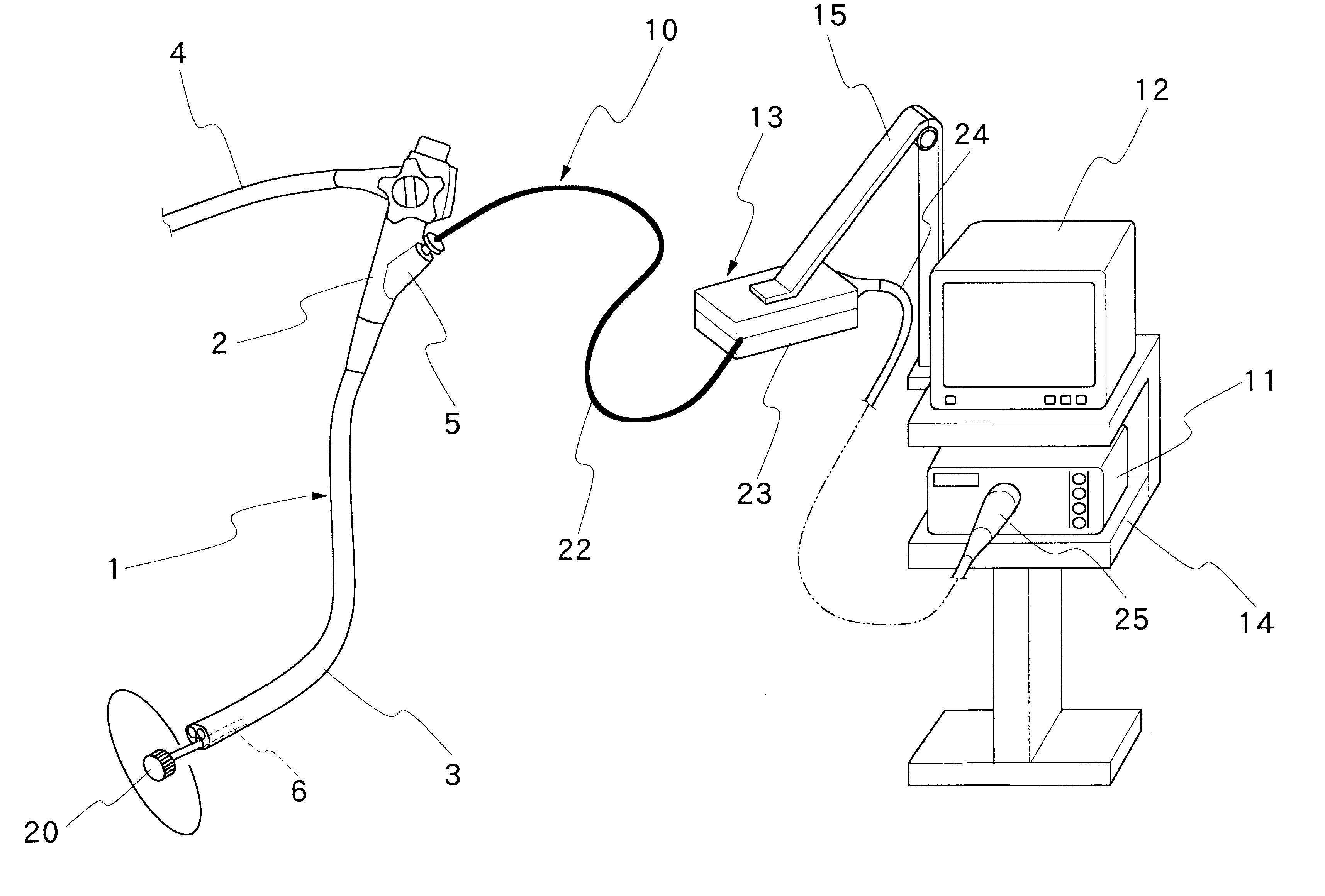
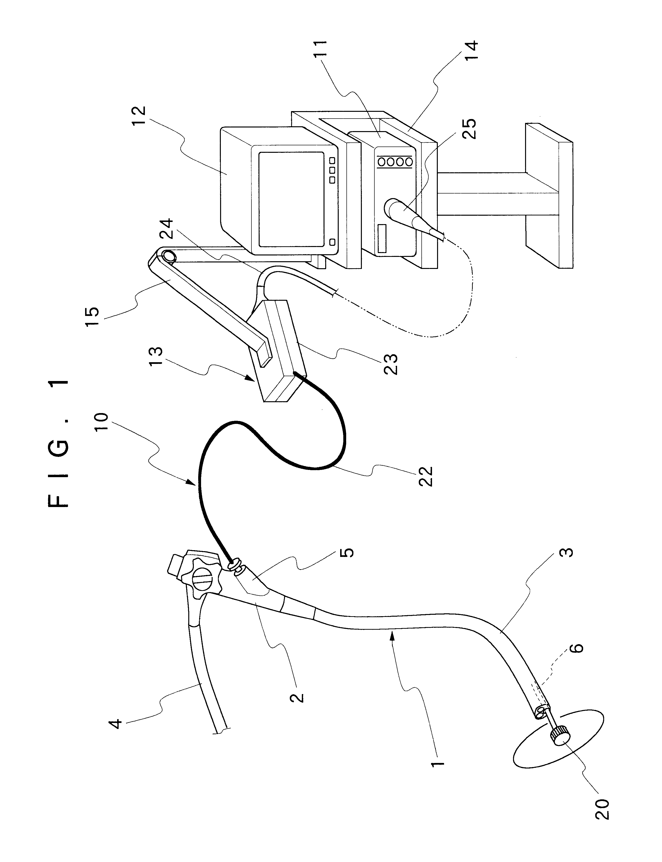
Hereafter, the present invention is described more particularly by way of preferred embodiments with reference to the accompanying drawings. Referring first to FIG. 1, there is shown general configuration of an ultrasound examination system. In the case of this embodiment, the ultrasound examination system employs by way of example an ultrasound probe which is adapted to be introduced into a body cavity of a patient by way of a biopsy channel of an endoscope or a similar guide means.

In that figure, indicated at 1 is an endoscope which has an insertion instrument 3 extended out from a manipulating head assembly 2 for insertion into a body cavity. A universal cable 4 which is led out from the opposite side of the manipulating head assembly 2 is for connection to a light source (or an image processor). An entrance housing 5 is provided on the manipulating head assembly 2 for insertion of biopsy or surgical instruments such as forceps, a high frequency tool and so forth. Although not sh...


PUM


Abstract

Attached to a proximal end portion of a flexible cord of an ultrasound probe of an ultrasound examination system, to be introduced into a body cavity through a narrow guide passage, is a flexible wiring film which can be coiled into a helical roll of a narrow cylindrical shape extending along an axial extension line of the flexible cord at the time of passing same through the narrow guide passage. Formed on a surface of the flexible wiring film are a plural number of wire connection points for connection of signal lines from transducer elements of an ultrasound transducer which is provided at a fore distal end of the flexible cord of the probe, and a corresponding number of electrodes which are electrically connected with the wire connection points through a wiring pattern. The electrodes on the flexible wiring film are disconnectibly connected to corresponding contact points on a contact assembly of a relay means.

Description

1. Field of the ArtThis invention relates to an ultrasound examination system, and more particularly to an ultrasound probe which can be introduced into a body cavity through a narrow guide passage like a biopsy channel of an endoscope or the like.2. Prior ArtUltrasound probes of the sort which are adapted to be inserted into a body cavity by way of an endoscope or a similar guide means have been well known in the art and widely used for ultrasound examination systems. For instance, Laid-Open Japanese Patent Application H11-56836 discloses an ultrasound examination system employing an ultrasound probe which is designed to be introduced into a body cavity through a biopsy channel of an endoscope. The ultrasound probe of this ultrasound examination system has an ultrasound scanner head with an ultrasound transducer element, attached to the fore distal end of a flexible cord to be inserted into an endoscopic biopsy channel. A connector is provided at the base or proximal end of the fle...

Claims


Application Information

Patent Timeline
Patent Type & Authority Patents(United States)
IPC IPC(8): A61B8/12A61B1/00H04R17/00
CPCA61B8/12
Inventor ITOI, HIROMU
Owner FUJI PHOTO OPTICAL CO LTD
Why Eureka
Try Eureka