Gamma camera and CT system

a gamma camera and ct system technology, applied in the field of nuclear medicine, to achieve the effect of reducing the sensitivity of the gamma camera head

Inactive Publication Date: 2005-04-12
ELGEMS
View PDF29 Cites 24 Cited by
  • Summary
  • Abstract
  • Description
  • Claims
  • Application Information

AI Technical Summary

Benefits of technology

This allows for three important advantages. Firstly, this allows for matching between the conditions under which the data is acquired, i.e., the same averaging of body motion is intrinsic for both acquisitions. Second, a slow rotation rate gantry may be used. Third, lower X-ray power may be used. This allows for a smaller power supply and for a smaller gantry, of the size and type normally suitable for gamma cameras alone.
An aspect of some preferred embodiments of the invention involves the alignment and calibration of a combined CT / NM imaging system. In preferred embodiments of the invention, the structure of the CT portion of the system is very simple as compared with dedicated CT systems, since the alignment and power requirements and the weight of the system are all greatly reduced. In order to simplify the adjustment of the X-ray system and especially the field replacement of the X-ray system, a method of alignment based on a standard alignment surface and position and a method of providing these surfaces without accurate machining of these surfaces. In a preferred embodiment of the invention, the alignment surfaces are mounted onto the gantry by screws, based on a position determined by an alignment jig centered at the center of rotation of the gantry. More preferably, the alignment surfaces are attached to the gantry by glue. Due to the relatively light weight of the X-ray system, these mounting methods are both efficient and secure.
reducing the sensitivity of gamma camera head while the X-rays are produced; and

Problems solved by technology

Third, lower X-ray power may be used.

Method used


Image

  • Gamma camera and CT system
  • Gamma camera and CT system
  • Gamma camera and CT system

Examples


Embodiment Construction

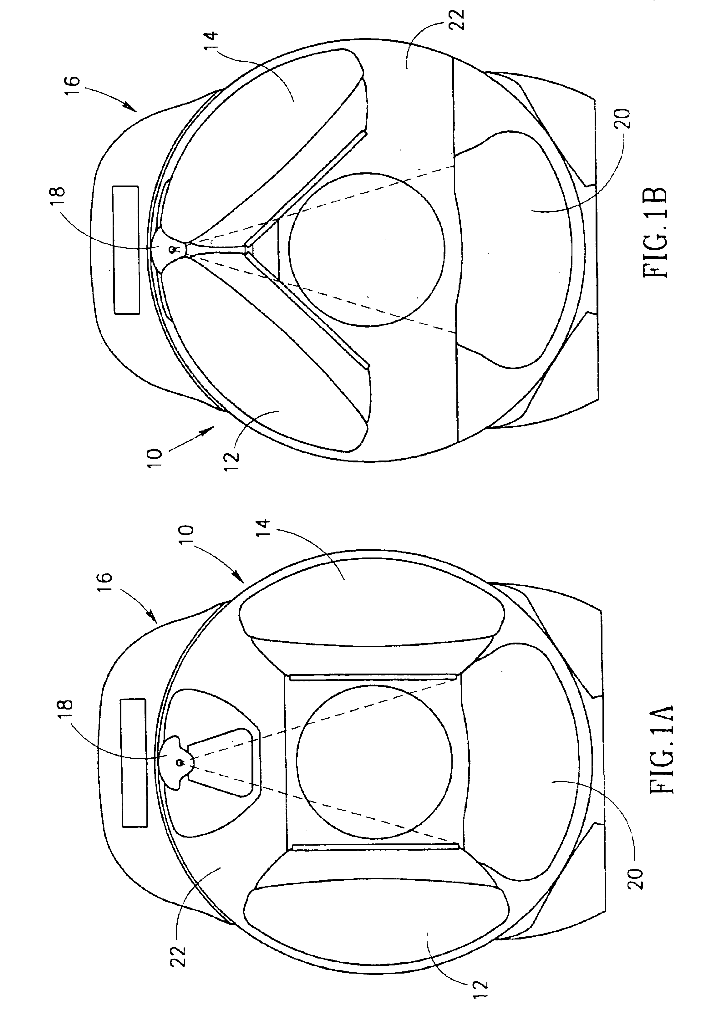
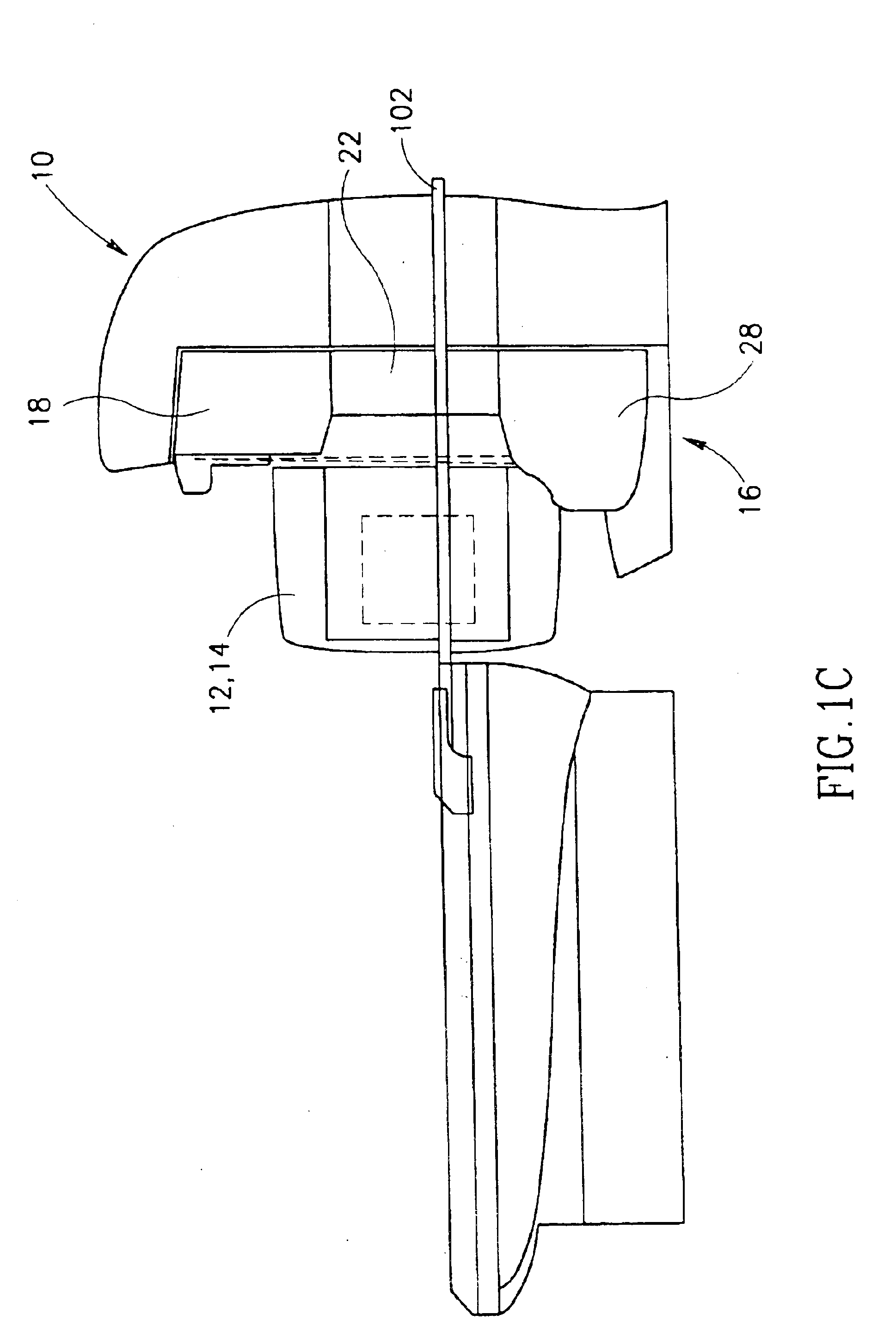
FIGS. 1A and 1B illustrate end views and FIG. 1C illustrates a lateral view of a gamma camera system 10, with attenuation correction, in accordance with a preferred embodiment of the invention. Camera system 10 preferably comprises a pair of gamma camera heads 12 and 14 and an X-ray imaging system 16. System 16 preferably comprises an X-ray source 18 and a plurality of X-ray detectors arranged in an array 20. Camera heads 12 and 14 and system 16 are preferably mounted on a same gantry 22, as shown in FIGS. 1A-1C. However, for some preferred embodiments of the invention (which may not embody all the above mentioned aspects of the invention) the camera heads and the X-ray system are mounted on different gantries. For some preferred embodiments of the invention, only a single gamma camera head is required. In others three or four heads, equally spaced circumferentially about the axis of rotation are used.

A patient (not shown in FIGS. 1A-1C) is preferably placed on a table 102 which is ...


PUM


Abstract

Apparatus for producing attenuation corrected nuclear medicine images of patients, comprising:at least one gamma camera head that acquires nuclear image data suitable to produce a nuclear tomographic image at a first controllable rotation rate about an axis;at least one X-ray CT imager that acquires X-ray data suitable to produce an attenuation image for correction of the nuclear tomographic image at a second controllable rotation rate about the axis; anda controller that controls the data acquisition and first and second rotation rates to selectively provide at least one of the following modes of operation:(i) a movement gated NM imaging mode in which the second rotation rate is substantially higher than the first rotation rate and the data from each view of the X-ray acquisition is associated with one of a plurality of respiration gated time periods;(ii) a cardiac gated NM imaging mode in which the second rotation rate is substantially higher than the first rotation rate and the data from each view of the X-ray acquisition for different rotations is averaged, wherein the X-ray data is not correlated with the cardiac cycle; and(iii) a cardiac gated NM imaging mode in which the second rotation rate is higher than the first rotation rate and the X-ray data is binned in accordance with a same binning as the NM data.

Description

FIELD OF THE INVENTIONThe present invention is related to the field of nuclear medicine and in particular to gamma cameras with x-ray transmission imaging for localization and attenuation correction of nuclear images.BACKGROUND OF THE INVENTIONAttenuation correction in nuclear medicine imaging is well known in the art. In particular, it is well known when producing SPECT or PET images to correct the images for the effect of attenuation of the gamma rays used for producing the image by intervening tissue and bone. In particular, it is known to generate an attenuation map (three dimensional image or a series of two dimensional slices) of the region being imaged by the gamma camera and correcting the counts of gamma events based on the attenuation of the tissue and bone between the source of the gamma ray and the detector.The attenuation image is produced in some prior art devices using a source of gamma rays to produce a nuclear CT (attenuation) image. X-ray based CT attenuation image...

Claims


Application Information

Patent Timeline
Patent Type & Authority Patents(United States)
IPC IPC(8): G01T1/161G01T1/00G01T1/164G21K1/12
CPCA61B6/4429A61B6/5235A61B6/584G01T1/1615A61B6/037A61B6/541
Inventor BALAN, ADISHREM, YIGALLONN, ALBERTHAJAJ, BENNYWAINER, NAORHEFETZ, YARONBERLAD, GIDEONYAKUBOVSKY, LEONID
Owner ELGEMS
Why Eureka
Try Eureka