
Impedance spectroscopy system and catheter for ischemic mucosal damage monitoring in hollow viscous organs

a hollow viscous organ and ischemic mucosal damage technology, which is applied in the field of impedance spectroscopy system and catheter for monitoring ischemic mucosal damage in hollow viscous organs, can solve the problems of no clinically acceptable method for directly monitoring ischemic mucosal damage, no clinically acceptable method for impedance spectroscopy measurements of inner walls, and no clinically acceptable method for ischemic mucosal damag

Inactive Publication Date: 2005-04-19
CRITICAL PERFUSION
View PDF34 Cites 82 Cited by
  • Summary
  • Abstract
  • Description
  • Claims
  • Application Information

AI Technical Summary

Benefits of technology

"The present invention is about a system for monitoring damage to the lining of hollow organs caused by reduced blood flow and oxygen delivery. The system uses a sensor catheter with electrodes that are placed on the end of the catheter and are used to measure the electrical properties of the tissue. This allows for the detection of ischemic tissue damage, which is different from simply measuring tissue ischemia. The system can be easily inserted into any hollow organ and the measured data can be used to quantify the level of damage and aid in the diagnosis and treatment of mucosal injuries."

Problems solved by technology

The gastrointestinal mucosa is at great risk of ischemia in the critically ill, and its disruption has been shown to be the motor of multiple organ failure, a leading cause of death.
Unfortunately, there exists no clinically suitable method to directly monitor ischemic mucosal damage in the gastrointestinal tract of the critically ill patient.
However, none of these references show or describe a clinically acceptable method for impedance spectroscopy measurements of the inner wall of hollow viscous organs such as the gastrointestinal mucosa, in vivo or in situ.

Method used


Image

  • Impedance spectroscopy system and catheter for ischemic mucosal damage monitoring in hollow viscous organs
  • Impedance spectroscopy system and catheter for ischemic mucosal damage monitoring in hollow viscous organs
  • Impedance spectroscopy system and catheter for ischemic mucosal damage monitoring in hollow viscous organs

Examples


third embodiment

[0034]FIGS. 4A-4C show the catheter 12c. The catheter 12c is generally similar to the catheter 12a, but has spacers (e.g., 42a, 42b, 42c), provided with annular internal sockets or spacer lips (e.g., 52a, 52b), into which flanged electrodes (e.g., 44a, 44b) lock into place. In this sample interlocking means, each flanged electrode (e.g., 44a, 44b) has a cylindrical central portion (e.g., 46a, 46b) having a first diameter and two annular side surfaces, two extensions (e.g., 48a, 48b) attached to the ends of the central portion and coaxial therewith, and an axial through-bore. The extensions (e.g., 48a, 48b) each have a second, reduced diameter, but instead of being purely cylindrical, the extensions (e.g., 48a, 48b) have annular electrode lips (e.g., 50a, 51a) that face towards the central portion 46. Additionally, the spacers (e.g., 42a-42c) are made of flexible plastic tubing (i.e., a relative non-conductor of electricity) or the like. Each spacer has two annular, inwardly-facing s...

fourth embodiment

[0037]FIGS. 5A-5C show the catheter 12d. The catheter 12d comprises: an injection-molded, plastic tip 60; two or four electrodes 62a-62d; one or three spacers 64a-64c (i.e., in the case of two electrodes, one spacer is needed, while three spacers are needed for a four electrode catheter); dual-lumen tubing 66 or the like; and the cables or leads 18a-18d. As best shown in FIG. 5B, the plastic tip 60 comprises a rounded fore portion 68 and a rounded, trough-like projection 70 that extends back from the fore portion 68. As indicated, the tip 60 can be injection molded, or it can be made via another suitable manufacturing process. The electrodes 62a-62d and spacers 64a-64c are generally similar in shape. Each has a small, cylindrical passageway (e.g., 72a, 72b, 72c, 72d) for the cables 18a-18d, as well as a rounded through-bore (e.g., 74a, 74b, 74c, 74d) through which the trough-like projection 70 of the tip 60 is dimensioned to fit (i.e., the rounded through-bores, e.g., 74a, 74b, 74c,...


PUM

PropertyMeasurementUnit
lengthaaaaaaaaaa
diameteraaaaaaaaaa
outer diameteraaaaaaaaaa

Abstract

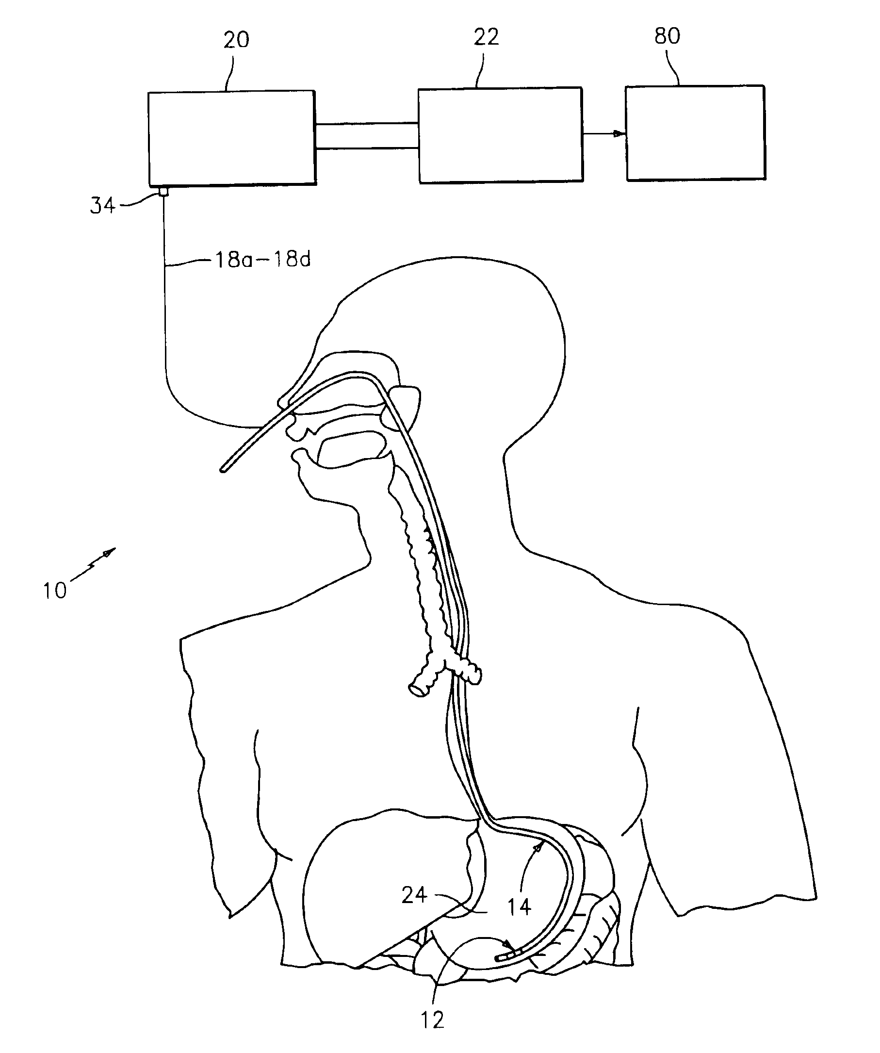
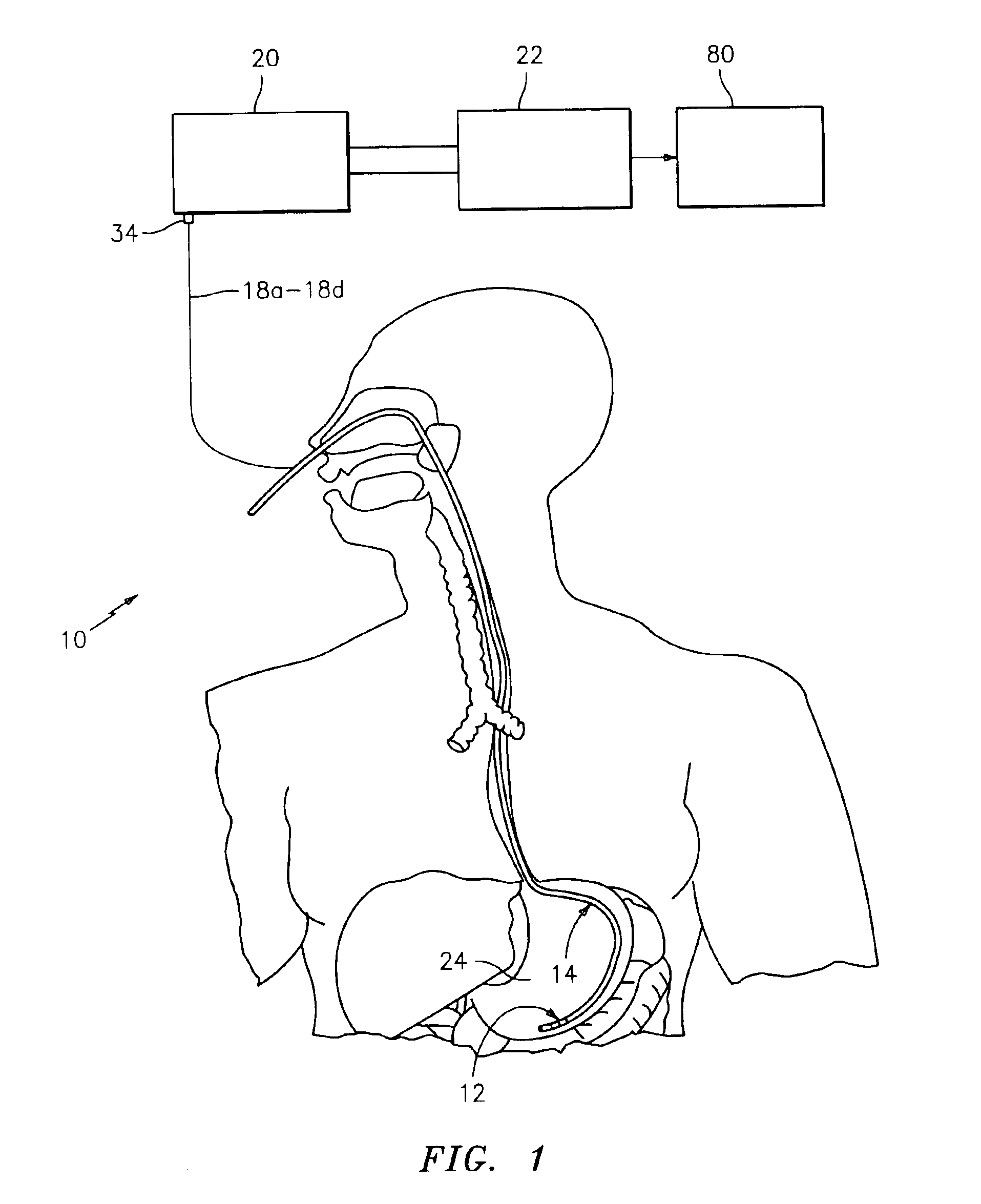
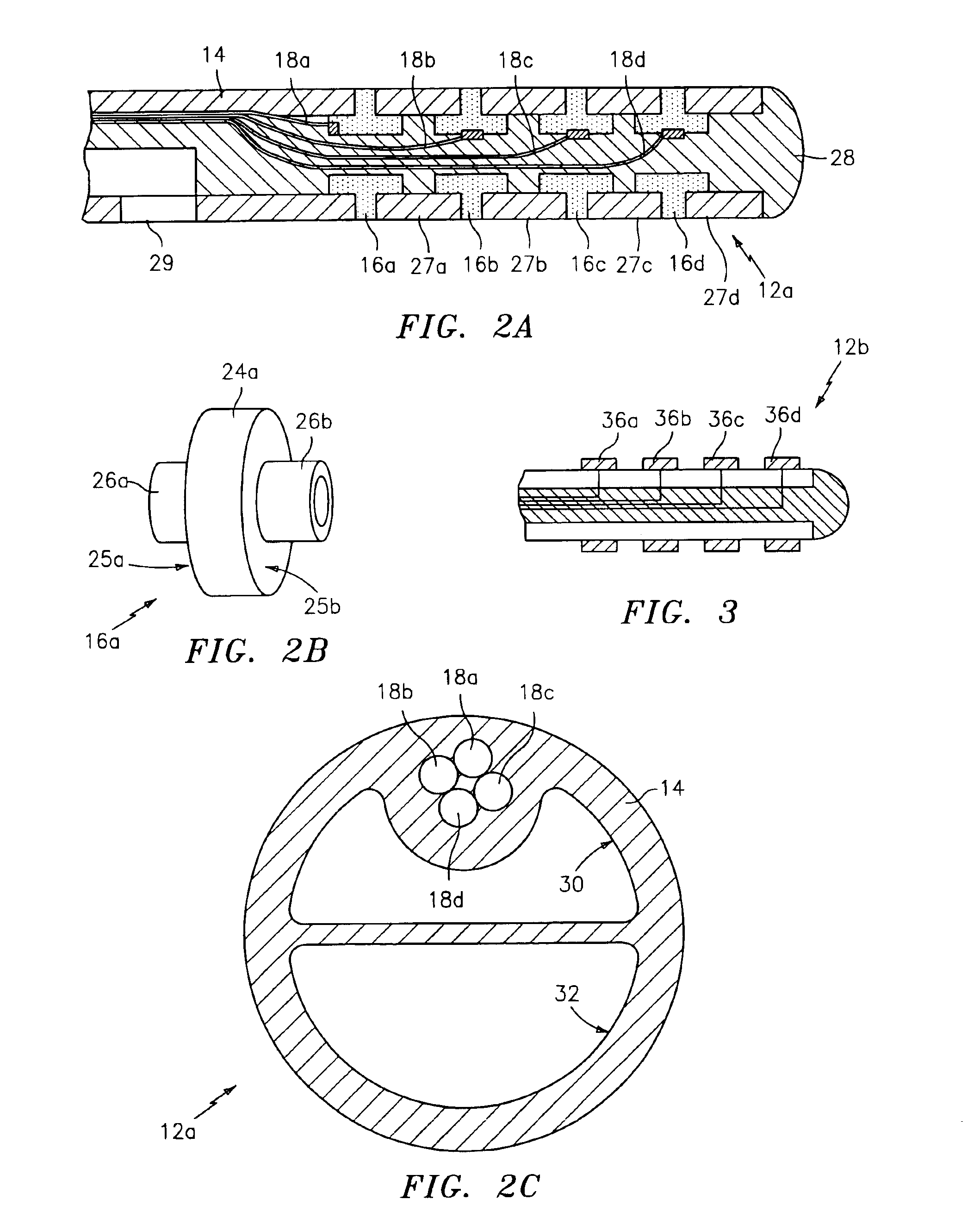
An impedance spectroscopy system for monitoring ischemic mucosal damage in hollow viscous organs comprises a sensor catheter and an impedance spectrometer for electrically driving the catheter to obtain a complex tissue impedance spectrum. Once the catheter is in place in one of a patient's hollow viscous organs, the impedance spectrometer obtains the complex impedance spectrum by causing two electrodes in the tip of the catheter to inject a current into the mucosal tissue at different frequencies, while two other electrodes measure the resulting voltages. A pattern recognition system is then used to analyze the complex impedance spectrum and to quantify the severity of the mucosal injury. Alternatively, the complex impedance spectrum can be appropriately plotted against the spectrum of normal tissue, allowing for a visual comparison by trained personnel.

Description

[0001]This application claims the benefit of a Provisional Application, Ser. No. 60 / 219,281 filed Jul. 19, 2000.FIELD OF THE INVENTION[0002]The present invention relates to systems and internal sensors for monitoring and quantifying ischemic damage in tissues.BACKGROUND OF THE INVENTION[0003]The gastrointestinal mucosa is at great risk of ischemia in the critically ill, and its disruption has been shown to be the motor of multiple organ failure, a leading cause of death. Knowledge of the level of damage can help guide therapy, reversing moderate damage and / or preventing further complications. For example, as indicated by path A in FIG. 6, the status of a healthy person's mucosa changes little, if at all, over time. Path C shows how the damage level of an ill person's ischemic mucosa greatly increases over the course of several hours if unchecked. However, as shown by path B, further damage can be arrested if the ischemic damage is detected and an appropriate course of treatment is u...

Claims


Application Information

Patent Timeline
Patent Type & Authority Patents(United States)
IPC IPC(8): A61B5/05G01N27/02A61B5/0408A61B5/042A61B5/053
CPCA61B5/05A61B5/0538A61B5/42A61B5/053A61B5/0422A61B5/7264A61B5/287
Inventor ROCK, EMILIO SACRISTAN
Owner CRITICAL PERFUSION
Learn More