Anti-HLA-DA antibodies and the methods of using thereof
a technology of anti-hla-da antibodies and antibodies, which is applied in the field of anti-hla-da antibodies, can solve the problems of death within a matter of months without effective treatmen
- Summary
- Abstract
- Description
- Claims
- Application Information
AI Technical Summary
Benefits of technology
Problems solved by technology
Method used
Image

Examples
example 1
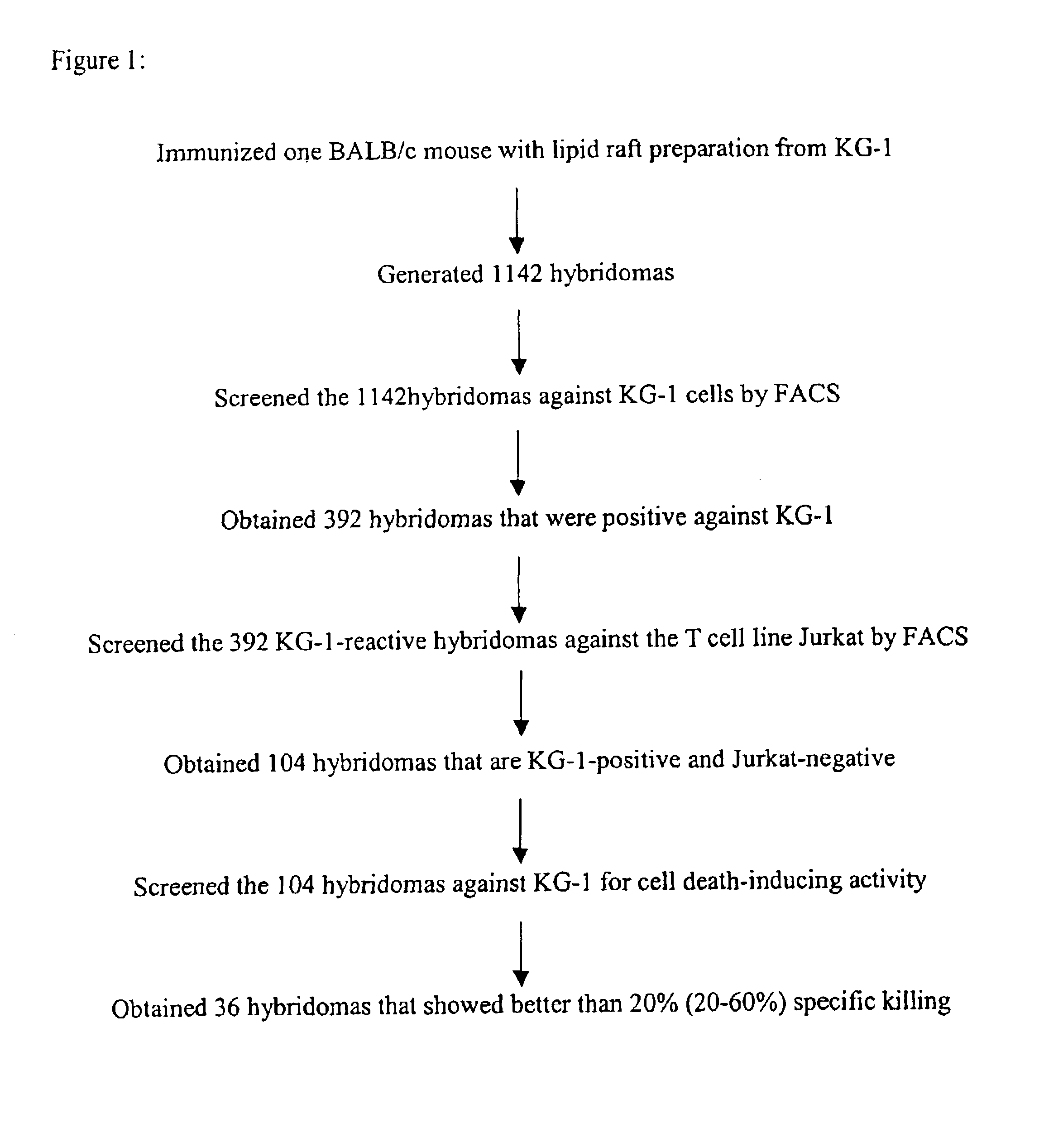
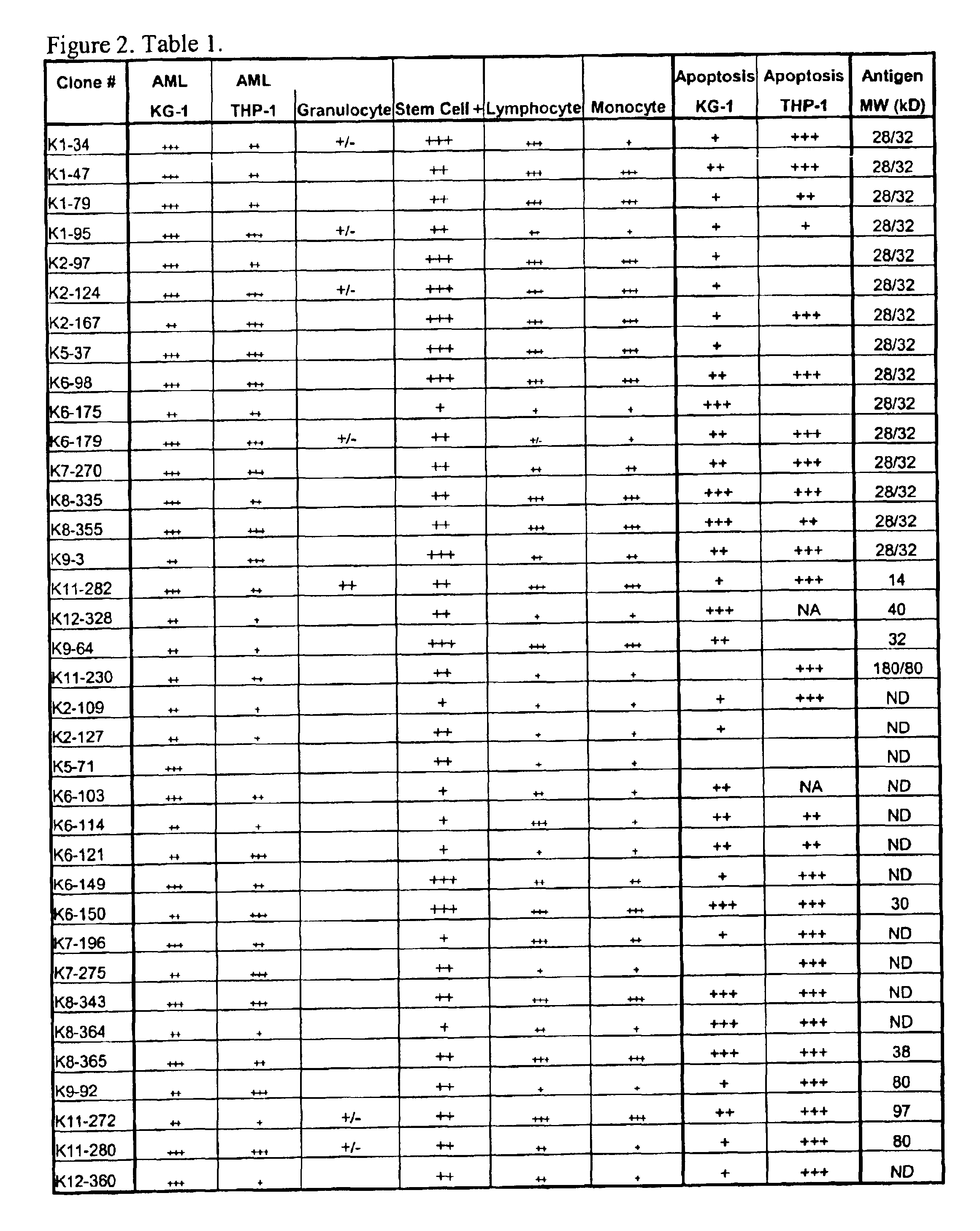
[0068]This example describes generation and characterization of anti-KG-1 lipid raft hybridomas.
Materials and Methods
[0069]a. Lipid Raft Preparation
[0070]Lipid rafts were prepared as described in Green et al, J. Cell Biol. 146, 673-682 (1999). Briefly, cells (8.0×10⁶ cells / sample) were lysed in 0.1% vol / vol Brij-58, 20 mM Tris HCl, pH 8.2, 140 mM NaCl, 2 mM EDTA, 25 μg / ml aprotinin, 25 μg / ml leupeptin, and 1 mM phenylmethylsulfonyl fluoride for 10 minutes on ice. Cells were homogenized using 10 strokes of a Dounce homogenizer, then lysed 20 minutes more on ice. The resulting lysate was adjusted to 40% wt / wt sucrose and applied onto a 60% wt / wt sucrose cushion. A sucrose step-gradient consisting of 25% wt / wt sucrose and 5% wt / wt sucrose were layered on top of the lysate. Gradients were centrifuged 18 hours at 170,000×g at 4° C. in a SW55 rotor. Fractions (0.2 ml) were taken from the top of the gradient. Lipid rafts float to the interface of the 25% and 5% sucrose layers (Fractions 7 ...
example 2
[0110]This example describes determining the identity of lipid raft tumor-associated antigens.
[0111]Monoclonal antibody that binds to each tumor-associated antigen is to be purified from hybridoma spent medium by protein-G affinity chromatography. The purified monoclonal antibody is then covalently linked to CNBr-activated Sepharose resin to generate an affinity column for a particular antigen. KG-1 cell lysate is to be prepared as in the immunoprecipitation experiment and passed onto the affinity column. Antigen retained in affinity column is eluted and subjected to protein sequence determination by the Edman degradation method. The determined N-terminal sequence is used to search for gene product identity against the Human Genome data bank. Alternatively, the eluted antigen can be subjected to MALDI-TOF peptide-mass profiling and the derived fingerprints be used to search for protein identity against the Human Genome data bank.
example 3
[0112]This example describes inhibition of tumor adhesion and spreading by cell adhesion and migration assay
[0113]Antibody inhibition of adhesion and spreading is evaluated. Tissue culture 12-well plates were coated 2 hours at room temperature with components of the extracellular matrix, i.e. vitronectin (VN), fibronectin (FN), collagen type I, type III and type IV, laminin (LA), or hyaluronic acid (HA) in Hanks buffered salt solution (HBSS). Plates are blocked for 2 hours with 1% BSA in PBS. Cells are plated in HBSS with 1 mM CaCl2 and 1 mM MgCl2 in the presence or absence of antibody. Cells are allowed to spread for 30 minutes to 2 hours at 37° C. prior to photography.
[0114]Inhibition of cancer cell migratory activity of anti-tumor agents is evaluated in a matrigel assay. Membranes with a pore size of 8 μm were coated with 50 μl matrigel. The membranes were inserted into 24 well plates that contain medium without supplements. Cancer cells are resuspended in medium with 10% FCS in ...
PUM
| Property | Measurement | Unit |
|---|---|---|
| pH | aaaaa | aaaaa |
| temperature | aaaaa | aaaaa |
| pH | aaaaa | aaaaa |
Abstract
Description
Claims
Application Information
 - R&D
- Intellectual Property
- Life Sciences
- Materials
- Tech Scout
- Unparalleled Data Quality
- Higher Quality Content
- 60% Fewer Hallucinations
Browse by: Latest US Patents, China's latest patents, Technical Efficacy Thesaurus, Application Domain, Technology Topic, Popular Technical Reports.
© 2025 PatSnap. All rights reserved.Legal|Privacy policy|Modern Slavery Act Transparency Statement|Sitemap|About US| Contact US: help@patsnap.com