Method for reconstruction of limited data images using fusion-aligned reprojection and normal-error-aligned reprojection

a limited data and reconstruction technology, applied in the direction of radiation beam directing means, radiation therapy, instruments, etc., can solve the problems of limited field of view of current patient imaging systems, especially those integrated into radiotherapy treatment systems, affecting applications, and affecting application, so as to reduce artifacts, quantitatively improve images, and increase dose calculation accuracy

Inactive Publication Date: 2005-07-05
TOMOTHERAPY INC
View PDF24 Cites 92 Cited by
  • Summary
  • Abstract
  • Description
  • Claims
  • Application Information

AI Technical Summary

Benefits of technology

[0020]A benefit of NEAR is that it may enable an iterative (two or more) variation of FAR (NEAR2FAR). It is possible to iterate these methods using multiple applications of FAR, or going from NEAR to FAR (NEAR2FAR) for a two-iteration process. NEAR can be followed by FAR iterations, or FAR can be tried multiple times with different registration results. After creating a NEAR image, the quantitatively improved voxel values in the FOV might enable an explicit fusion with the planning image, and a FAR image could be generated. NEAR and NEAR2FAR may be particularly beneficial when a LFOV causes severe quantitative and qualitative degradation of the images, whether because of a large patient, a small detector or MLC, or because a ROIT strategy is being pursued. NEAR may also be quicker than FAR, as no time is required to do an explicit fusion.
[0021]NEAR, FAR, and NEAR2FAR utilize planning CT data or other images as imperfect prior information to reduce artifacts and quantitatively improve images. These benefits can also increase the accuracy of dose calculations and be used for augmenting CT images (e.g. megavoltage CT) acquired at different energies than planning CT images.

Problems solved by technology

However, many current patient imaging systems, especially ones that are integrated into radiotherapy treatment systems suffer from a limited field-of-view (LFOV) in that collected imaging data does not encompass the patient's complete cross-section.
This LFOV can cause visibility problems with the images, images with artifacts, images with distorted values, and affect applications that use these images, including dose calculations, delivery verification, deformable patient registration, deformable dose registration, contouring (automatic, manual, or template-based).
Uncertainty in the positioning of the patient with respect to the planning CT image can undermine the conformality of the radiation delivery.
Unfortunately, the imaging data sets obtained on the day of treatment to be used for preparing the patient model are often incomplete or limited.
These limitations may be caused by limited FOVs set by the field size of the multi-leaf collimator (MLC) attached to the linear accelerator and the detector size of the radiotherapy system.
The limitations may also be caused by patients that are too large to fit within the FOV of the CT imaging system associated with the radiotherapy equipment applying the radiation dose, yielding a LFOV image as shown in FIG. 3, which shows only a portion of the image shown in FIG.
For example, in FIG. 3, not only is there a LFOV, but the data around the edges contains significant artifacts so that the image has an irregular border and internal values that are distorted.
As mentioned above, the LFOV of radiotherapy images creates problems of impaired visibility and degraded dose calculations.
The most common reasons for impaired visibility are the limited field size of the MLC attached to the linear accelerator and the limited detector size.
These limitations prevent the CT imaging system from collecting complete FOV data for all sizes of patients at all sites.
The problem of degraded dose calculations is caused by distorted electron densities and the loss of peripheral information for attenuation and scatter from the LFOV images.
This distortion of image values and loss of peripheral information can likewise affect other applications that utilize these images.
This is often not possible.
Further, the problem of artifacts is still present due to the significant degree of mismatch between such data sets, while the additional handling of the patient is more costly, time intensive and can be difficult for frail patients.
Reconstruction of incomplete imaging data sets using available techniques results in images that do not show the complete extent of the patient's body, can have artifacts and incorrect voxel values, and thus, limit the extent to which the images can be used for applications including delivery verification, dose reconstruction, patient set-up, contouring, deformable patient registration and deformable dose registration.

Method used


Image

  • Method for reconstruction of limited data images using fusion-aligned reprojection and normal-error-aligned reprojection
  • Method for reconstruction of limited data images using fusion-aligned reprojection and normal-error-aligned reprojection
  • Method for reconstruction of limited data images using fusion-aligned reprojection and normal-error-aligned reprojection

Examples


Embodiment Construction

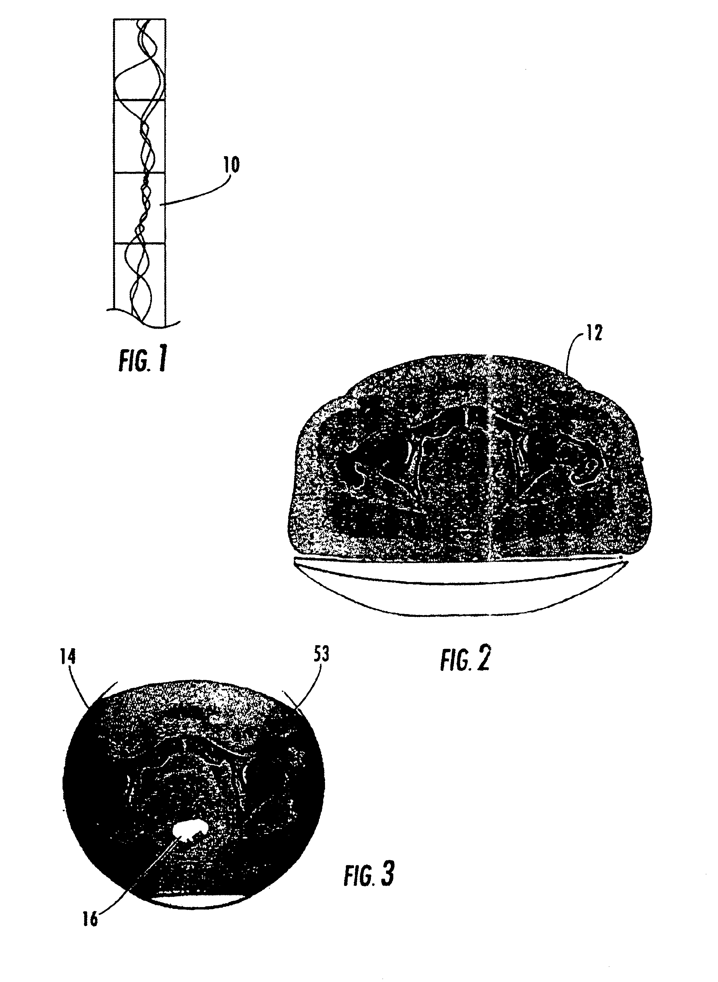
[0046]Referring now to the drawings, FIG. 1 is an example of a sinogram 10 obtained from the CT image of a patient. FIG. 2 is an example of a planning CT image obtained from a sinogram similar to that shown in FIG. 1, and FIG. 3 is an example of a LFOV image from an online CT scan of the patient just prior to radiotherapy treatment.

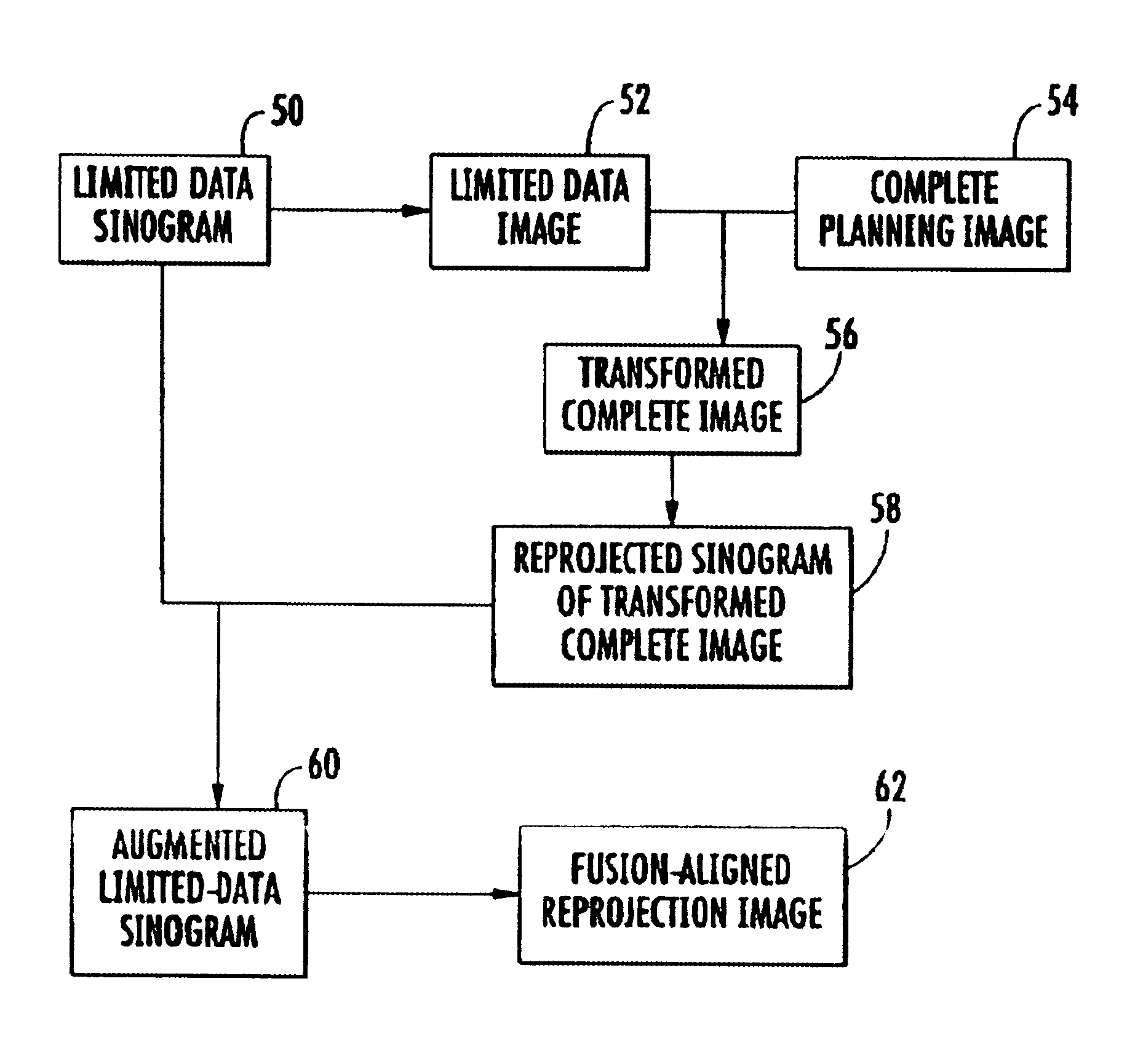
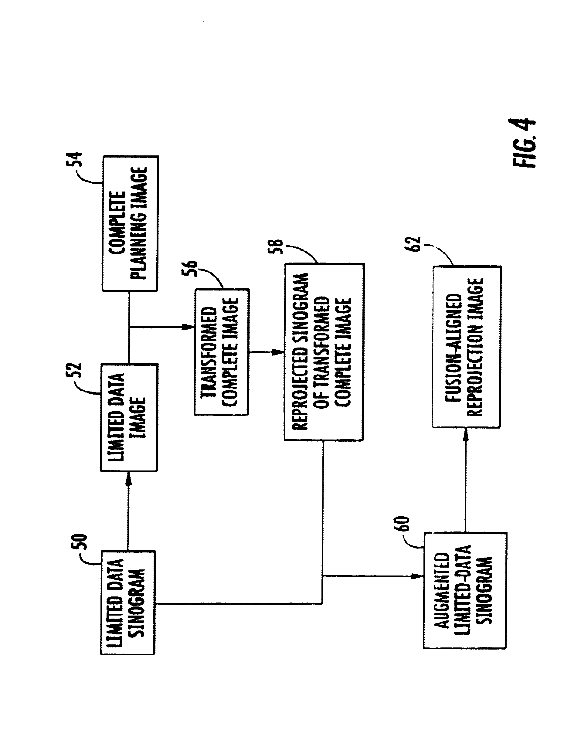
[0047]A preferred method in accordance with a first embodiment of the present invention is shown in the flow diagram of FIG. 4. FIG. 4 represents the first embodiment process involved in creating a fusion-aligned reprojection (FAR) image from a limited data image and a complete planning image. The process begins by obtaining a limited data sinogram 50 typically representing the treatment area from a patient. The limited data sinogram 50 is preferably obtained near the time that the patient is receiving his or her radiation treatment, but may be obtained at any time. The limited data sinogram 50 is reconstructed to a limited data image 52, as seen in the e...


PUM


Abstract

The present invention provides methods of using current but incomplete data to prepare an approximated complete image of a patient potentially undergoing radiation therapy. A complete image of the patient is fused or aligned with a limited patient image using image registration techniques. The aligned image is converted to sinogram data. This sinogram data is compared to sinogram data corresponding to the limited patient image to determine what data exists beyond the scope of the limited sinogram. Any additional data is added to the limited data sinogram to obtain a complete sinogram. This complete sinogram is then reconstructed into an image that approximates the complete image that would have been taken at the time the limited image was obtained.

Description

CROSS-REFERENCE TO RELATED APPLICATIONS[0001]This application is a continuation in part of U.S. application Ser. No. 09 / 802,468, filed Mar. 9, 2001, entitled “System and Method for Fusion-Aligned Reprojection of Incomplete Data,” the disclosure of which is incorporated herein by reference.BACKGROUND OF THE INVENTION[0002]The present invention relates generally to radiation therapy equipment for the treatment of tumors, and more particularly to methods for reconstructing incomplete patient data for radiation therapy and treatment verification.[0003]Medical equipment for radiation therapy treats tumorous tissue with high energy radiation. The amount of radiation and its placement must be accurately controlled to ensure both that the tumor receives sufficient radiation to be destroyed, and that the damage to the surrounding and adjacent non-tumorous tissue is minimized.[0004]External source radiation therapy uses a radiation source that is external to the patient to treat internal tumo...

Claims


Application Information

Patent Timeline
Patent Type & Authority Patents(United States)
IPC IPC(8): A61B6/08G06T11/00G06F19/00A61N5/10A61B6/03G06T1/00
CPCA61B6/08A61B6/5235G06T11/005A61N5/1049A61B6/508
Inventor RUCHALA, KENNETH J.OLIVERA, GUSTAVO H.MACKIE, THOMAS R.KAPATOES, JEFFREY M.RECKWERDT, PAUL J.
Owner TOMOTHERAPY INC
Why Eureka
Try Eureka