Ultrasonic cellular tissue screening system

a cellular tissue and ultrasonic technology, applied in the field of ultrasonic scanning and diagnostics of cellular tissue, can solve the problems of misrepresentation of overall findings, process is open to errors, and the coverage of scanned tissue is somewhat haphazard, and achieves rapid sequential display of images and different ultrasonic characteristics

Inactive Publication Date: 2005-08-23
SONOCINE INC
View PDF34 Cites 11 Cited by
  • Summary
  • Abstract
  • Description
  • Claims
  • Application Information

AI Technical Summary

Benefits of technology

[0013]The present invention is directed to a system for screening cellular tissue, in particular breast tissue. The system employs an ultrasound probe for generating image data representing images of cellular tissue. A pad which is ultrasonically conductive is placed over the patient's nipple. The pad has different ultrasonic characteristics than the cellular tissue to be scanned. A fabric covering is placed over the breast tissue to be scanned. The fabric covering includes an ultrasonic coupling agent and is used to hold the breast tissue in place. Further, the fabric of the covering and the coupling agent transmit ultrasonic energy with minimal interference. With the pad and fabric covering in place, the ultrasound probe is scanned over the tissue of interest, while one or more sensors are employed to determine the probe's location. The images representing the scanned tissue are displayed by a viewer which is capable of providing a rapid sequential display of the images.

Problems solved by technology

Because the operator must choose a few frames from the large number generated during the scan, the process is open to error.
The operator may fail to select an image of an important finding, or may select an image that misrepresents the overall findings.
In addition, since the operator is manipulating the probe by hand, and the speed of the probe over the tissue cannot be correlated with the image capture rate of the probe, the coverage of the scanned tissue is somewhat haphazard.
As a result, the operator does not record a series of images that represent a contiguous and complete set of images for the entire scanned tissue.
Nor does the manual operation of the probe allow for entirely uniform coverage of the tissue, even if multiple passes are used.
Unfortunately, this analog method is not matched to the digital sonographic recording of individual frames.
Consequently, there is a great loss of detail that prevents the evaluation of individual frames, which limits the usefulness of the videotape for diagnosing tissue anomalies.
The interpreting physician has no way to vary the speed of playback or to vary the size of the images.
Nor can the physician vary the inherent contrast and brightness of the images, only the monitor settings.
These difficulties lengthen the review time and prevent optimum viewing.
In addition, the use of separate videotapes for individual patients is expensive, and creates a storage problem.
Both of these methods are imperfect.
Physical examination usually cannot detect cancers smaller than ½ inch in diameter.
Mammography is unable to detect as many as 30 percent of cancers smaller than ½ inch.
Mammograms also use radiation and necessitate painful compression of the breasts, which discourage women from having routine mammograms.
When using ultrasound as a screening method for the entire breast, however, malignancies are usually difficult to pick out of the background tissue.
In the past there have been two schemes to use ultrasound for breast screening, but they failed to gain acceptance due to their unacceptably low success rate in finding cancers.
This method had the disadvantage of being somewhat haphazard in breast coverage.
The variable speed of manual motion does not allow the tissue to be uniformly imaged because the speed is not synchronized to the frame capture rate of the ultrasound probe.
Videotaping also results in a degradation of the images for the reasons described above.
To date, no method has been developed to uniformly and reliably use ultrasound probes to create a contiguous and complete set of scan images for an entire area of cellular tissue, such as a human breast.

Method used


Image

  • Ultrasonic cellular tissue screening system
  • Ultrasonic cellular tissue screening system
  • Ultrasonic cellular tissue screening system

Examples


Embodiment Construction

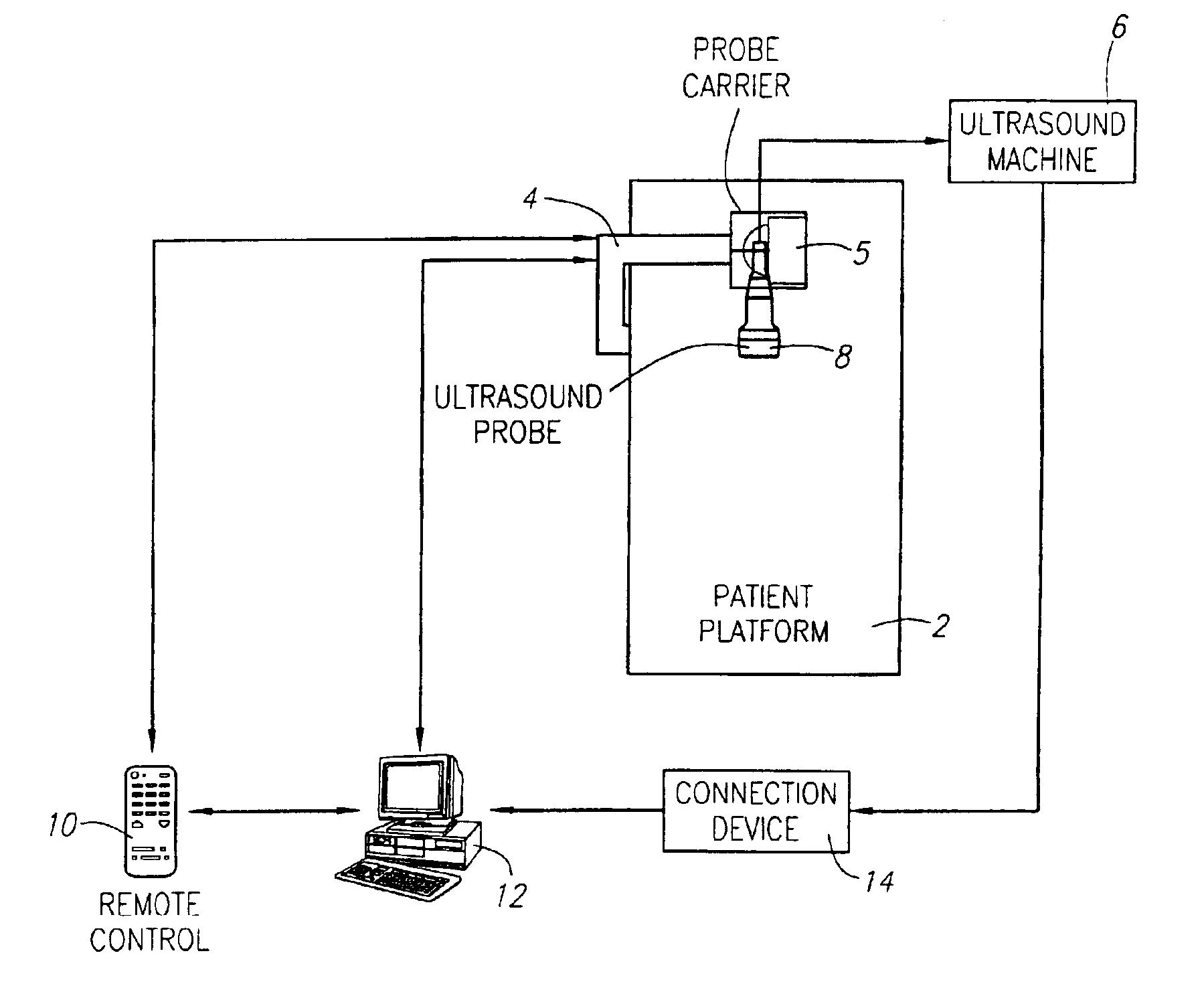
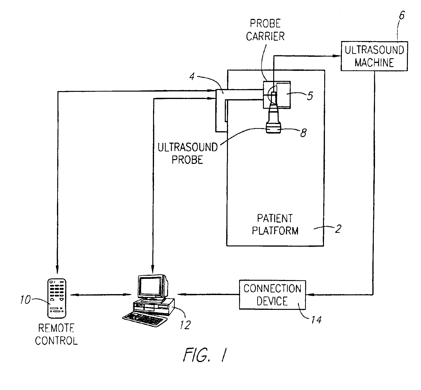
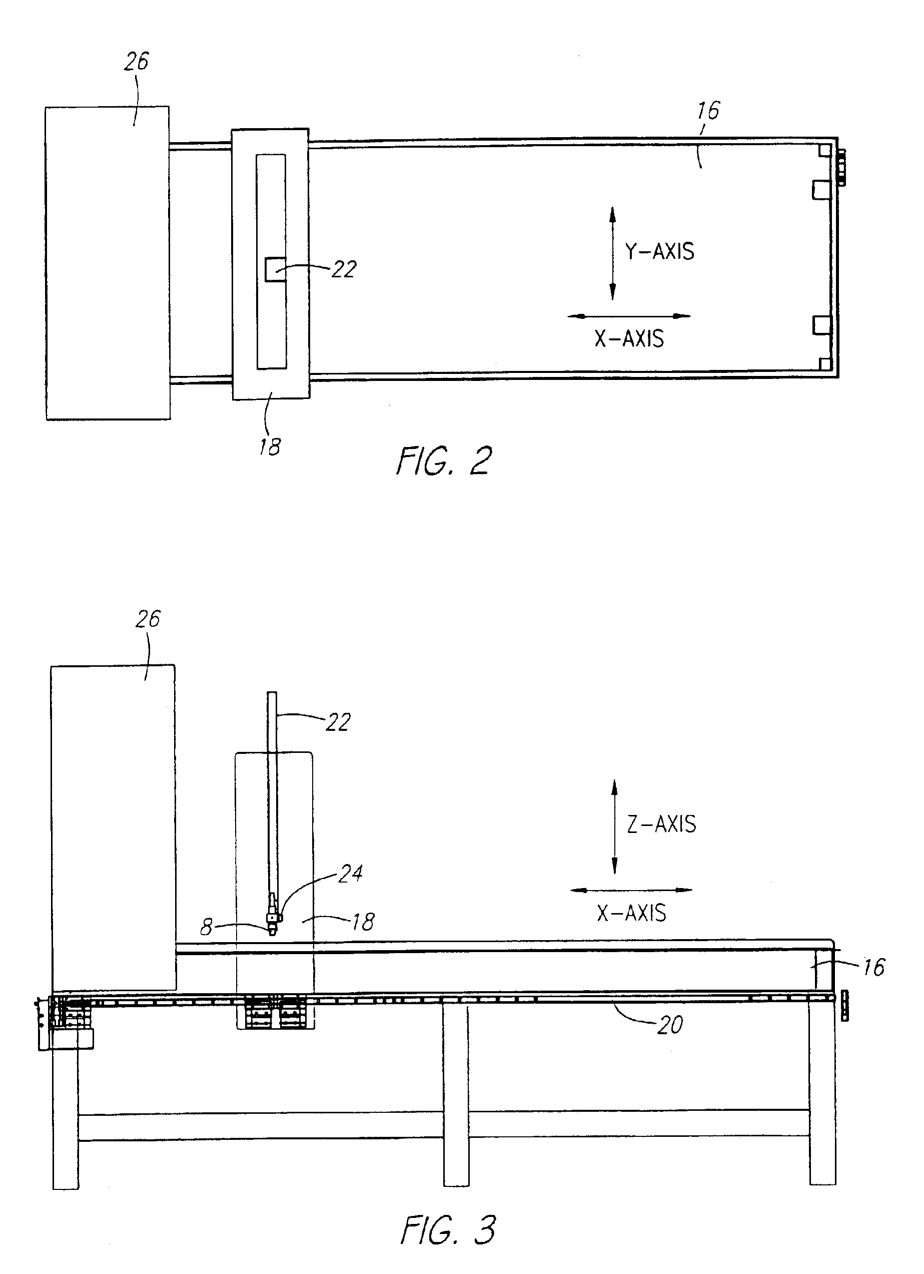
[0035]As shown in FIG. 1, a preferred embodiment is comprised of a patient platform 2 to steady the patient and provide a base for the support member 4, the probe carrier 5 connected with the support member 4 that is capable of translational movement to guide the probe across the tissue to be scanned, a standard medical ultrasound scanning device 6 with an associated probe 8, a remote control device 10 that operates the probe carrier 5, a standard computer 12, a connection device 14 between the ultrasound device 6 and the computer 12, and a viewing program that obtains image data from the ultrasound device and converts it into image data compatible with the viewing program and displays the images. The medical ultrasound scanning device 6 is a machine that sends and receives signals from the associated ultrasound probe 8, both of which are usually sold as a single unit. The ultrasound scanning device 6 with associated probe 8, computer 12, and connection device 14 are commercially av...


PUM


Abstract

A system for screening cellular tissue, in particular breast tissue. The system comprises an ultrasound probe that is capable of generating image data representing images of cellular tissue, one or more sensors to determine the probe's location, an image viewer, a pad, which is ultrasonically conductive and placed over the patient's nipple, and a fabric covering is placed over the breast tissue to be scanned. The pad has different ultrasonic characteristics than the cellular tissue to be scanned. The fabric covering includes an ultrasonic coupling agent and is used to hold the breast tissue in place. The fabric of the covering and the coupling agent transmit ultrasonic energy with minimal interference. The ultrasound probe is scanned over the tissue, and the one or more sensors are employed to determine the probe's location. The images representing the scanned tissue are displayed by a viewer which is capable of providing a rapid sequential display of the images.

Description

PRIORITY[0001]This application is a continuation of International Application PCT / US01 / 32392, with an international filing date of Oct. 15, 2001, published in English under PCT Article 21(2), which is a continuation of U.S. Ser. No. 09 / 687,128, now U.S. Pat. No. 6,524,246 B1, filed Oct. 13, 2000.BACKGROUND OF THE INVENTION[0002]1. Field of the Invention[0003]The field of the present invention is ultrasonic scanning and diagnostics for cellular tissue.[0004]2. Background of the Invention[0005]Ultrasonic probes have been used for scanning cellular tissue for many years. Presently, any medical ultrasound examination, whether of the heart, pelvis, abdomen, soft tissues or any other system, is usually displayed as a number of individual frames or pictures from a study performed in a dynamic movie-like manner. The usefulness of the scan, however, is dependent on the skill of the operator, who manipulates the probe by hand while watching the scan images on a monitor to identify areas of in...

Claims


Application Information

Patent Timeline
Patent Type & Authority Patents(United States)
IPC IPC(8): A61B8/08A61B8/00G01S15/89
CPCA61B8/0825A61B8/406A61B8/4209A61B8/565A61B8/488A61B8/54B82Y15/00A61B8/4281A61B8/4245A61B8/4218A61B8/085A61B8/145A61B8/4254A61B8/4444A61B8/463A61B8/5223
Inventor KELLY, KEVIN M.ROYCE, ROGERPETERSON, RICHARD J.PONCE, LUIS E.UNDERBRINK, CHRISTOPHER M.SMITH, MATTHEW W.GOSS, DONALD C.
Owner SONOCINE INC
Why Eureka
Try Eureka