Methodology of using raman imaging microscopy for evaluating drug action within living cells

a technology of living cells and imaging microscopy, which is applied in the direction of optical radiation measurement, separation process, instruments, etc., can solve the problems of resistance of some cells to drugs, pharmaceutical companies spending millions of dollars, and no cost effective way to understand the details of how these potential drugs work at the cellular level, so as to achieve convenient and cost-effective effects

Inactive Publication Date: 2005-09-06
SOUTHWEST RES INST
View PDF8 Cites 22 Cited by
  • Summary
  • Abstract
  • Description
  • Claims
  • Application Information

AI Technical Summary

Benefits of technology

[0028]Another objective of the present invention is to provide a convenient and cost effective method to evaluate the efficacy of drugs at the cellular level.

Problems solved by technology

The determination of drug action at the cellular level is a problem of great importance to drug evaluation and development.
However, currently there is no cost effective way to understand the details of how these potential drugs work at the cellular level.
This lack of methodology requires pharmaceutical companies to spend millions of dollars in animal and clinical studies to evaluate a candidate drug.
Another major obstacle of drug efficacy is the resistance of some cells to a drug.
Using methods in molecular biology to study drug actions at the cellular level is difficult if only conventional optical microscopes are used.
However, the fluorescent approach has several disadvantages and limitations: (1) the sample preparation procedure is complicated and time consuming, (2) the fluorescent markers used in the specimen may cause undesireable pharmacological or toxicological effects, (3) suitable markers are not available for all biomolecules, (4) the fundamental problems of fluorophore photon bleaching during measurement severely limit the use of fluorescence microscopy, and (5) the relatively short wavelength used in fluorescence microscopy can easily cause photo-damage to the specimen.
Consequently, it is often difficult, and sometimes impossible to obtain images of many molecular groups of interest by infrared microscopy.
Unfortunately, the signal for a Raman spectrum is inherently weak compared to the strength of the fluorescent signals, and therefore, can be difficult to detect.
Consequently, Raman spectroscopy, especially Raman imaging, was not practical until the recent development of a number of new signal generation, processing and detection tools.

Method used


Image

  • Methodology of using raman imaging microscopy for evaluating drug action within living cells
  • Methodology of using raman imaging microscopy for evaluating drug action within living cells
  • Methodology of using raman imaging microscopy for evaluating drug action within living cells

Examples


Embodiment Construction

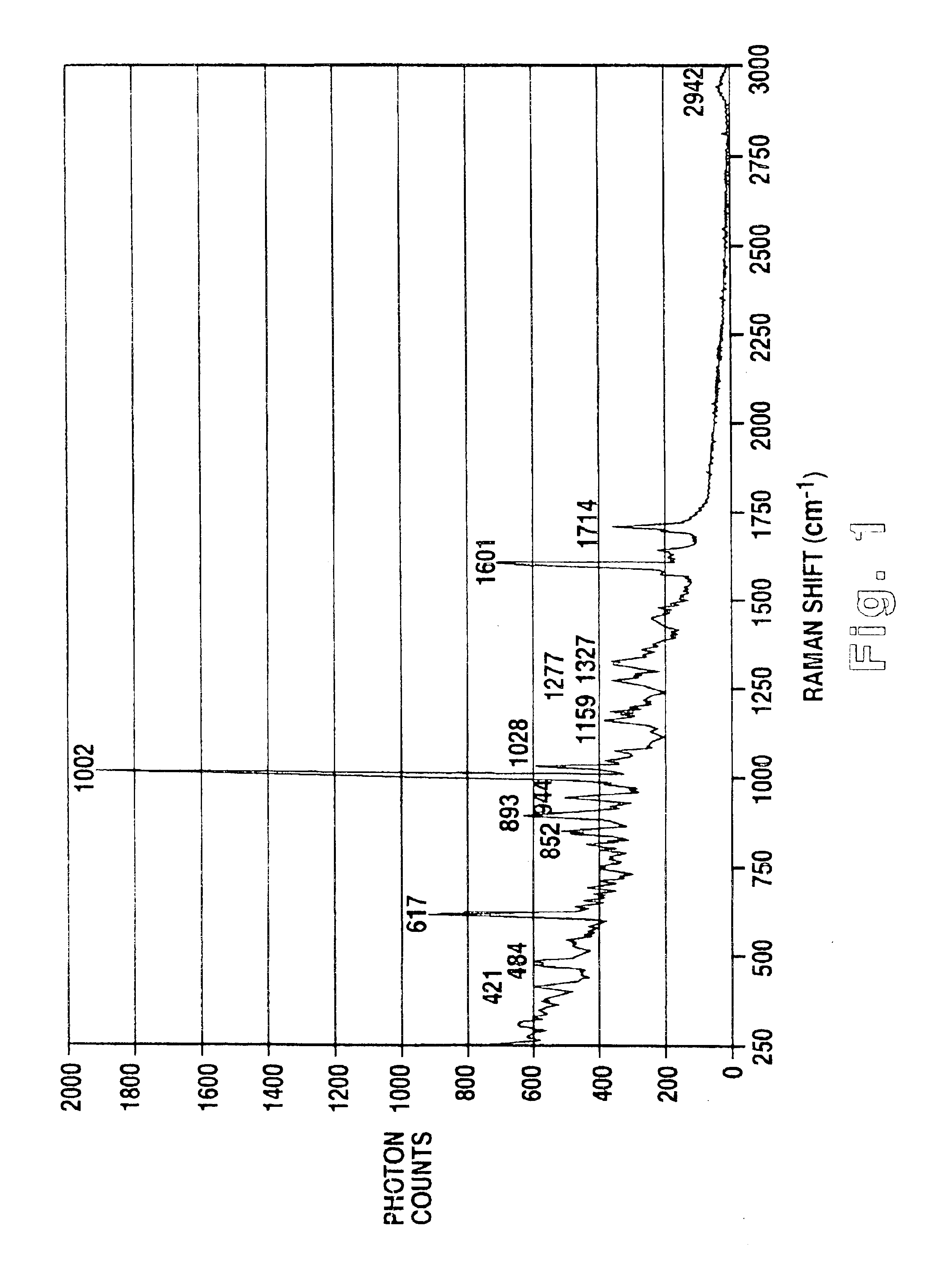
[0037]FIGS. 1-7 represent the results obtained using Raman imaging microscopy in the study of interactions between the anticancer drug taxol and MDA435 breast cancer cells. While the present description speaks to this preferred embodiment, this technique could be used in the study of the interactions of any type of drug in any type of cell.

[0038]Raman imaging of the cell-drug interactions consists of several steps. First, the Raman spectrum of the drug is measured. From the Raman spectrum, the locations and relative intensities of the Raman peaks (or Raman modes) is determined. The combination of the multiple Raman peaks and their relative intensities provides a unique fingerprint of the drug. In the preferred embodiment, the Raman spectrum of the anticancer drug taxol was measured as illustrated in FIG. 1. From the spectrum the most significant Raman mode is 1002 cm−1.

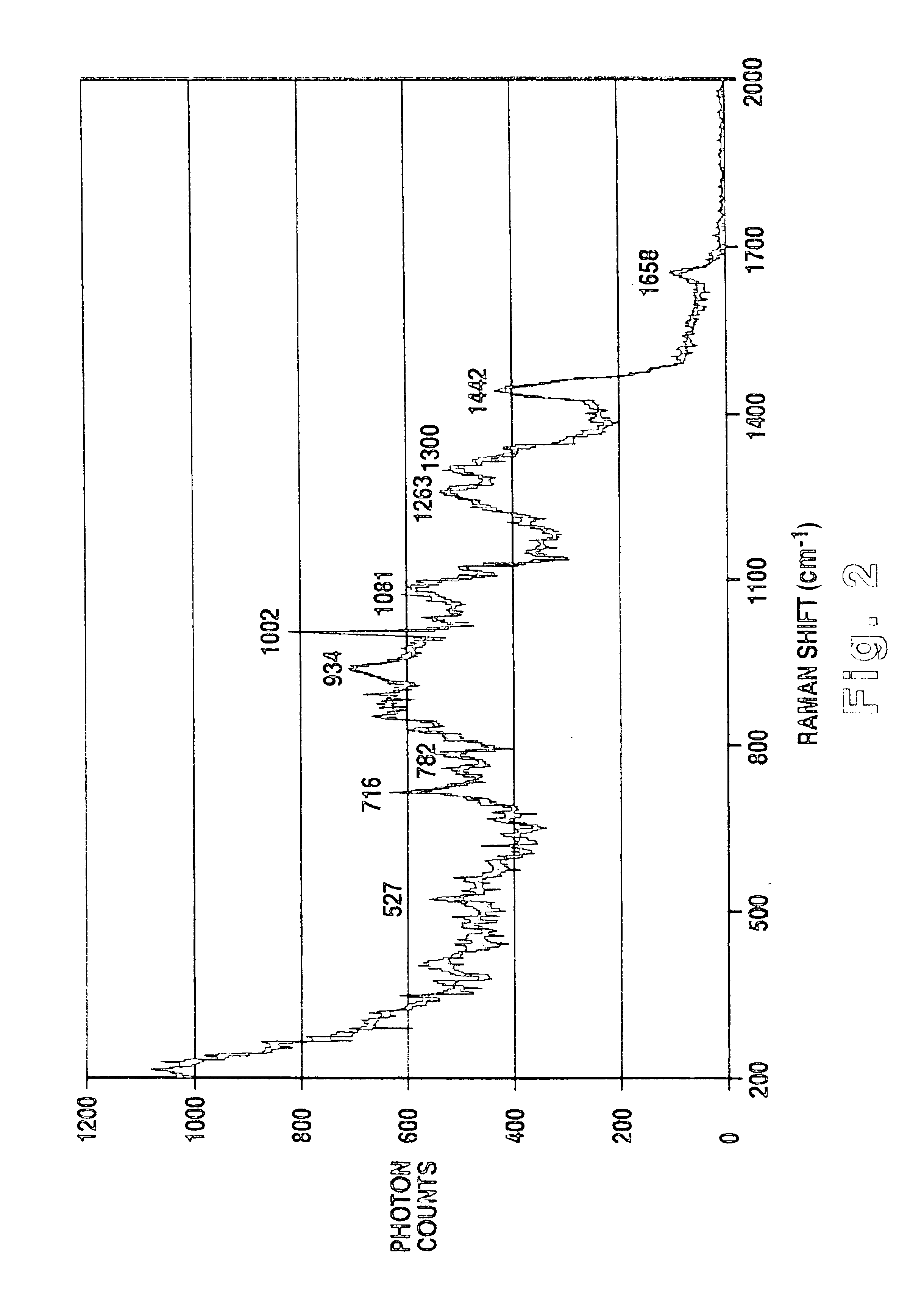
[0039]Next, a Raman spectrum is obtained for the cells to determine their fingerprint and in order to ultimately d...


PUM

PropertyMeasurementUnit
timeaaaaaaaaaa
heightaaaaaaaaaa
pixel sizeaaaaaaaaaa

Abstract

A method of using Raman imaging microscopy to evaluate drug actions in living cells is disclosed. Specifically the invention describes the methods of using Raman imaging microscopy to detect drug uptake, distribution, binding, and metabolism in a single cell, and to study drug pharmacokinetics at the cellular level. The method involves measuring the Raman image of both the drug and the cell. Control images and post-treatment images of the cell were studied. Ratio images were calculated and the requisite information was obtained from a study of the intensity of the bright areas in the ratio images.

Description

[0001]This is a continuation of U.S. patent application Ser. No. 09 / 804,774 filed on Mar. 13, 2001, now abandoned which claims benefit of 60 / 189,123 filed on Mar. 14, 2000.BACKGROUND OF THE INVENTION[0002]1. Field of the Invention[0003]The present invention relates to a method of using an optical apparatus for drug development and evaluation. More specifically, the present invention provides a convenient and cost effective method to evaluate the action of a drug at the cellular level, including its uptake, distribution, binding characteristics, etc.[0004]2. Background Information[0005]The determination of drug action at the cellular level is a problem of great importance to drug evaluation and development. Recently, the implementation of rational drug design, combinatorial chemistry techniques, and high throughput screening have led to large numbers of new potential drugs. However, currently there is no cost effective way to understand the details of how these potential drugs work a...

Claims


Application Information

Patent Timeline
Patent Type & Authority Patents(United States)
IPC IPC(8): G01N21/65G01N21/63
CPCG01N21/65Y10T436/24
Inventor LING, JIANWEITMAN, STEVEN D.MILLER, MICHAEL A.
Owner SOUTHWEST RES INST
Why Eureka
Try Eureka