Digital X-ray scanning apparatus

a scanning apparatus and digital technology, applied in the field of x-ray imaging, can solve the problems of inability of digital sensors to deliver useful x-ray images of patients' bodies, inability to maintain constant distance source-detector distance, and inability to achieve large-scale parallel data processing. , to achieve the effect of high image quality, avoiding massive parallel data processing, and high image quality

Inactive Publication Date: 2005-09-06
DDI DIRECT TIGITAL IMAGING AG
View PDF15 Cites 35 Cited by
  • Summary
  • Abstract
  • Description
  • Claims
  • Application Information

AI Technical Summary

Benefits of technology

[0012]Embodiments refer to translational means for moving the X-ray detector on a linear path. By choosing a linear scanning movement along the patient's body the distance patient-detector can be kept constant which leads to a uniform and high image quality throughout the X-ray scan.
[0013]Alternatively the X-ray detector can be moved along a curved or circular path about the X-ray source such that the distance source-detector is kept constant. Rotational means for orienting the X-ray detector can be implemented such that an aspect ratio of the X-ray detector as viewed from the X-ray source is maintained throughout a scan. A single-line or few-or multi-line digital detector is well suited geometrically for linewise scanning an extended object such as a patient's body and serially reading out the detector in order to generate a two-dimensional image. As well, a single- or multi-line digital X-ray detector is advantageous over a large two-dimensional detector array since massive parallel data processing is avoided and only rather modest computing power is required.
[0014]In specific embodiments a carriage having a rotatable plate for receiving an elongated single- or multi-line x-ray detector is provided, wherein the carriage is movable in a direction essentially perpendicular to its lateral extension. In this set-up large area scans, as required for digital partial or full body X-ray imaging, can be realized with unprecedented image quality and little computing power. The means for scanning and orienting the X-ray detector can be mounted inside a housing that remains stationary during a scan. The patient can then lean to the housing in order to maintain a constant distance to the detector. Thereby the quality and repeatability of digital X-ray imaging is further improved.
[0015]Moreover, a swiveling mounting of the X-ray source, a mechanical or electrical coupling between the scanning and orienting movement of the X-ray detector and the swiveling or translational movement of th...

Problems solved by technology

However, the digital sensor by its own cannot deliver a useful X-ray image of the patient's body.
The digital image quality, in particular the spatial resolution, scattering, dynamic range and sensitivity, is inappropriate for medical X-ray imaging.
The disclosed digital scanning system shows insufficient performance in several respects: the detector is positioned in a rather large distance from the patient's body and behind the loaded film cassette, thereby enhancing X-ray scattering; the vertical scanning range of the detector is limited by a decrease of the effective aspect ratio and hence sensitivity when the detector is moved out of a position facing the X-ray source; as detector an image intensifier plate is proposed that suffers from low X-ray sensitivity and large optical scattering; furthermore the optical readout of the image intensifier plate is accomplished by a plurality of moving parts that add considerable mechanical complexity to the digital detector.
A major problem is the fact that the patient cannot lean still at the X-ray apparatus, because the collimator in the front and the detector...

Method used


Image

  • Digital X-ray scanning apparatus
  • Digital X-ray scanning apparatus
  • Digital X-ray scanning apparatus

Examples


Embodiment Construction

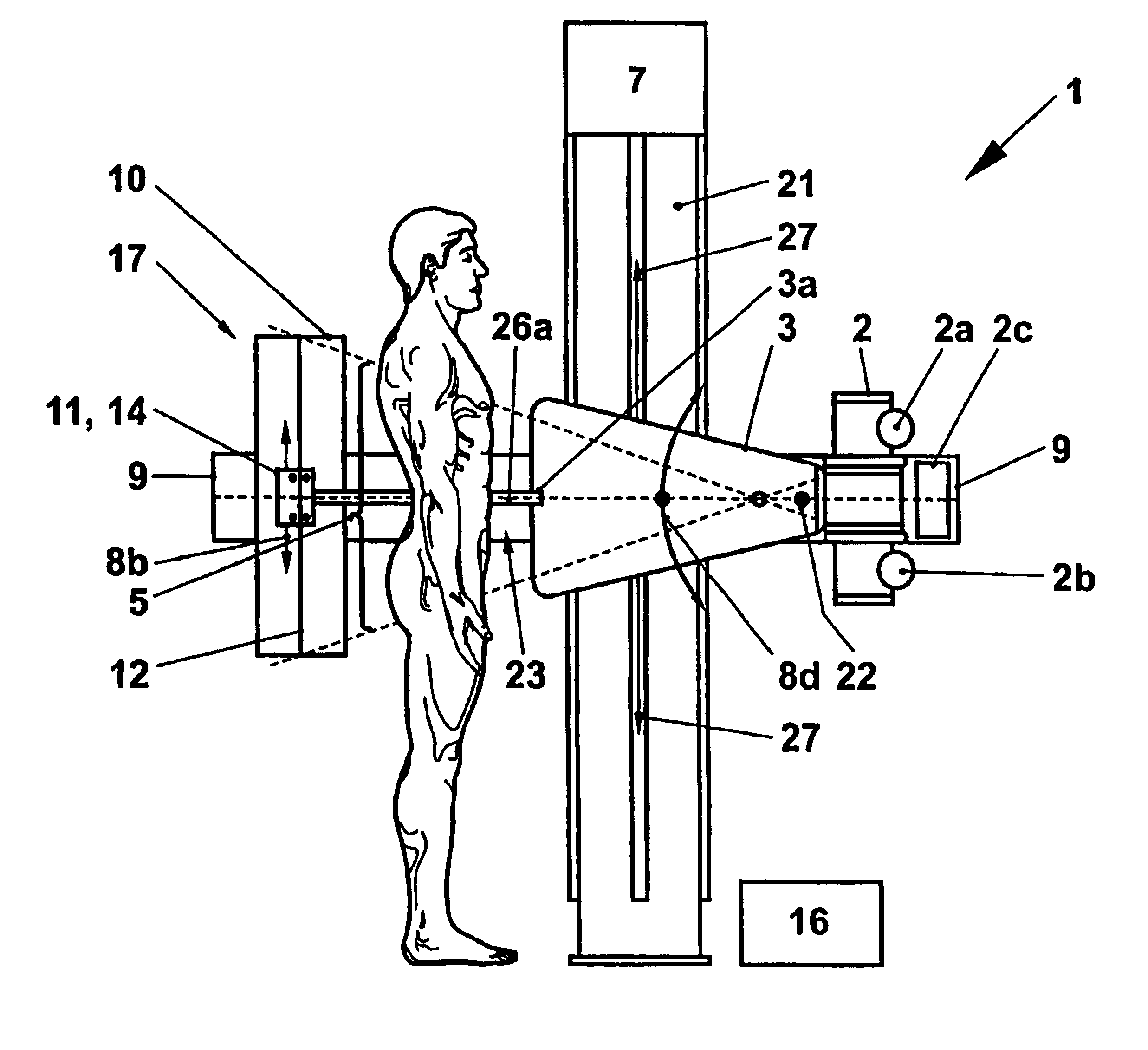
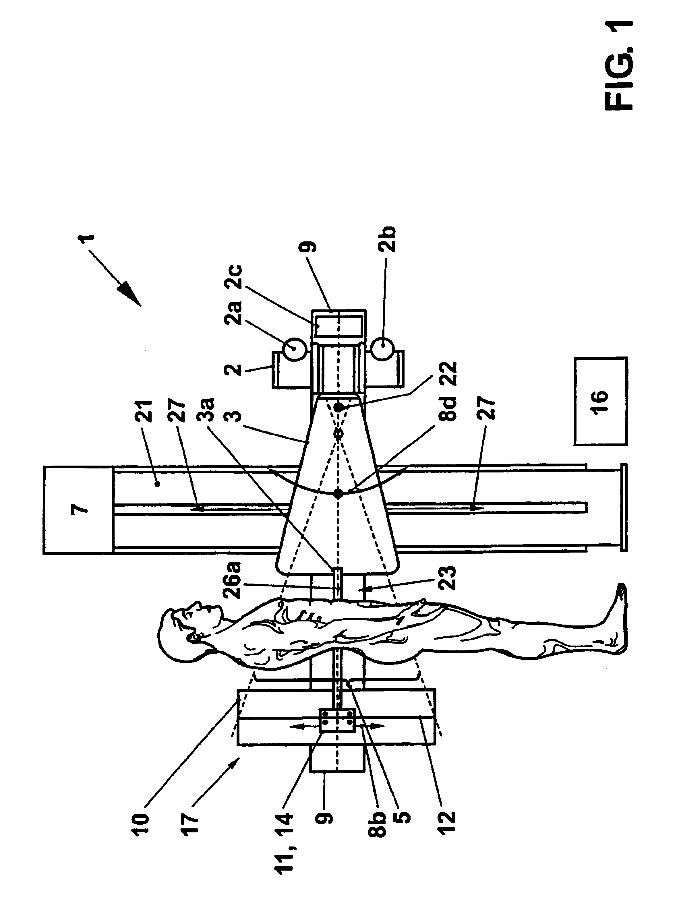
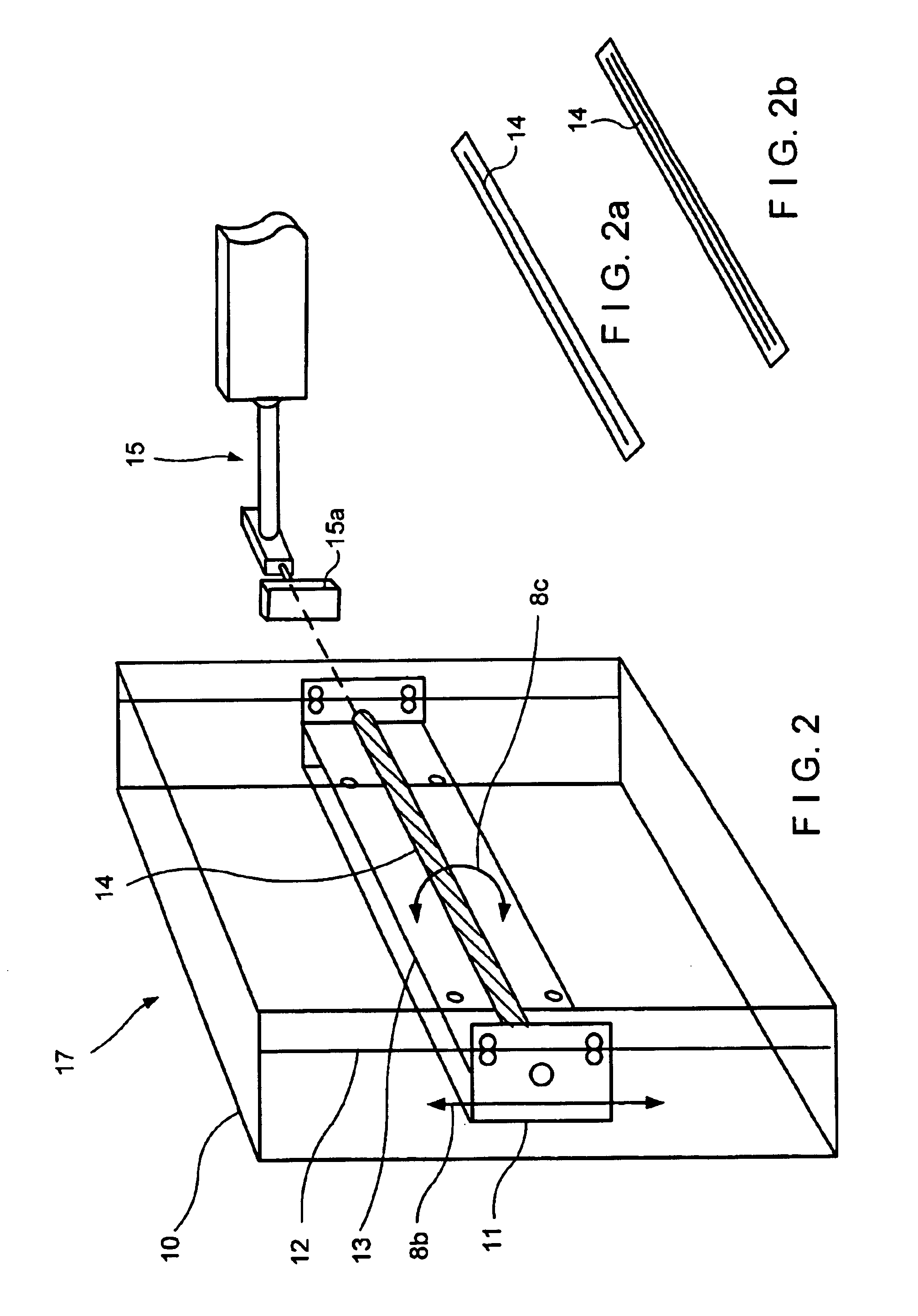
[0035]The invention refers to an X-ray apparatus 1 as exemplified in FIG. 1. The apparatus 1 comprises an X-ray source 2 with an anode 2a and a cathode 2b (indicated schematically), a collimator 3 with a collimator silt 3a and an X-ray detector arrangement 17, a supporting arm 9 for holding the X-ray source 2 and the detector arrangement 17 and a structure or mount 21 for suspending the supporting arm 9. The detector arrangement 17 includes mounting and scanning means 10-12 for an X-ray detector 14. These means 10-12 comprise a detector housing 10 and a carriage 11 that is movable along guiding rails 12. The scanning means further comprise at least one motor 7. Means 16 for digital data acquisition from the X-ray detector 14 and a control unit 2c for steering the X-ray apparatus 1 are provided. A digital image of a patient's body is generated by scanning a collimated X-ray beam 26a over an area 5 and coordinately moving the carriage 11 with the detector 14 along a path 8b and / or 27....


PUM


Abstract

In order to provide an improved X-ray imaging quality an X-ray detector (14) is equipped with means (13, 15) for orienting it towards an X-ray source (2) during a scan (8a, 8b, 27). Means (9, 10, z) for reorienting a housing (10) comprising the X-ray detector (14) and a conventional cassette holder (4) and detector positioning means (9-12) cooperate to receive an X-ray beam (26a) with improved collimation quality. Thus the detection efficiency is increased, the image resolution is enhanced, and the beam exposure of patients (5) can be minimized. Embodiments relate to a linear X-ray detector (14) designed for a serial readout of image pixels, a collimator (3, 3a, 3b) for both scanning and wide-perture X-ray imaging, and a supporting arm (9) carrying the X-ray source (2), collimator (3) and detector arrangement (17).

Description

CROSS REFERENCE TO RELATED APPLICATION[0001]This application claims the priority of the European patent application No. 99112248.2, filed on Jun. 25, 1999, the disclosure of which is incorporated herein by reference in its entirety.[0002]1. Technical Field[0003]The invention refers to the field of X-ray imaging for medical and other purposes. It is based on the subject-matter as set forth in the preamble of the independent claims.[0004]2. Background Art[0005]The invention refers to a state of the art as known from the U.S. Pat. No. 4,773,087, in which a conventional photographic X-ray imaging system for full body or partial body imaging is disclosed. The system is equipped with a scanning digital detector for monitoring primary and scattered X-ray radiation. The laterally extended detector is positioned behind a film cassette and is addressed linewise and linearly shifted up and down for providing a two-dimensional scan of the film cassette area. The digital detector signal is used ...

Claims


Application Information

Patent Timeline
IPC IPC(8): A61B6/00G21K1/02G01T1/00G01T7/00G21K1/04G21K5/02G21K5/10
CPCA61B6/00
Inventor TRETIAKOV, VJATCHESLAVSKOK, ANDREIPORTMANN, MARKUSGEISSER, ALBERT
Owner DDI DIRECT TIGITAL IMAGING AG
Why Eureka
Try Eureka