Magnetic resonance method for assesing amide proton transfer effects between amide protons of endogenous mobile proteins and peptides and tissue water in situ and its use for imaging ph and mobile protein/peptide content

a technology of endogenous mobile proteins and amide protons, which is applied in the field of magnetic resonance imaging apparatus and methods for magnetic resonance imaging, can solve the problems of difficult to determine the history of specific ms lesions, unobservable lesions, and presently difficult to assess the viability of ischemic penumbra

Inactive Publication Date: 2005-09-13
THE JOHN HOPKINS UNIV SCHOOL OF MEDICINE
View PDF0 Cites 51 Cited by
  • Summary
  • Abstract
  • Description
  • Claims
  • Application Information

AI Technical Summary

Benefits of technology

[0031]In more particular embodiments, such assessing includes irradiating said pool of amide protons in said at least one region of said sample or subject that is in exchange with another pool of water protons in said at least one region of said sample or subject with sufficient electromagnetic radiation to magnetically label said pool of amide protons; determining a given amide proton transfer effect corresponding to the transfer of saturation between said pool of amide protons and said another pool of water protons; and assessing effects of one of labile amide proton content,

Problems solved by technology

The evolution and history of specific MS lesions is difficult to resolve with conventional T1-weighted or T2-weighted MRI, and so some lesions are unobservable.
Despite many available MRI modalities, it is presently difficult to assess the viability of the ischemic penumbra (i.e., a zone of reduced flow around the ischemic core).
Also, impaired oxygen metabolism and concomitant pH changes are crucial in the progress of the ischemic cascade, however, pH effects cannot be ascertained using the water signal.
As is known to those skilled in the art, phosphorous magnetic resonance spectroscopy (MRS) can be used to assess absolute pH and pH changes, however, this particular technique has low spatial resolution (e.g., 20-30 ml) in part because the strength of the NMR signal from phosphorous is significantly less than that for the water signal.
Phosphorous MRS, however, is not available on standard clinical equipment,

Method used


Image

  • Magnetic resonance method for assesing amide proton transfer effects between amide protons of endogenous mobile proteins and peptides and tissue water in situ and its use for imaging ph and mobile protein/peptide content
  • Magnetic resonance method for assesing amide proton transfer effects between amide protons of endogenous mobile proteins and peptides and tissue water in situ and its use for imaging ph and mobile protein/peptide content
  • Magnetic resonance method for assesing amide proton transfer effects between amide protons of endogenous mobile proteins and peptides and tissue water in situ and its use for imaging ph and mobile protein/peptide content

Examples


example 1

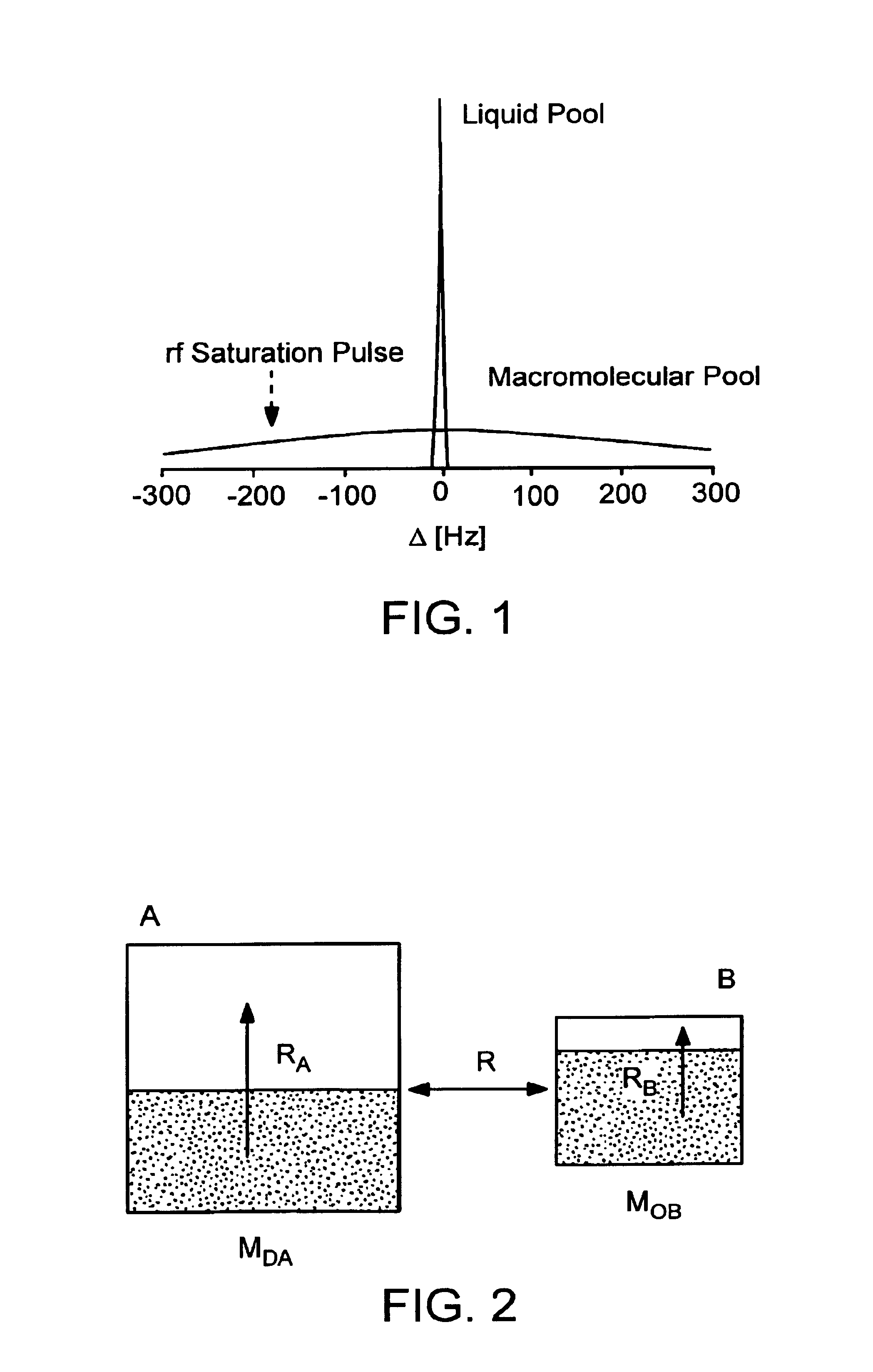
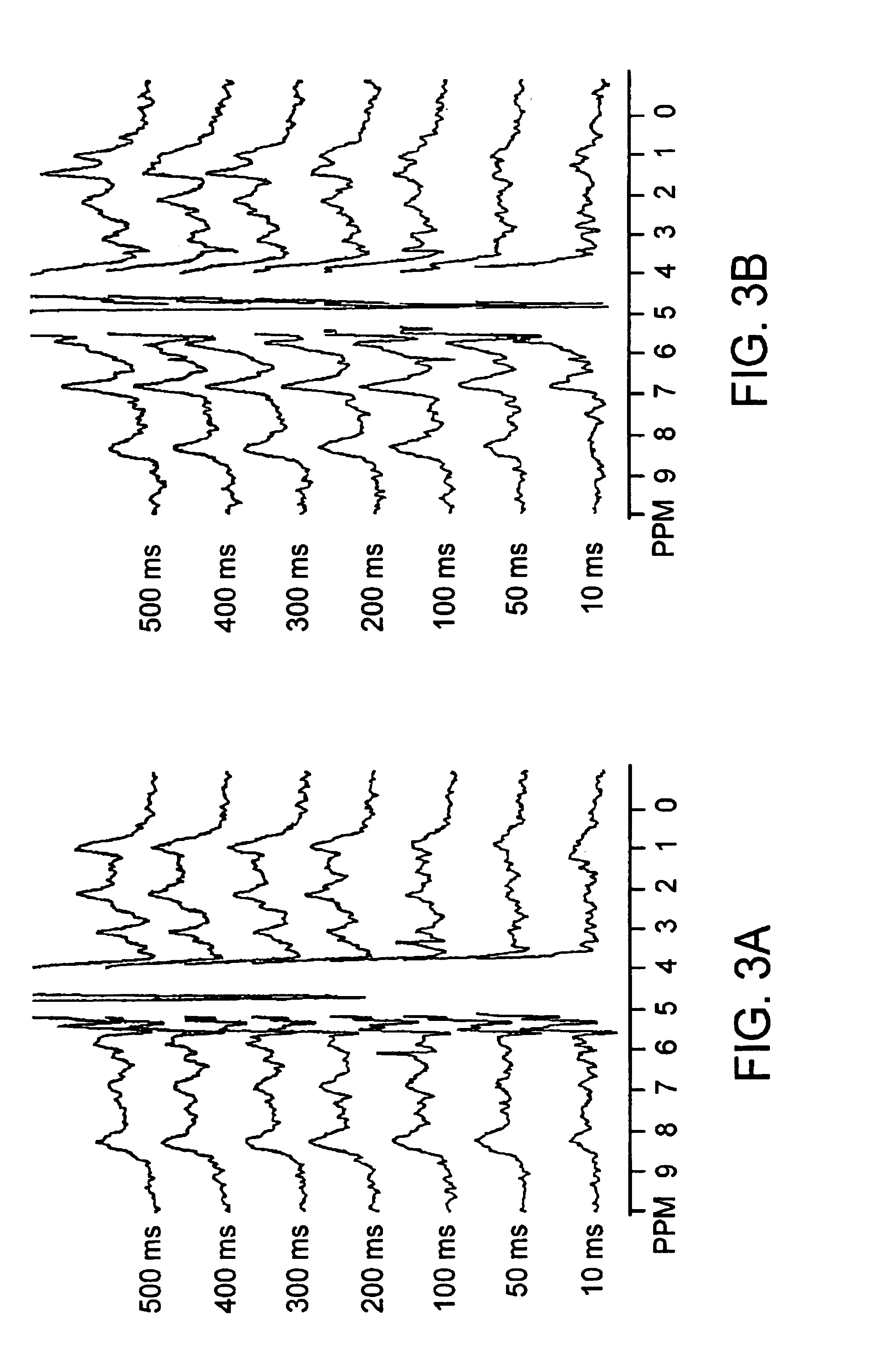
[0067]An in situ rat brain spectroscopy experiment was performed in which the opposite of a typical MT experiment was done. Namely, the water magnetization was selectively labeled and its transfer properties monitored as a function of time after labeling (mixing time tm). In such water exchange (WEX) experiments, the amide protons of proteins and peptides (6-10 ppm) and the aliphatic signals (0-3.5 ppm) from mobile macromolecules (proteins, larger peptides, some lipids) are visible, while those of smaller molecules, such as brain metabolites, are not. On the other hand, the solid-like spectrum of importance in conventional magnetization transfer (MT) imaging experiments is too broad to be detected (120-200 kHz). This experiment illustrates the difference between the properties of protons of mobile marcomolecules versus solid-like macromolecules and small metabolites.

[0068]Referring to FIGS. 3A,B there is shown normocapnic (3A) and postmortem (3B) water exchange (WEX) spectra for rat...

example 2

[0089]Referring to FIG. 7 there is shown a water exchange spectra (WEX spectra) for perfused RIF-1 cancer cells as a function of magnetization transfer time Tm. The illustrated WEX spectra (256 scans, 9.4T, sweep width 5000Hz, TE =8 ms, TR =2s) includes 5 curves (curves A-E) illustrating the WEX spectra for different magnetization transfer times. For purposes of comparisbn, FIG. 7 also includes a normal water suppressed spectrum (64 scans); curve F. Calculations based on signal to noise ratio (including a correction for the number of scans) show that the intensity of the amide resonance around 8.3 ppm at Tm =502.3 is about 75% of that in the reference spectrum. The chemical shifts and intensities for certain of the peaks between about 0.5 ppm to about 3.5 ppm, have striking resemblance with mixed protein spectra assigned to brain literature. The large resonance at 8.3 ppm is attributed to the protein peptide amide proton groups.

[0090]Referring now to FIG. 8 there is shown a graph of...

example 3

[0094]This example illustrates a specific modality in the clinical MRI examination of cancers. Referring now to FIGS. 11 A-C there are shown various graphs illustrating MT spectra, MTRasym spectra, and ΔPTR spectra for a 9L rat brain tumor and collateral region. Similar to the in vivo / postmortem system and ischemic / contralateral system, the tumor / contralateral imaging signal intensities are substantially reduced due to the effects of direct water saturation and conventional MT. However, it is surprising that the MTRasym curves in the tumor / contralateral case show something quite different, namely an increase in the tumor MTRasym spectra over a range of offsets between 2-5 ppm. Another different point based on these two tumor-implanted animals (6 measurements) is that there is a positive magnitude of ΔPTR at this offset range, and the maximum change in the tumor MTRasym spectra was found to be at an offset of 2.5-3 ppm. This is attributed to high pHi, high labile amide proton content...


PUM

PropertyMeasurementUnit
Fractionaaaaaaaaaa
Concentrationaaaaaaaaaa
Ratioaaaaaaaaaa

Abstract

Featured is an MRI/NMR methodology or process to detect amide protons of endogenous mobile proteins and peptides via the water signal. Such methods and processes can be used for the purposes of detection of pH effects and labile amide proton content of mobile proteins/peptides or content changes thereto using MRI Also featured are methods whereby assessment of determined pH effects and amide proton content or content changes and related mobile protein and/or peptide content or content changes can be used in connection with diagnosis, program and treatment of brain related disorders and diseases, cardiac disorders and diseases, and cancer and to use such methods for monitoring, detecting and assessing protein and peptide content in vivo and pathologically for any of a number of diseases or disorders of a human body, including but not limited to cancers, ischemia, Alzheimers and Parkinsons.

Description

[0001]This application claims the benefit of U.S. Provisional Application Ser. No. 60 / 339,666 filed Dec. 13, 2001, the teachings of which are incorporated herein by reference.STATEMENT REGARDING FEDERALLY-SPONSORED RESEARCH[0002]Funding for the present invention was provided in part by the Government of the United States by virtue of Grant No. NIH / NINDS (NS31490). Thus, the Government of the United States has certain rights in and to the invention claimed herein.FIELD OF INVENTION[0003]The present invention generally relates to apparatus and methods for magnetic resonance (MR) imaging (MRI), also known as nuclear magnetic resonance (NMR) imaging (NMRI). More particularly the present invention relates to methods for magnetic resonance imaging and spectroscopy relating to exchange of magnetization between protons and more specifically methods for detecting, assessing and imaging pH effects as well as amide proton content using the water signal. The present invention also features MRI ...

Claims


Application Information

Patent Timeline
IPC IPC(8): G01R33/563G01R33/48G01R33/54G01R33/485G01R33/483G01R33/465
CPCG01R33/4804G01R33/4833G01R33/563G01R33/465Y10T436/24G01R33/5605G01R33/485
Inventor VAN ZIJL, PETER C. M.ZHOU, JINYUAN
Owner THE JOHN HOPKINS UNIV SCHOOL OF MEDICINE
Why Eureka
Try Eureka