Methods, compositions, and automated systems for separating rare cells from fluid samples

a technology of rare cells and fluid samples, applied in the field of bioseparation, can solve the problems of difficult automation, high labor intensity of separation, and small filtration area of filters, and achieve the effect of facilitating diagnosis and prognosis

Inactive Publication Date: 2005-09-27
AVIVA BIOSCI
View PDF15 Cites 147 Cited by
  • Summary
  • Abstract
  • Description
  • Claims
  • Application Information

AI Technical Summary

Benefits of technology

[0011]The present invention recognizes that diagnosis, prognosis, and treatment of many conditions can depend on the enrichment of rare cells from a complex fluid sample. Often, enrichment can be accomplished by one or more separation steps. In particular, the separation of fetal cells from maternal blood samples, can greatly aid in the detection of fetal abnormalities or a variety of gen

Problems solved by technology

Such separations are often labor intensive and difficult to automate.
While the membrane filters can be used for a number of separation applications, the variation in the pore size and the irregular shapes of the pores prevent them being used for precise filtration based on particle size and other properties.
While these microfabricated filters have precise geometries, their limitations are that the filtration area of the filter is small, limited by the geometries of these filters, so that these filters can process only small volumes of the fluid sample.
Blood samples provide special challenges for sample preparation and analysis.
However, the great abundance of non-nucleated red blood cells, and their major component hemoglobin, can be an impediment to gene

Method used


Image

  • Methods, compositions, and automated systems for separating rare cells from fluid samples
  • Methods, compositions, and automated systems for separating rare cells from fluid samples
  • Methods, compositions, and automated systems for separating rare cells from fluid samples

Examples


example 1

Isolation of Fetal Nucleated Red Blood Cells from a Maternal Blood Sample

Preparation of Magnetic Beads

[0389]Magnetic beads for the capture of nucleated red blood cells were prepared during the processing (centrifugation) of collected blood. For each 1 milliliter of anticipated final cell preparation, twenty microliters of commercially available streptavidin magnetic beads (Bang's Laboratories) were used, to give a ratio of 10 to 30 beads per target cell, or 5 to 20 micrograms of iron per milliliter of solution. The beads were diluted ten-fold with PBE (PBS containing 0.5% BSA and 5 mM EDTA) and pipeted into a 12×75 mm polypropylene tube. The beads were collected with a magnet placed along the side of the tube for ten minutes. The supernatant was removed, and the washing process was repeated twice. The beads were finally resupended in ten times their original volume of PBE.

Preparation of DEP Chip

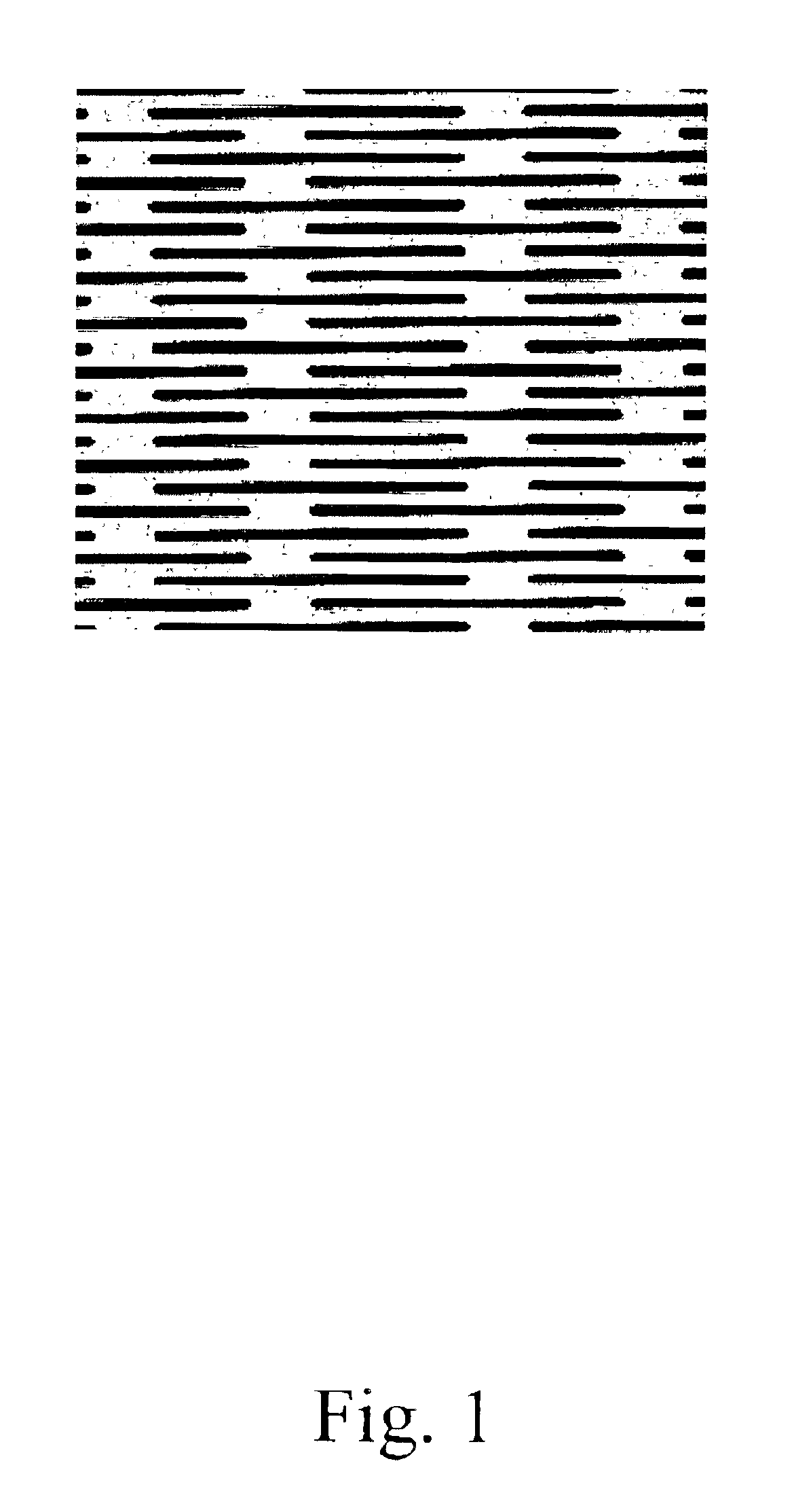
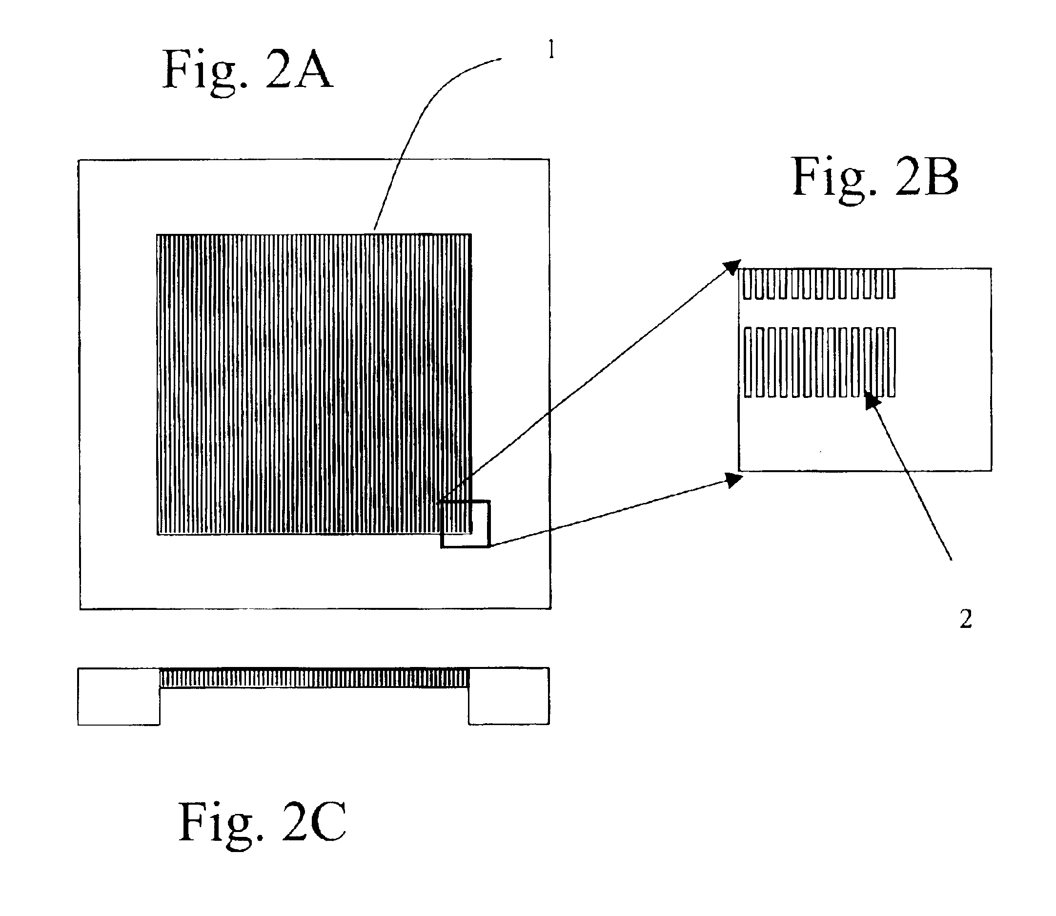
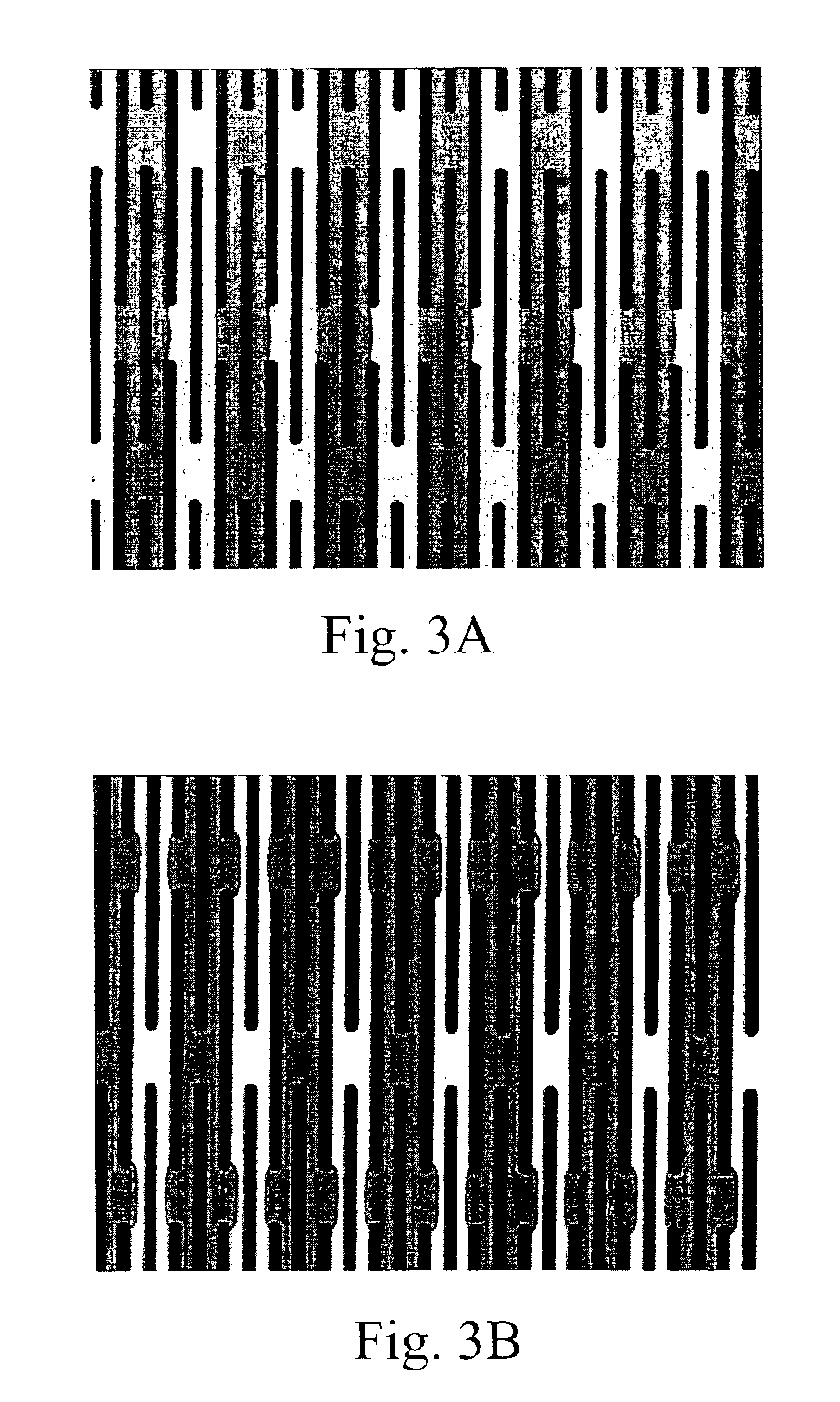
[0390]During antibody enrichment of nucleated red blood cells (below) a parallel electrod...

example 2

Comparison of Commercially Available Antibodies for the Selective Removal of White Blood Cells from Maternal Blood Samples

Preparation of Magnetic Beads

[0405]Preparation of the 0.8 micron diameter magnetic beads was performed during the processing (centrifugation) of collected blood. For each 1 milliliter of anticipated final cell preparation, fifteen microliters of streptavidin-coated magnetic beads, or approximately ten to thirty beads per target cell, were used. The beads were diluted ten-fold with PBE (PBS containing 0.5% BSA and 5 mM EDTA) and pipeted into a 12×75 mm polypropylene tube. The beads were collected with a magnet placed along the side of the tube for ten minutes. The supernatant was removed, and the washing process was repeated twice.

Density Gradient Centrifugation of Blood Samples

[0406]Twenty to forty milliliter post-surgery peripheral blood samples from women who had undergone clinical abortion at the sixth to the sixteenth week of pregnancy were collected in tubes...

example 3

Isolation of Fetal Nucleated Red Blood Cells from a Blood Sample

Preparation of Magnetic Beads

[0413]Bead preparation was performed during the centrifugation of collected blood. For each 1 milliliter of anticipated final cell preparation, twenty to forty microliters of streptavidin-coated 0.8 micron magnetic beads, or approximately ten beads per target cell, were used. The beads were diluted ten-fold with PBE and pipeted into a 12×75 mm polypropylene tube. The beads were collected with a magnet placed along the side of the tube for ten minutes. The supernatant was removed, and the process was repeated twice.

Density Gradient Centrifugation of Blood Samples

[0414]A ten milliliter peripheral blood samples from a pregnant woman was collected in a tube rinsed with PBE (PBS containing 0.5% BSA and 5 mM EDTA). Approximately four thousand fetal liver cells that include nucleated red blood cells, dissected from an eighteen week gestational age male human abortus, were added to the maternal bloo...


PUM


Abstract

The present invention recognizes that diagnosis and prognosis of many conditions can depend on the enrichment of rare cells from a complex fluid sample. In particular, the enrichment of fetal cells from maternal samples, such as maternal blood samples, can greatly aid in the detection of fetal abnormalities or a variety of genetic conditions. In addition, the present invention recognizes that the enrichment of rare malignant cells from patient samples, can aid in diagnosis, prognosis, and development of therapeutic modalities for patients.
A first aspect of the present invention is a microfabricated filter for filtering a fluid sample. A microfabricated filter of the present invention comprises at least one tapered pore, and preferably comprises at least two tapered pores whose variation in size is 20% or less. The present invention also includes a method of enriching rare cells of a fluid sample using a microfabricated filter of the present invention.
Another aspect of the invention is solutions for the selective sedimentation of red blood cells (RBCs) from a blood sample comprising a red blood cell aggregating agent and at least one specific binding member that selectively binds RBCs. Solutions of the present invention include a combined solution for rare cell enrichment that comprise RBC aggregating agents, at least one specific binding member that selectively binds RBCs, and at least one additional specific binding member for the removal of undesirable sample components other than RBCs. The invention also includes methods of using selective RBC sedimentation solutions and combined solutions for enriching rare cells of a fluid sample.
Yet another aspect of the invention is an automated system for processing a fluid sample that includes: at least one filtration chamber that comprises or engages one or more microfabricated filters of the present invention; automated means for directing fluid flow through the one or more filtration chambers of the automated system, and means for collecting enriched rare cells. The present invention also includes methods of using an automated system for separating rare cells from a fluid sample. Preferred fluid samples are effusion, blood, or urine samples, and rare cells that can be enriched from such sample include nucleated red blood cells and cancer cells.

Description

CROSS-REFERENCE TO RELATED APPLICATIONS[0001]This application claims benefit of priority to the following patent applications: U.S. Provisional Patent application No. 60 / 348,228, filed on Oct. 29, 2001, entitled “Methods and automated systems for separating rare cells from fluid samples”, U.S. Provisional Patent application No. 60 / 328,724, filed Oct. 11, 2001, entitled “Methods and automated systems for separating rare cells from fluid samples”, and U.S. Provisional Patent application No. 60 / 394,517, filed on Jul. 9, 2002, entitled “Methods and automated systems for separating rare cells from fluid samples”, each of which is incorporated by reference herein in its entirety.BACKGROUND OF THE INVENTION[0002]The present invention relates generally to the field of bioseparation, and in particular to the field of biological sample processing.[0003]Sample preparation is a necessary step for many genetic, biochemical, and biological analyses of biological and environmental samples. Sample ...

Claims


Application Information

Patent Timeline
IPC IPC(8): A61M1/36C12N5/06G01N15/02G01N15/04G01N15/05G01N1/40G01N15/06C12N5/00C12N5/08
CPCA61M1/36A61M1/3679G01N1/40G01N15/0272G01N15/05A61M1/3618A61M1/362G01N2015/0288G01N2015/0294G01N2015/0681G01N33/491A61M1/3616
Inventor YAMANISHI, DOUGLASHUJSAK, PAULYANG, ZHAOHUAIXU, JUNQUANWANG, XIAOBOWU, LEIHUANG, MINGXIANTAO, GUOLIANGCHENG, JING
Owner AVIVA BIOSCI
Why Eureka
Try Eureka