Methods and compositions for detecting non-hematopoietic cells from a blood sample

a technology of blood sample and non-hematopoietic cells, applied in the field of bioseparation, can solve the problems of inefficiency, time-consuming and labor-intensive procedures for fluid sample processing, and the inability to detect non-hematopoietic cells in the slots or pores, so as to improve diagnosis and prognosis.

Inactive Publication Date: 2014-03-13
AVIVA BIOSCI
View PDF1 Cites 5 Cited by
  • Summary
  • Abstract
  • Description
  • Claims
  • Application Information

AI Technical Summary

Benefits of technology

[0012]The present invention recognizes that diagnosis, prognosis, and treatment of many conditions can depend on the enrichment of rare cells from a complex fluid sample. Often, enrichment can be accomplished by one or more separation steps. In particular, the separation of fetal cells from maternal blood samples, can greatly aid in the detection of fetal abnormalities or a variety of genetic conditions. In addition, the present invention recognizes that the enrichment or separation of rare malignant cells from patient samples, such as the isolation of cancerous cells from patient body fluid samples, can aid in the detection and typing of such malignant cells and therefore aid in diagnosis and prognosis, as well as in the development of therapeutic modalities for patients.
[0013]A first aspect of the present invention are methods of enriching rare cells from a peripheral blood sample in which during washing of a blood sample, the blood sample is centrifuged at a speed that enhances the recovery of a rare cell type of interest. In one embodiment of this aspect, a blood sample (obtained from a pregnant female) is washed by centrifuging at a speed that enhances recovery of mononucleated fetal cells. In another embodiment of this aspect, a maternal blood sample is washed by centrifuging at a speed that enhances recovery of fetal polynucleated or less healthy fetal cells (e.g. trophoblasts, apoptotic or dying cells).
[0014]In a related aspect of the present invention, methods of enriching rare cells from a maternal blood sample are provided in which recovery of a rare cell type of interest is enhanced by use of a maternal blood sample of a particular gestational age or window. In one embodiment of this aspect, a maternal blood sample of a particular gestational age or window is harvested for enrichment of nucleated fetal cells. In another embodiment of this aspect, a maternal blood sample of a particular gestational age or window is harvested for enrichment of fetal trophoblasts or nucleated red blood cells.
[0017]A fifth aspect of the present invention is a magnet configuration for efficient separation of magnetic particles from a sample. In preferred embodiments, the magnet configuration comprises multiple magnets positioned around a vessel such as a tube. The present invention also includes methods of separating sample components from a fluid sample using magnetic particles and a magnet configuration comprising multiple magnets positioned around the sample vessel.
[0019]The present invention also comprises methods of treating or modifying (e.g. chemically) a filter of the present invention to increase the efficiency of filtering a fluid sample, such as a fluid sample that comprises cells. The present invention also includes filters treated using the methods of the present invention.

Problems solved by technology

Sample analysis can be even more problematic when the target of the analysis is a rare cell type, for example, when the target cells are fetal cells present in maternal blood or malignant cells present in the blood or urine of a patient.
Procedures for the processing of fluid samples are often time consuming and inefficient.
For example, when an electric signal is applied, acoustic elements can cause mixing of the components within the chamber, thereby dislodging nonfilterable components from the slots or pores.
As the sample flows through the column, undesirable components that are bound to the magnetic beads adhere to one or more walls of the tube adjacent to the magnet or magnets.
In many cases, the undesirable components of a blood sample will be white blood cells.
Fluid flow through the chamber removes additional residual red blood cells and further reduces sample volume, resulting in a sample having enriched rare cells.
As the sample flows through the column, undesirable components that are bound to the magnetic beads adhere to one or more walls of the tube adjacent to the magnet or magnets.
In many cases, the undesirable components of a blood sample will be white blood cells.

Method used


Image

  • Methods and compositions for detecting non-hematopoietic cells from a blood sample
  • Methods and compositions for detecting non-hematopoietic cells from a blood sample
  • Methods and compositions for detecting non-hematopoietic cells from a blood sample

Examples


example 1

Fabrication of a Filter for Removing Red Blood Cells from a Blood Sample

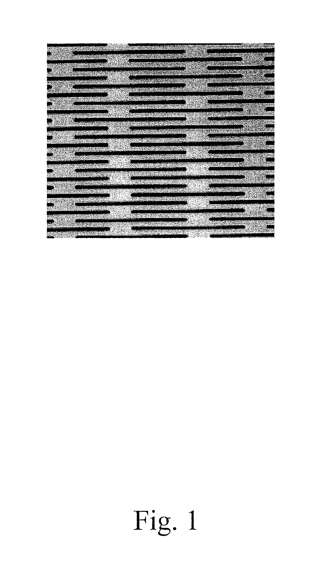
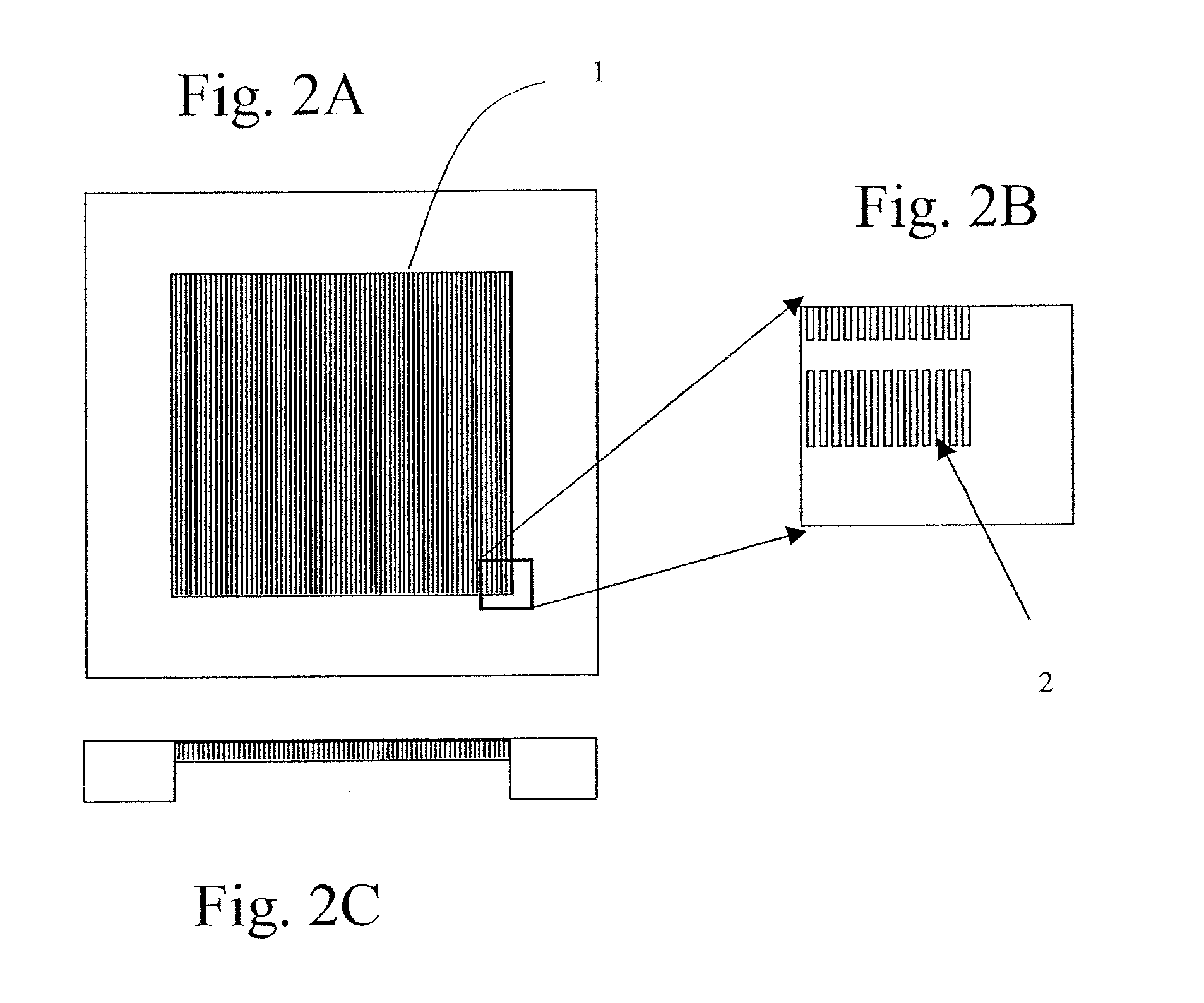
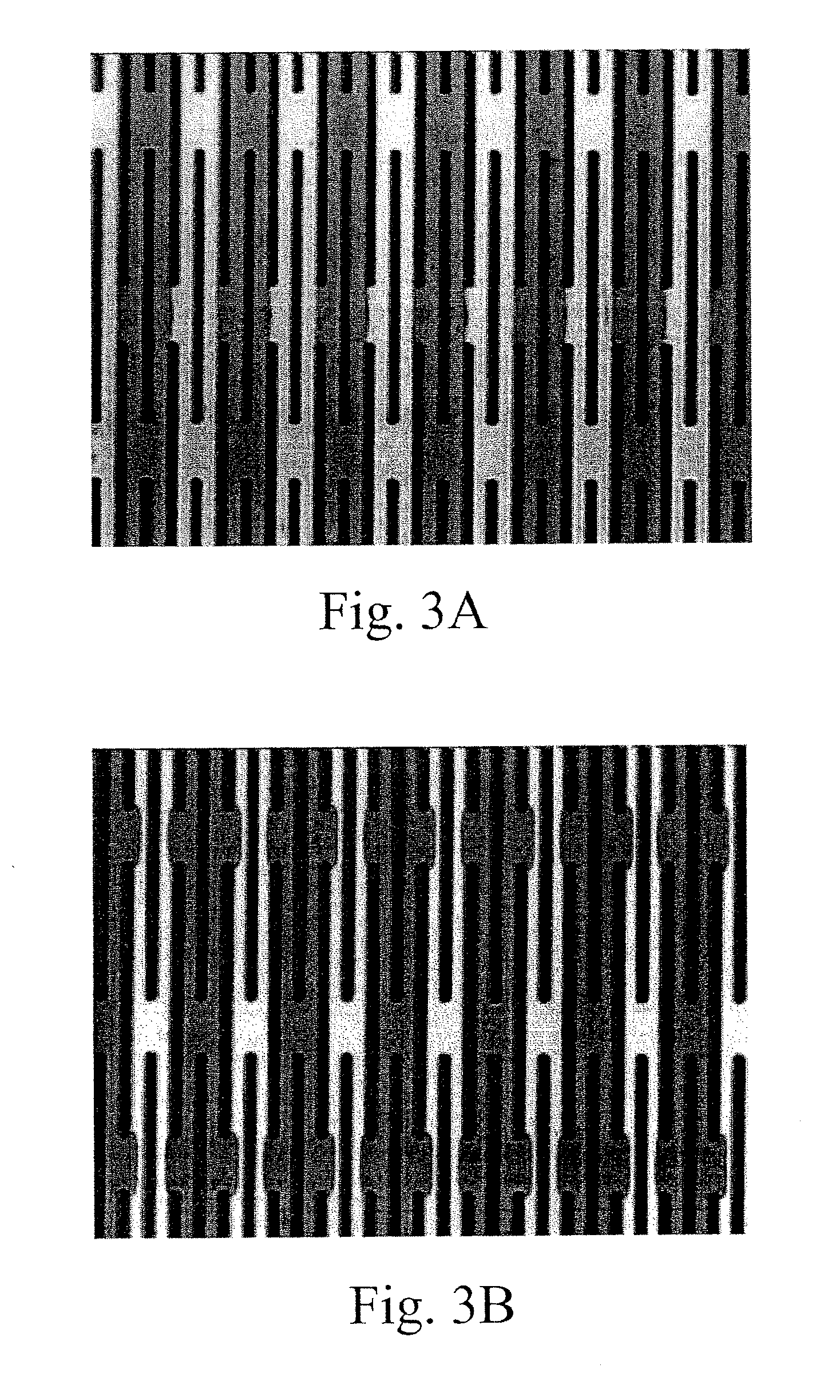
[0417]A silicon chip of dimensions (1.8 cm by 1.8 cm×500 micron) was used to fabricate a filtration area of 1 cm by 1 cm by 50 micron with slots having dimensions from about 0.1 micron to about 1000 microns, preferably from about 20 to 200 microns preferably from about 1 to 10 microns, more preferably 2.5 to 5 microns. The slots were vertically straight with a maximum tapered angle of less than 2%, preferably less than about 0.5% with an offset distance between neighboring columns of the filter slots were 1 to 500 microns, preferably from 5 to 30 microns.

[0418]Manufacturing included providing a silicon chip having the above referenced dimensions and coating the top and bottom of the silicon chip with a dielectric layer. A cavity along the bottom portion of the chip was then created. The cavity was formed by removing an appropriate cavity pattern from the dielectric layer then etching the silicon chip generally f...

example 2

Chemical Treatment of a Microfabricated Filter

[0419]A filter chip made as described in Example 1 was placed on a ceramic heating plate in an oven and heated at 800 degrees Celsius for 2 hours in oxygen containing gas (e.g. air). The heating source was then turned off the chips are slowly cooled overnight. This results in a thermally grown layer on the surface of the chip.

[0420]A nitride layer could also be deposited onto the filter surface. An oxide layer is put on the surface of the chip by low-pressure chemical vapor deposition (LPCVD) in a reactor at temperatures up to ˜900° C. The deposited film is a product of a chemical reaction between the source gases supplied to the reactor. The process is typically performed on both sides of the substrate at the same time to form a layer of Si3N4.

example 3

Polyvinylpyrrolidone (PVP) and Polyvinyl Alcohol (PVA) Filter Coatings

[0421]Filter chips made by the method of Example 1 were coated with either PVP or PVA. For coating the chips with either PVP or PVA, the chips were pre-treated as follows: The filter chips were rinsed with deionized water and then immersed in 6N nitric acid. The chips were placed on a shaker for 30 minutes at 50 degrees Celsius. After acid treatment, the chips were rinsed in deionized water.

[0422]For PVP coating, chips were immersed in 0.25% polyvinylpyrrolidone (K-30) at room temperature until the chips were ready for use. Chips were then rinsed with deionized water and dried by pressurized air.

[0423]For PVA coating, after acid treatment and rinsing in water, the chips were stored in water prior to coating. To make the 0.25% PVA (Mn 35,000-50,000) solution, dissolve the PVA in water under slow heating to 80 degrees Celsius and gentle stirring. To coat, the chips were immersed in a hot PVA solution and heated for ...


PUM

PropertyMeasurementUnit
areaaaaaaaaaaa
areaaaaaaaaaaa
areaaaaaaaaaaa

Abstract

The present invention recognizes that diagnosis and prognosis of many conditions can depend on the enrichment of rare cells, especially tumor cells, from a complex fluid sample such as a blood sample. In particular, the present invention is directed to methods and compositions for detecting a non-hematopoietic cell, e.g., a non-hematopoietic tumor cell, in a blood sample via, inter alia, removing red blood cells (RBCs) from a blood sample using a non-centrifugation procedure, removing white blood cells (WBCs) from said blood sample to enrich a non-hematopoietic cell, if any, from said blood sample; and assessing the presence, absence and / or amount of said enriched non-hematopoietic cell.

Description

CROSS-REFERENCE TO RELATED APPLICATIONS[0001]This application is a continuation-in-part of U.S. patent application Ser. No. PCT / US2004 / 030359, entitled ““Methods, compositions, and automated systems for separating rare cells from fluid samples,” filed Sep. 15, 2004 and a continuation-in-part of U.S. patent application Ser. No. 10 / 701,684, entitled “Methods, compositions, and automated systems for separating rare cells from fluid samples,” filed Nov. 4, 2003, now pending, which is a continuation-in-part of U.S. patent application Ser. No. 10 / 268,312, entitled “Methods, Compositions, and Automated Systems for Separating Rare Cells from Fluid Samples” filed Oct. 10, 2002, now U.S. Pat. No. 6,949,355 B2, which claims benefit of priority to the following patent applications: U.S. Provisional Patent application No. 60 / 348,228, filed on Oct. 29, 2001, entitled “Methods and automated systems for separating rare cells from fluid samples”, U.S. Provisional Patent application No. 60 / 328,724, f...

Claims


Application Information

Patent Timeline
Patent Type & Authority Applications(United States)
IPC IPC(8): G01N1/34
CPCG01N1/34A61M1/36A61M1/3679B01D57/02B01D61/18B01D67/0034B01D67/0062B01D71/02B01D71/027B01D71/04B01L3/502B01L3/5027B01L3/502753B01L3/502761B01L2300/0681B01L2300/0816B01L2300/0861B01L2400/0409B01L2400/0457B01L2400/0478B01L2400/0644B01L2400/065G01N1/40G01N15/0272G01N15/05G01N33/56966G01N33/57407G01N2001/4016G01N2015/0073G01N2015/0288G01N2015/0294G01N2015/0681G01N2333/4724B01D2325/021A61M1/3618A61M1/362Y10T436/25375C12Q1/6881G01N1/4077A61M1/3616A61B5/150755B01L2200/10B01L2300/0627B01L2400/0415C12Q1/6841G01N35/026G01N35/04G01N35/1002G01N2001/4083G01N2001/4088G01N2035/0406
Inventor LIN, PINGYAMANISHI, DOUGLAS T.WALKER, GEORGEXU, JUNQUANHUANG, MINGXIANTAO, GUOLIANGWU, LEIWANG, XIAOBOOUYANG, JOECHENG, JINGXU, JIA
Owner AVIVA BIOSCI
Why Eureka
Try Eureka