Human inflammatory breast carcinoma xenograft capable of lymphovascular invasion and methods for its use

a technology of lymphovascular invasion and xenograft, which is applied in the field of human inflammatory breast carcinoma xenograft, can solve the problems of severe hampered process and lack of appropriate and clinically relevant modeling systems, and achieve the effect of preventing lymphovascular invasion

Inactive Publication Date: 2006-02-14
RGT UNIV OF CALIFORNIA
View PDF1 Cites 3 Cited by
  • Summary
  • Abstract
  • Description
  • Claims
  • Application Information

AI Technical Summary

Benefits of technology

[0011]Another embodiment of the invention consists of methods for evaluating at least one agent for treating inflammatory breast cancer by utilizing a immunocompromised host animal inoculated with a human inflammatory breast cancer xenograft, administering at least one agent to said inoculated immunocompromised host animal and evaluating the effects of the agent(s) on the human inflammatory breast cancer xenograft. Optionally, the agent that is being evaluated targets a molecule that is identified as being associated with the inflammatory phenotype. Yet another related embodiment of the invention consists of methods for evaluating the potential of an agent, or a combination of agents, for the prevention of lymphovascular invasion of carcinoma cells by utilizing a immunocompromised host animal inoculated with a human inflammatory breast cancer xenograft, administering at least one agent to said inoculated immunocompromised host animal and evaluating the effectiv...

Problems solved by technology

This process has been severely hampered by the lack...

Method used


Image

  • Human inflammatory breast carcinoma xenograft capable of lymphovascular invasion and methods for its use
  • Human inflammatory breast carcinoma xenograft capable of lymphovascular invasion and methods for its use
  • Human inflammatory breast carcinoma xenograft capable of lymphovascular invasion and methods for its use

Examples


example 1

Generation and Propagation of Cell lines and Xenografts

[0133]Informed patient consent and certification from the UCLA Human Subject Protection Committee was obtained prior to all studies. Approval from the Chancellor's Animal Research Committee was requested and obtained, certification ARC #95–127-11. The MARY-X xenograft was established directly from a 45 year old female who presented with a warm and erythematous breast and ill-defined mass. The mass was biopsied and diagnosed as inflammatory carcinoma exhibiting florid invasion of dermal lymphatics. Minced 1 mm3 portions of the biopsy were washed in Hank's balanced salt solution, placed in RPMI media and subsequently implanted subcutaneously in several female scid and athymic (nude) mice (nu / nu mutants on a BALB / c background). The tumoral xenografts which grew were subsequently transplanted when they reached 1 cm in diameter. Following this procedure, an illustrative stable serial transplantable xenograft designated MARY-X was suc...

example 2

Analysis of MARY-X Xenograft

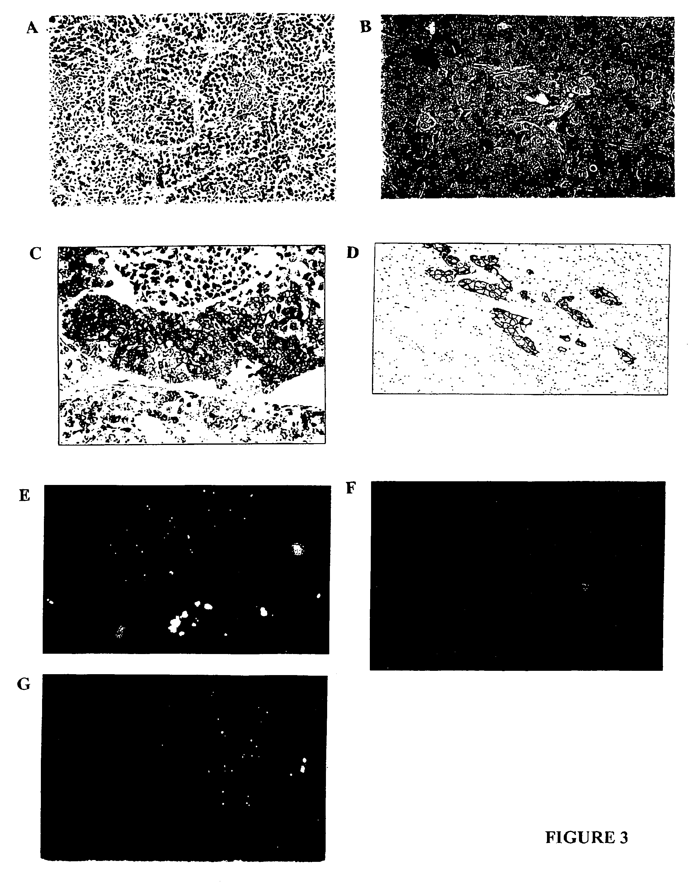
[0135]Histopathological studies. Xenografts were processed for routine light microscopic, immunocytochemical and ultrastructural studies according to standard protocols. Human cases of inflammatory and non-inflammatory breast cancer were retrieved from archival material and studied immunocytochemically.

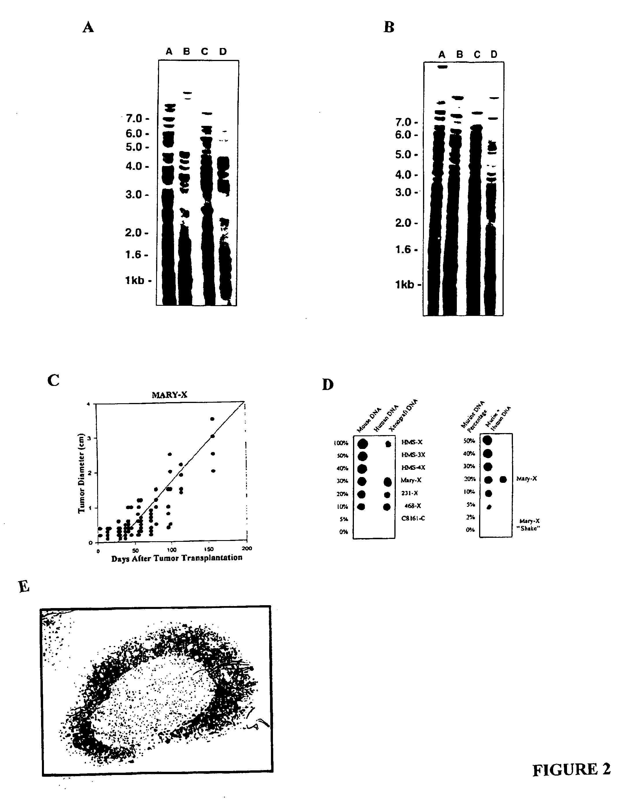
[0136]DNA profiling. High molecular weight DNA was extracted from the xenografts and host murine tissues by standard procedures (Sambrook, J., Fritsch, E. F., and Maniatis, T. Molecular cloning: a laboratory manual. 2nd ed. Cold Spring Harbor, N.Y.: Cold Spring Harbor Laboratory Press. 1989), digested with HaeIII and HinfI and probed with the multilocus 33.6 Jeffeys probe (Jeffreys, A. J., Wilson, V., and Thein, S. L. Hypervariable “minisatellite” regions in human DNA. Nature, 314: 67–73, 1985) (Cellmark Diagnostics, Germantown Md.). This probe recognizes both human as well as murine DNA. A human-specific [α-32P] dCTP-labeled human Cot-1 DNA probe (GIBCO-BR...

example 3

MARY-X In Vivo Cancer Model

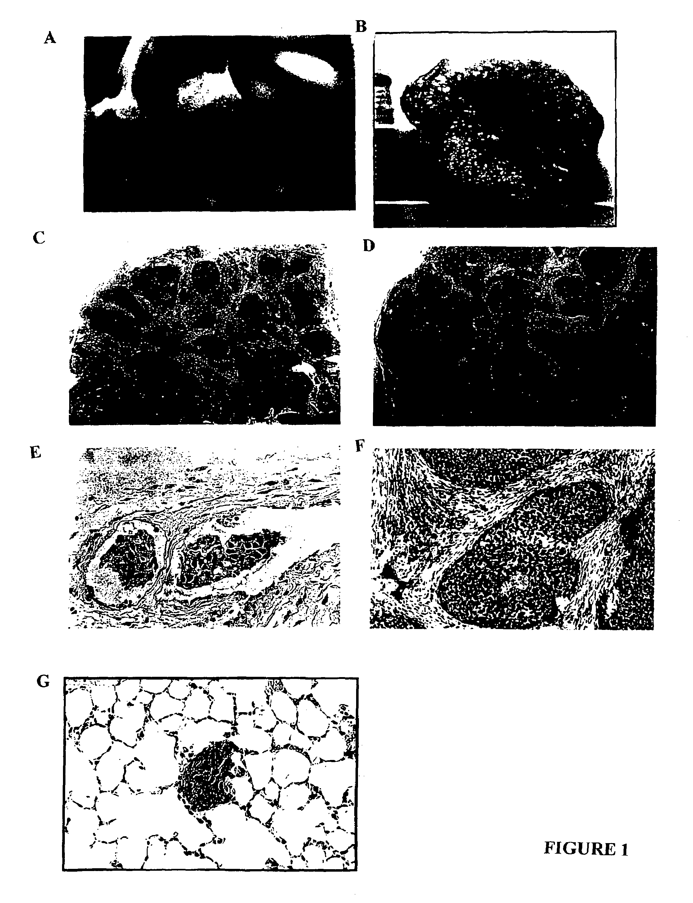
[0149]The xenograft disclosed herein exhibits striking lymphovascular invasion in an immunocompromised host. In fact it does not grow as an isolated tumor nodule but grows exclusively within lymphovascular channels. Some of these channels are lymphatics and some are blood vessels confirmed by anti-vWf factor and anti-CD31 endothelial staining. Interestingly the skin overlying the inflammatory xenograft is intensely erythematous just as it is in humans presumably from the lymphovascular obstruction. In both the athymic (nude) mouse and the Scid, the inflammatory carcinoma xenograft exhibits a high degree of spontaneous metastasis as early as 6 weeks following local subcutaneous implantation. In contrast the non-inflammatory xenografts, 231 and 468 grow as isolated tumor nodules exhibiting no lymphovascular invasion and no metastasis.

[0150]MARY-X induced erythema in the overlying mouse skin (FIG. 1A) mimicking the clinical presentation of inflammatory carcin...


PUM

PropertyMeasurementUnit
Atomic weightaaaaaaaaaa
Levelaaaaaaaaaa

Abstract

The present invention provides human transplantable inflammatory breast carcinoma xenografts. Such xenografts exhibit a number of unique characteristics which allows their use in experimental models of inflammatory carcinoma in order to dissect out the molecular basis of this phenotype. This experimental model of inflammatory carcinoma can be used to identify molecular targets for therapeutic intervention and to assess the efficacy of a broad spectrum of diagnostic and therapeutic agents. Specific animal models of inflammatory breast cancer are described as well as methods for evaluating diagnostic and therapeutic agents for treating inflammatory breast cancer. Methods for identifying molecules whose expression is modulated in inflammatory breast cancer are provided. In addition, methods for diagnosing and inhibiting the growth of inflammatory breast cancer metastases in vivo are provided.

Description

[0001]This application claims the benefit of U.S. provisional patent application Ser. No. 60 / 154,408, filed Sep. 17, 1999. The entire content of this provisional patent application is incorporated herein by reference.FIELD OF THE INVENTION[0002]The present invention relates to the use of inflammatory breast carcinoma xenografts as models for both the evaluation of the cancer phenotype as well as the generation of novel diagnostic and therapeutic methods for the clinical management of this pathology.BACKGROUND OF THE INVENTION[0003]Cancers of the breast are one of the leading causes of death among women, with the cumulative lifetime risk of a woman developing breast cancer estimated to be 1 in 9. Consequently, understanding the origins of these malignancies as well as models for the identification of new diagnostic and therapeutic modalities is of significant interest to health care professionals.[0004]Most women that die from breast cancer succumb not to the original primary disease...

Claims


Application Information

Patent Timeline
IPC IPC(8): A01K67/00A01K67/033A61K49/00C12N5/00C12N5/08A01K67/027C07K16/18C07K16/30C12N5/09
CPCA01K67/0271A61K49/0008C07K16/18C07K16/30C12N5/0693A01K2227/105A01K2267/0331C12N2503/00A61K2039/505
Inventor BARSKY, SANFORD H.ALPAUGH, MARY L.TOMLINSON, JAMES S.
Owner RGT UNIV OF CALIFORNIA
Why Eureka
Try Eureka