
Positive power anterior chamber ocular implant

a technology of ocular implants and anterior chambers, which is applied in the field of positive power anterior chamber ocular implants, can solve the problems of concomitant increase in damage to the eye, immediate or latent inflammation, and damage to the corneal endothelium

Inactive Publication Date: 2006-03-21
NOVARTIS AG
View PDF72 Cites 17 Cited by
  • Summary
  • Abstract
  • Description
  • Claims
  • Application Information

AI Technical Summary

Benefits of technology

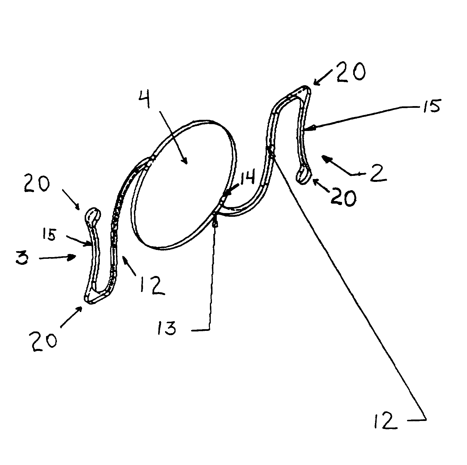
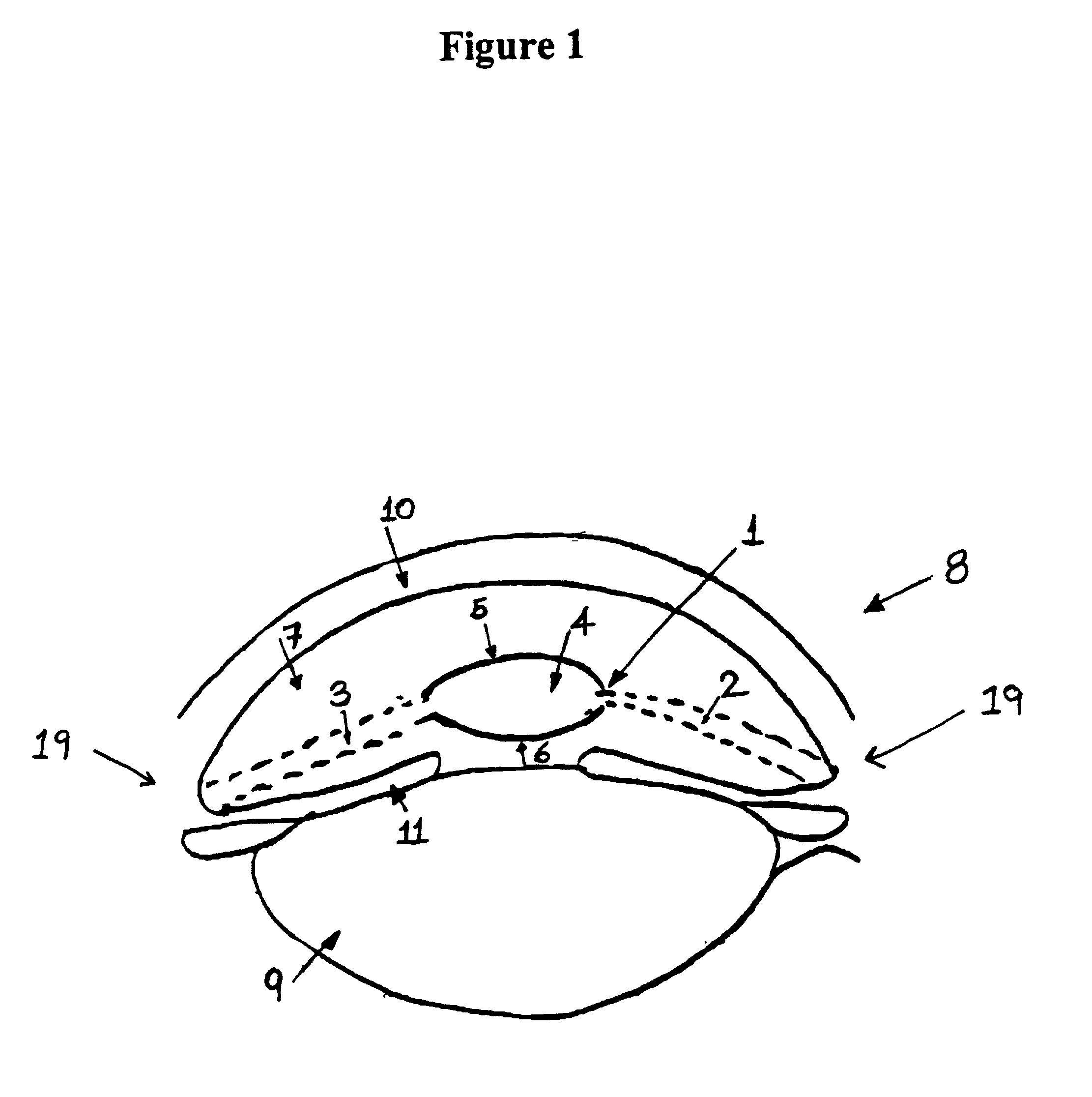
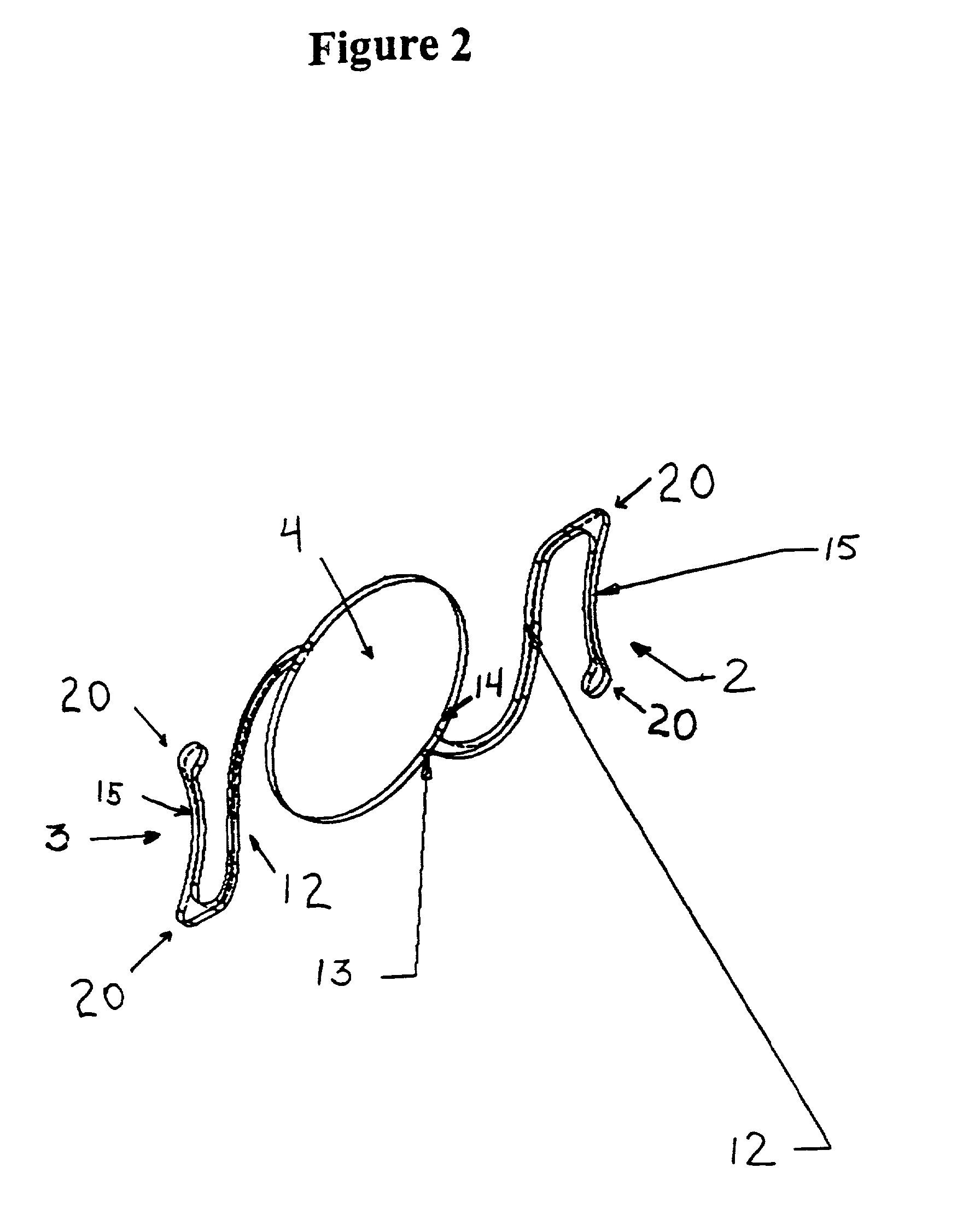
The invention is a positive power ocular implant that can be placed in the eye to correct refractive errors. The implant is designed to avoid contact with other anatomical bodies in the eye, such as the iris and corneal endothelium. It is flexible and can compress when pressure is applied during surgery, allowing for proper placement without damaging the eye's blood supply. The implant can also be coated with a medicament containing a compatible sulfated polysaccharide. The invention provides a safe and effective method for correcting refractive errors in patients.

Problems solved by technology

However, although the implantation of intraocular lenses has constituted an appreciable surgical advance, such implantation has been known to cause immediate or latent damage to the corneal endothelium, immediate or latent inflammatory responses in the anterior and / or posterior segments of the eye, immediate or latent secondary fibrosis and / or neovascularization, and other problems.
Certain difficulties are associated with implanting an intraocular lens in the phakic eye that are not encountered when implanting a lens in the aphakic eye.
Inflammatory reactions tend to be greater in the phakic eye resulting in a concomitant increase in damage to the eye caused by implanting intraocular lenses.
In contrast, implanting intraocular lenses in patients having cataract removal cannot induce such an effect since the natural lens has been removed.
However, significant drawbacks were discovered.
Furthermore, if posterior chamber implants were to cause an inflammatory reaction in the phakic eye, cataract formation may occur in the natural lens which remains in situ.
However, a drawback existed for correcting refractive errors in patients with high myopia, extreme nearsightedness, with posterior chamber ocular lenses.
Because of the high degree of corrective refraction of the optic, which causes the optical zone to be quite small, a myopic posterior chamber lens may irritate the iris and potentially touch the natural lens.
The prior art does not disclose implanting a positive power implant into the anterior chamber of a phakic eye for the treatment of hyperopia.
Moreover, due to the shallowness of the anterior chamber in hyperopic eyes, surgical location of a positive power implant in the anterior chamber of the phakic eye for the treatment of hyperopia would be in contravention of, and counterintuitive to, the state of the art.
It has been generally acknowledged by those skilled in the art that there are significant risks involved with the use of anterior chamber angle supported implants in the aphakic eye (reviewed in Apple et al., in Intraocular Lenses: Evolution, Designs, Complications and Pathology (William and Wilkens, Baltimore) 1989, Chapter 4, pp.
For example, when such an implant is inserted into the eye, temporary or permanent adhesions of the implant to iris tissue may result, causing damage to these structures to ensue either immediately or over the long term affecting pupillary mobility and contour.
In addition, once the implant is in position, it may cause similar angle adhesions due to mechanical and / or chemical inflammation which may lead to fibrosis of a progressive nature.
This would make subsequent removal of the implant a complex, dangerous surgical procedure.
Other problems associated with such implants are cataract formation, secondary glaucoma, corneal edema, hyphema, and progressive endothelial cell loss, in addition to other complications.
41–42, Comment on page 42, several American manufacturing companies that were preparing to begin clinical trials of phakic anterior chamber ocular implants in the United States are now likely to abandon these studies, due to the attendant risks associated with anterior chamber implants in the phakic eye and the difficulty of obtaining approval of the U.S. Food and Drug Administration (FDA) for the use of the implants.
A significant risk involved in the use of such anterior chamber implants in the phakic eye is the potential for the implanted lens to contact the corneal endothelium, the anatomic lens or the iris with resultant complications, even with the enhanced vault design theorized in the studies discussed in Ophthalmology Alert because such design would bring the lens optic edge quite close to the midperipheral corneal endothelium.
One alternative to avoid contact with the corneal endothelium is to reduce the diameter of the optic of the minus power lens, however, such a modification creates significant drawbacks, including glare and haloing under low light conditions, ovalization of the pupil, and, as reported in Perez-Santonja et al., J. Cataract Refract Surg 22:183–187 (1996) (discussing Baikoff ZB5M), induces an inflammatory response.
Although such positioning on the iris may provide for safe clearance away from vital structures such as the natural lens and the corneal endothelium, the implant has a number of significant drawbacks.
Damage may occur to the sensitive iris tissue and the damage is increased when adjusting the position of the implant in cooperation with the existing lens.
Also, deterioration of the iris tissue occurs, thereby decreasing the ability to place future replacement implants on the iris.
The '363 patent does not appreciate the problems associated with positive power ocular implants actually implanted into the phakic eye.
The '792 patent does not appreciate the complications associated with a planar or convex posterior surface of the optic in combination with a convex or planar anterior surface.
To Applicant's knowledge, there has not been a successful implantation of a positive power anterior chamber ocular implant angle supported in the phakic eye for the treatment of hyperopia.

Method used


Image

  • Positive power anterior chamber ocular implant
  • Positive power anterior chamber ocular implant
  • Positive power anterior chamber ocular implant

Examples


example 1

[0053]Representative uncoated positive power ocular implants were made from polymethylmethacrylate (“PMMA”) each comprising an optic having a diameter of 6 mm and an omega value of 13 mm. The center thickness (TC) of the optical portion of each implant is shown in Table 1 below. The edge thickness (TE) of each optic was held constant at 0.220 mm. The posterior surface (RP) of each optic was kept planar. The anterior surface (RA) of each optic was convex, each having a degree of curvative shown in Table 1 below. The vault of each optic was set at 1.0 mm. The resulting degree of positive refraction is shown for each optic as indicated by its diopter value. The sagitta values of each provided optic are provided in Table 1. The haptics of each optic are made of PMMA and are in a four point “S” configuration. Each haptic has an intermediate beam length of 5.25 mm. The transition between the haptic and optic is normal to the optic, having a blend radius ranging in the horizontal and verti...

example 2

[0055]Representative implants of Table 1 were then tested for compression force, axial displacement, decentration, contact angle and optic tilt. Eighteen implants were made according to the specifications of Implant 1, described above in Example 1. Implants were exposed to a 0.5 mm diametral compression and a 1.0 mm diametral compression. The resulting mean and standard deviation values measured in grams are shown in Table 2 below.

[0056]

TABLE 2DiametralDiametralCompressionCompressionof 0.5 mmof 1.0 mmmeanstd devmeanstd devCompression Force0.1580.0310.4590.082Axial Displacement0.1120.0180.2450.030Decentration0.0820.0430.1220.077Contact Angle6.6570.0516.6940.085Optic Tilt0.9870.5060.7190.447

example 3

[0057]The present invention is further directed to a method of correcting refractivc errors caused by hyperopia by way of surgically implanting and anchoring a positive power anterior chamber ocular implant in the anterior chamber of the phakic eye in a subject in need thereof, wherein the positive artificial refracting lens has at least one convex surface and a means for positioning the positive refracting lens in the anterior chamber of the eye. By this method contact between the positive refractings lens and other anatomic bodies is avoided and the means for positioning the lens in the anterior chamber avoids contact with the iris and corneal endothelium.

[0058]Implants made in accordance with the present invention were surgically implanted into subjects having a natural lens in situ and anchored in the anatomic angle of the anterior chamber of the eye, according to traditional surgical techniques. The subjects were studied for inflamation and damage to the eye post insertion. No ...


PUM

PropertyMeasurementUnit
thicknessaaaaaaaaaa
lengthaaaaaaaaaa
anterior chamber depthaaaaaaaaaa

Abstract

A positive power anterior chamber ocular implant for placement in a phakic eye is disclosed to correct refractive errors caused by hyperopia which includes at least one convex surface and haptics for positioning the lens in the anterior chamber of the eye. Contact between the lens and other anatomic bodies, such as the anatomical lens, the corneal endothelium and iris is avoided, and the haptics avoid contact with the iris.

Description

CROSS REFERENCE TO RELATED APPLICATIONS[0001]This application is a continuation of application Ser. No. 09 / 282,476, filed Mar. 31, 1999, now U.S. Pat. No. 6,171,337.FIELD OF THE INVENTION[0002]This invention relates to a positive power anterior chamber ocular implant for placement in a phakic eye to correct refractive errors such as hyperopia, hyperopic or mixed astigmatism, a method for treating hyperopia in a patient in need thereof comprising surgically implanting and anchoring in the anterior chamber angle of a phakic eye a positive power anterior chamber ocular implant comprising a positive artificial refracting lens having at least one convex surface and a means for positioning the lens in the anterior chamber of the eye, wherein contact between the positive refracting lens and other anatomic bodies is avoided and wherein the means for positioning the lens avoids contact with the iris.BACKGROUND OF THE INVENTION[0003]It is well known to those skilled in the art that intraocula...

Claims


Application Information

Patent Timeline
Patent Type & Authority Patents(United States)
IPC IPC(8): A61F2/16A61L27/16A61L27/18A61L27/34
CPCA61F2/16A61L27/16A61L27/18A61L27/34C08L5/00A61F2002/1605A61F2002/1683A61L2430/16A61F2/1605A61F2002/1681
Inventor GALIN, MILES A.
Owner NOVARTIS AG
Learn More