
Methods of treating and detecting cancer using viruses

a virus and cancer technology, applied in the field of mammals' methods of treating and detecting cancer, can solve the problems of not carrying out work to address direct tumor cytotoxicity of the virus, and not fully exploring the mechanism of any anti-cancer

Inactive Publication Date: 2006-06-06
WELLSTAT BIOLOGICS CORP
View PDF50 Cites 41 Cited by
  • Summary
  • Abstract
  • Description
  • Claims
  • Application Information

AI Technical Summary

Benefits of technology

[0019]The invention also provides a genetically engineered Paramyxovirus comprising heterologous nucleic acid encoding a biologically or chemically active peptide, polypeptide or protein. The heterologous nucleic acid is operatively linked to one or more control sequences.
[0020]The invention further provides an article of manufacture and kit containing materials useful for treating and detecting cancer. The article of manufacture comprises a container, a label on the cont

Problems solved by technology

The overall five-year survival approximates fifty percent for all patients, and the prognosis remains particularly poor for those with advanced solid tumors.
Furthermore, the mechanism of any anti-cancer effect was never fully explored by the investigators.
Since the mechanism of the observed tumoricidal activity was thought to be immunologic, no work was carried out to address direct tumor cytotoxicity of the virus.
There are disadvantages in using retroviruses because the viruses are rendered incapable of producing progeny viruses, i.e., virus amplification cannot take place.
Also, the use of retroviruses to deliver genetic material to cancer cells is not desirable because retroviral vectors tend to infect tumor cells at a relatively low percentage, thereby precluding efficient in-situ transfer of the desired genetic material into the tumor site.
Furthermore, production of the gene of interest delivered by the retrovirus occurs at a rate determined by the cell cycle of the host cell which can severely limit the amount of gene product being translated.

Method used


Image

  • Methods of treating and detecting cancer using viruses
  • Methods of treating and detecting cancer using viruses
  • Methods of treating and detecting cancer using viruses

Examples


example # 1

Example #1

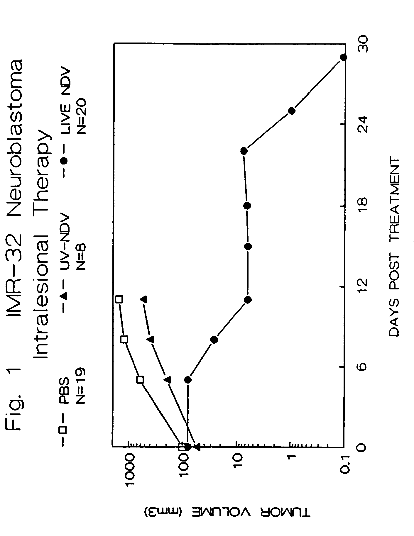
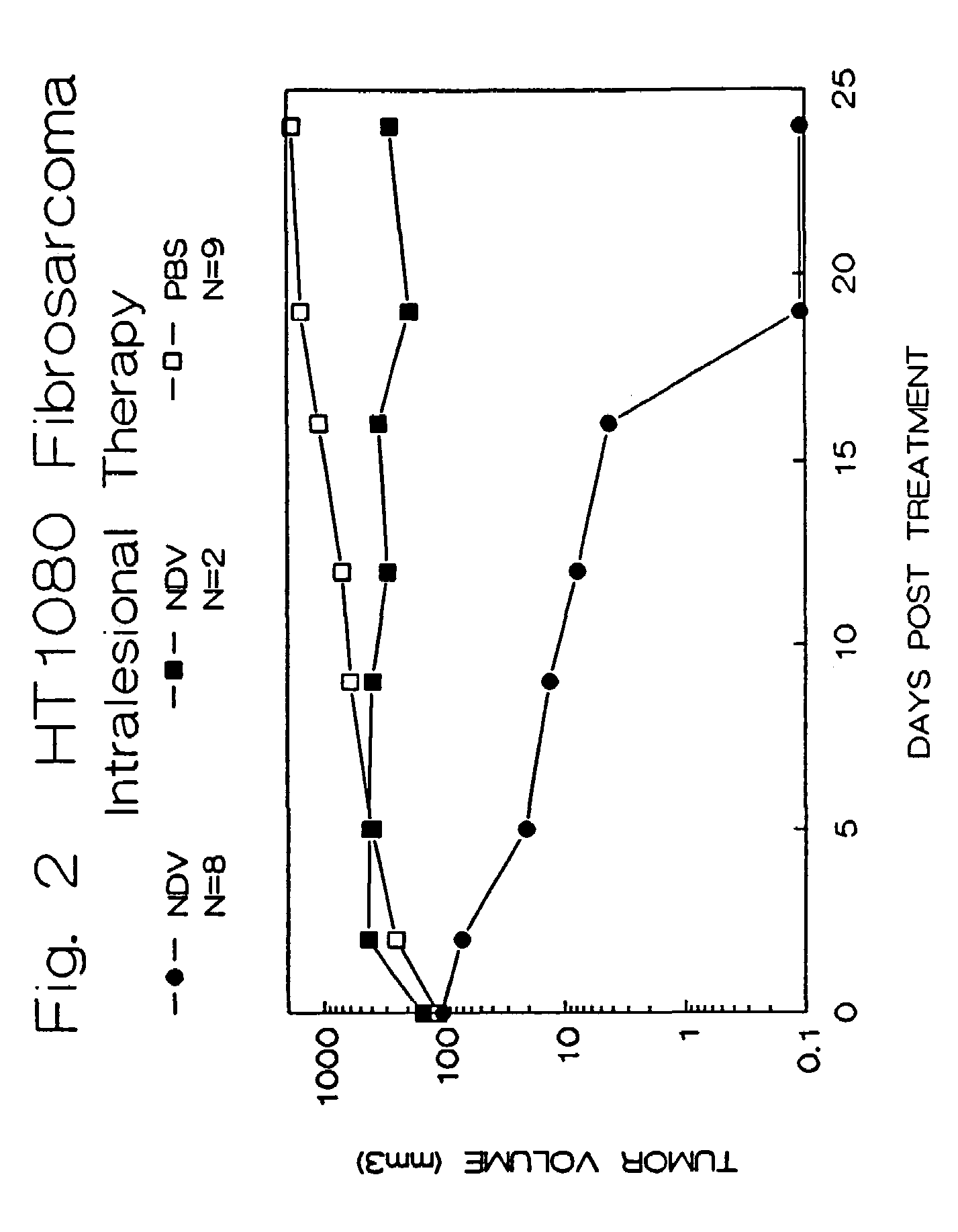
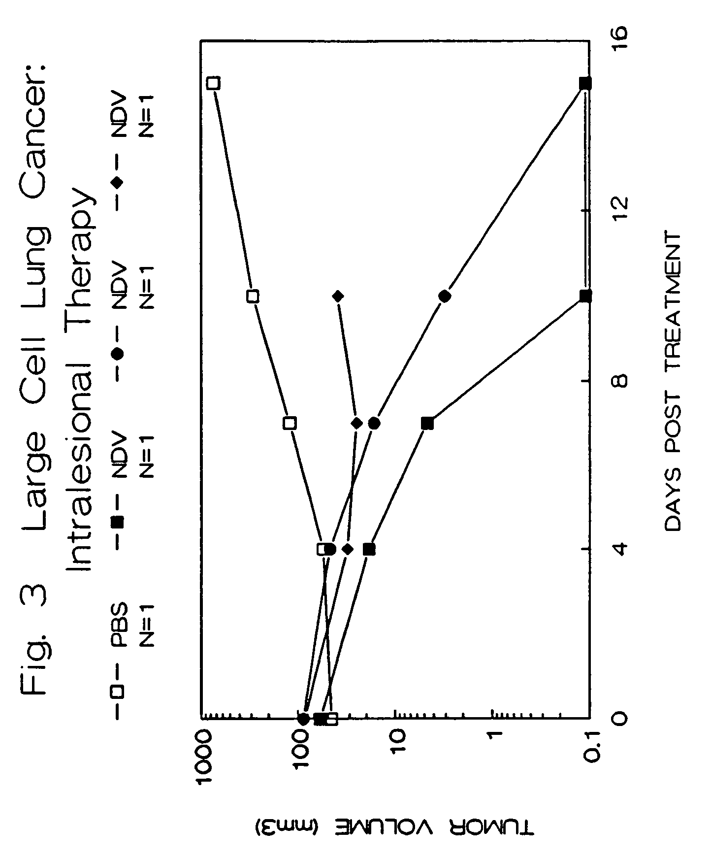
Treatment of Human Tumors in Athymic Mice with Intralesional NDV

[0072]Female athymic Balb-c mice were obtained from Life Sciences, Incorporated, St. Petersburg, Fla. and Charles River Laboratories, Wilmington, Mass. They were housed in an isolated room in sterilized, filtered cages and given autoclaved-sterilized chow and water ad libitum. The care of these animals and the experimental protocols were conducted according to guidelines established by the Institutional Animal Care Committees, Cook County Hospital and Rush-Presbyterian-St. Luke's Medical Center, Chicago, Ill. Mice were obtained at approximately four weeks of age, and allowed to acclimate to their new environment for one week.

[0073]Tumor cell lines were obtained frozen from the American Type Culture Collection, Rockville, Md., and were derived from human tumors. The cell lines were maintained in Opti-MEM (Gibco-BRL, Grand Island, N.Y.) containing 2.4 g / L sodium bicarbonate, 10% heat-inactivated fetal calf serum...

example # 2

Example #2

Treatment of Human Tumors in Athymic Mice Using Systemic NDV Therapy

[0086]Athymic mice were injected with human tumor xenografts as described in Example #1. When tumors greater than 6 mm in diameter were achieved, the animals were divided into three groups. The NDV was prepared (see Example #1) in concentrations of 1×10⁸ PFU / 0.3 ml or 1×10⁹ PFU / 0.3 ml of saline. The control solution was an equal volume of saline alone.

[0087]The abdomens of the animals were washed thoroughly with alcohol, and the NDV was injected intraperitonealy using a 25 gauge needle. One set of experimental mice (N=3) received a single injection of NDV at the higher dose (109 PFU), while the other set of 4 mice received injections of 108 PFU three times per week. Control animals received injections of saline. The animals were then observed and the tumor sizes were followed as described for Example #1.

[0088]The results are summarized in FIG. 4. Six out of 7 animals treated with intraperitoneal live NDV e...

example # 3

Example #3

Treatment of Human Tumors in Athymic Mice Using Attenuated Strains of NDV

[0090]NDV strain 73-T was obtained and prepared as described in Example #1. NDV strain M (Mass-MK107) is a strain less virulent than 73-T and was obtained from Dr. Mark Peeples, Rush-Presbyterian-St. Luke's Medical Center, Chicago, Ill. Strain M was replicated in a similar manner to that described in Example #1 for NDV strain 73-T.

[0091]Athymic animals were injected with human HT1080 tumor cells subcutaneously as described above. They were allowed to develop tumors of at least 6.5 mm in diameter at which time they were randomly divided into three groups, and their tumor sizes were recorded.

[0092]The animals from one group were injected with 1.0×10⁸ PFU of NDV strain 73-T in 0.1 ml saline intralesionally, as was described in Example #1. The second group was injected with 1.0×10⁸ PFU of NDV strain M in 0.1 ml saline intralesionally. The last group was injected with 0.1 ml of saline alone. The animals we...


PUM

PropertyMeasurementUnit
Diameteraaaaaaaaaa
Massaaaaaaaaaa
Cytotoxicityaaaaaaaaaa

Abstract

The invention provides a method of treating cancer in a mammal comprising administering to the mammal an effective amount of virus, particularly Newcastle Disease Virus or other Paramyxovirus. The invention also provides a method of treating cancer in a mammal comprising administering such viruses to the mammal in combination with another agent such as a chemotherapeutic compound, immunoadjuvant, cytokine, or immunosuppressive agent. The invention further provides a method of detecting cancer cells in a mammal using Paramyxovirus as an imaging agent and as an indicator of cancer cell growth in the mammal. The invention further provides genetically engineered Paramyxoviruses, and kits containing the viral compositions disclosed by the invention.

Description

RELATED APPLICATIONS[0001]This application is a continuation-in-part application of U.S. application Ser. No. 08 / 055,519, filed Apr. 30, 1993, incorporated herein by reference, now abandoned.[0002]This invention was made with government support under Grant No. RRO5477 awarded by the National Center for Research Resources. The government has certain rights in the invention.FIELD OF THE INVENTION[0003]The present invention relates generally to methods of treating and detecting cancer in mammals. More particularly, the invention relates to methods of treating and detecting cancer using certain viruses. The invention also relates to methods of producing viral compositions, to genetically engineered viruses containing heterologous nucleic acid and to kits containing the viral compositions and genetically engineered viruses.BACKGROUND OF THE INVENTION[0004]1. Cancer Treatment[0005]Despite recent advances in chemotherapy and radiation, cancer remains the second deadliest disease in the Uni...

Claims


Application Information

Patent Timeline
IPC IPC(8): G01N33/574A61K35/76A61K35/768A61K38/19A61K39/17A61K49/00A61P35/00C12N7/00C12Q1/70
CPCA61K39/17C12N7/00A61K35/768C12Q1/70A01K2267/0331C12N2760/18132A61K2300/00A61K39/12A61P35/00A61P35/04A61P43/00
Inventor LORENCE, ROBERT M.REICHARD, KIRK W.
Owner WELLSTAT BIOLOGICS CORP
Learn More