
Methods for multiple acquisitions with global inversion cycling for vascular-space-occupancy dependant and apparatuses and devices related thereto

a global inversion cycle and multi-shot acquisition technology, applied in the field of methods, apparatuses and systems for magnetic resonance imaging (mri), can solve the problems of limited approach to single slice or multi-shot 3-d acquisition techniques with limited readout time, complex multi-slice problems,

Active Publication Date: 2006-07-04
THE JOHN HOPKINS UNIV SCHOOL OF MEDICINE
View PDF6 Cites 26 Cited by
  • Summary
  • Abstract
  • Description
  • Claims
  • Application Information

AI Technical Summary

Benefits of technology

This approach enables efficient multi-slice MR imaging with minimal contamination from nulled species, maintaining signal quality and reducing residual blood signal, thus enhancing the application potential of MRI in neuroscience and clinical studies.

Problems solved by technology

In MRI, one of the most common problems or issues to solve relates to the selective imaging of a particular tissue type (i.e., T1 species) or body component or the like of interest, without contamination or too much influence from other tissue types or body components of a specific location of the body.
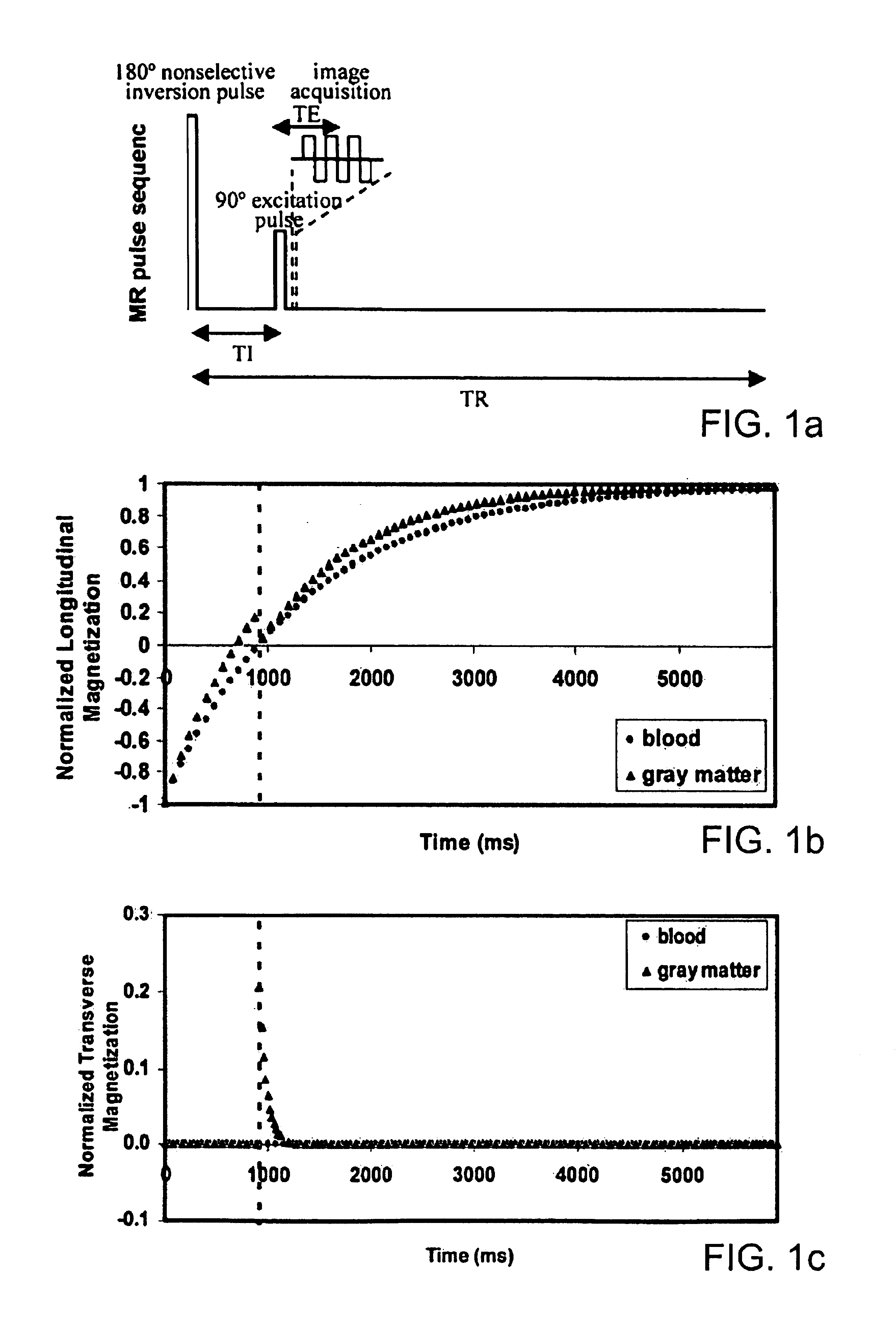
The above-described inversion-recovery sequence technique, however, is limited in that as a practical matter the approach is limited to a single slice or multi-shot 3-D acquisition techniques with limited readout time due to the fact that there is only one zero-crossing point on the T1 relaxation curve as is illustrated in FIG.
The problem is more complex in multi-slice black-blood imaging, for which very complicated schemes of interleaved inversion pluses have been proposed for only limited volume coverage.
The problem is then that the available signal in the slice decays with the transverse relaxation time (T2), which is one order of magnitude lower than the longitudinal relaxation times (T1).
Furthermore, in a large number of MR clinical imaging sequences besides the three examples above, it is desired to suppress the large signal coming from lipids, which can produce artifacts, especially in fast sequences, such as spiral or echo-planar imaging.
The weakness of such methods, however, is that they rely on a very good homogeneity of the main magnetic field Bo, which can be hard to achieve at high fields or in the part of the body placed away from the isocenter of the magnet (e.g., breast imaging), or in areas with strong susceptibility differences (e.g., in cartilage imaging).

Method used


Image

  • Methods for multiple acquisitions with global inversion cycling for vascular-space-occupancy dependant and apparatuses and devices related thereto
  • Methods for multiple acquisitions with global inversion cycling for vascular-space-occupancy dependant and apparatuses and devices related thereto
  • Methods for multiple acquisitions with global inversion cycling for vascular-space-occupancy dependant and apparatuses and devices related thereto

Examples


Embodiment Construction

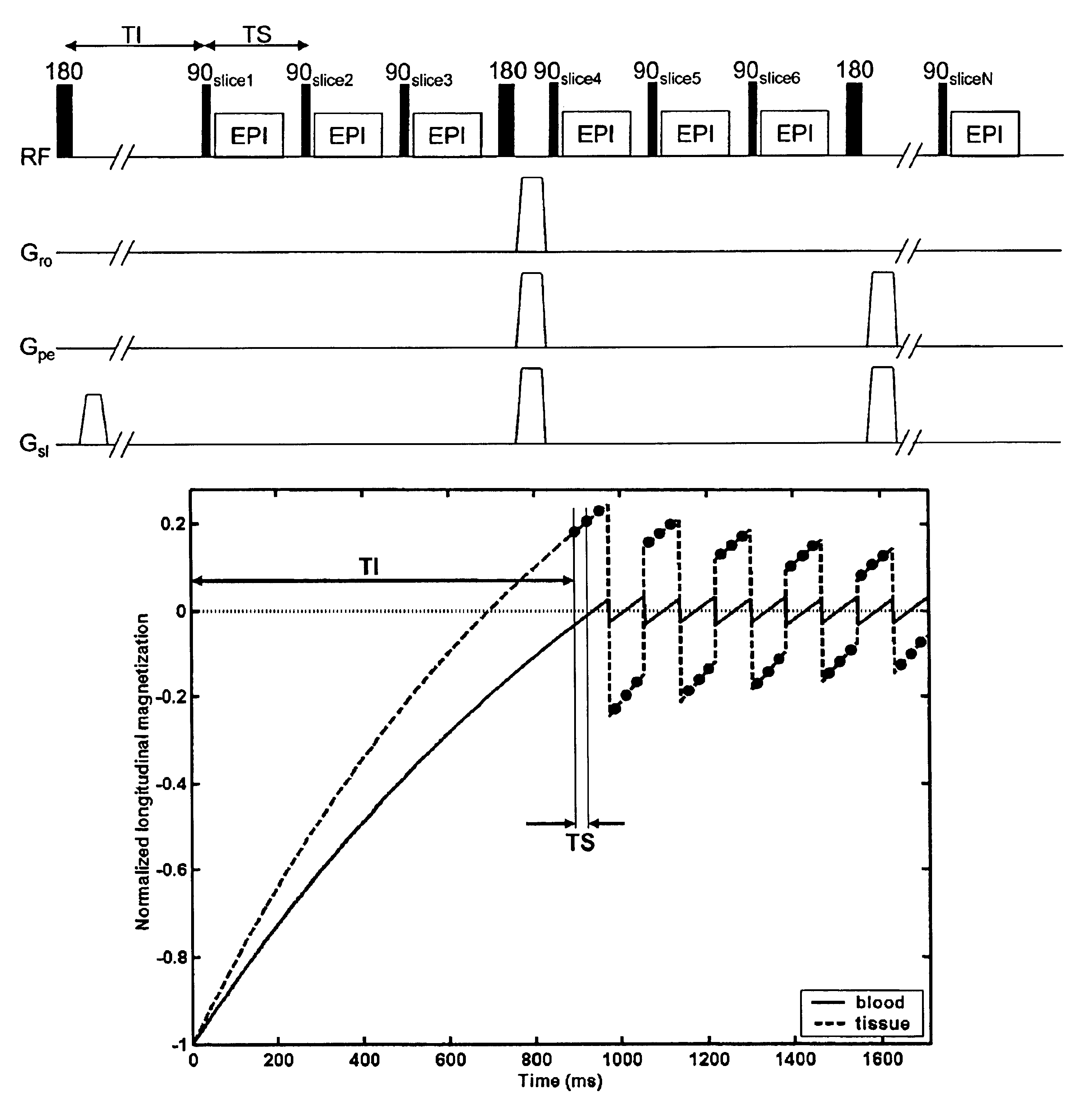
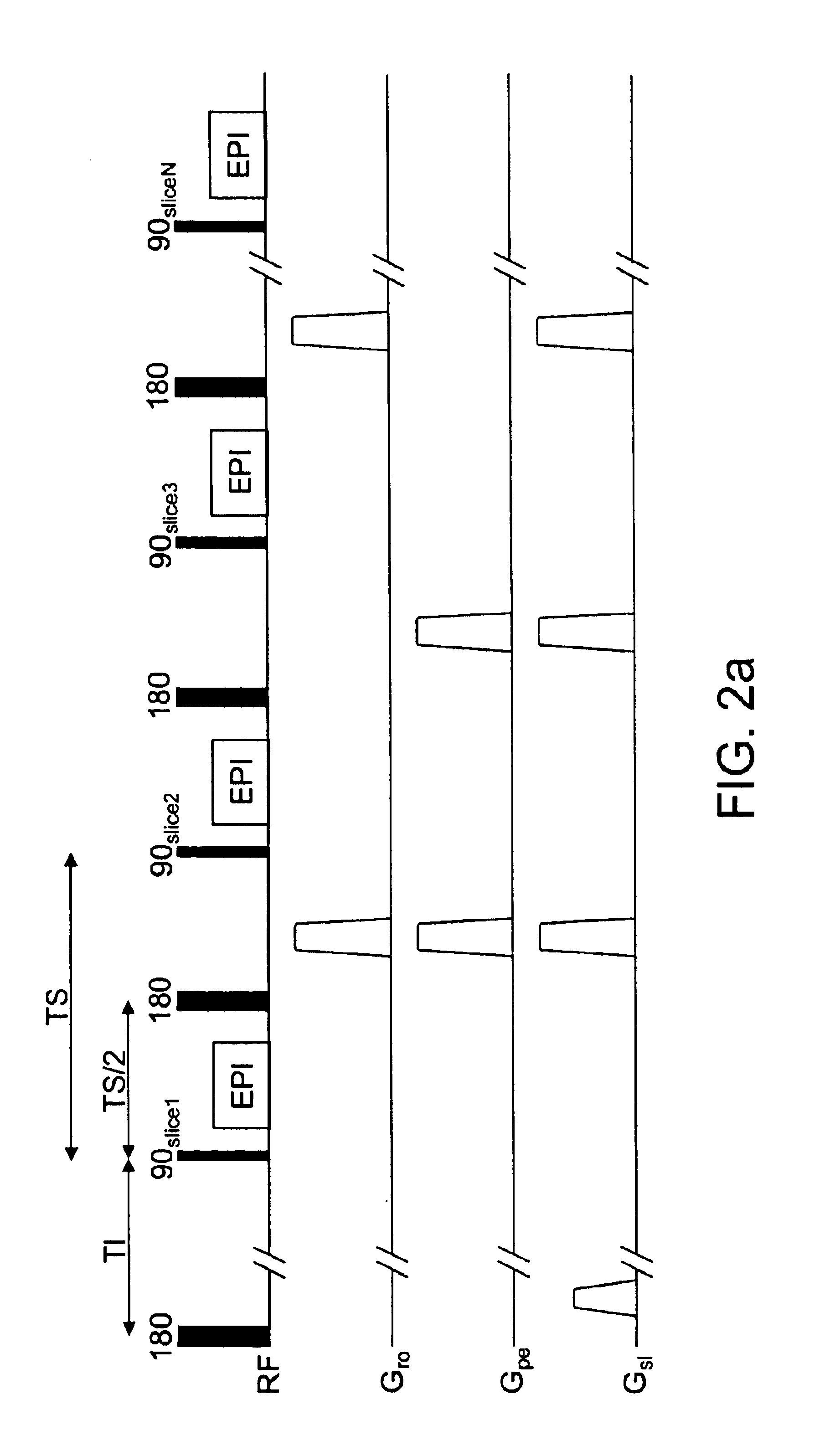
[0042]The present invention features a method for MR imaging particularly MR imaging in which RF inversion pulses are applied to a target area for essentially nulling one or more particular species, tissues or body components of the object to be imaged such as a mammalian body and acquisition of MR image data in one or more slices while keeping the signal from the one or more species, tissues or the like essentially nulled. The image data being acquired also can be 3-dimensional (3-D) image data. In its broadest aspects, and also with reference to the pulse sequence illustrated in FIG. 2A, the methodology of the present invention applies an initial preparation sequence (i.e., inversion pulse, a plurality or more of inversion pulses, a saturation pulse or other T1-prepration scheme) to the target area to null the magnetization of the selected tissues and at a time thereafter, preferably corresponding to the time that is optimal for the elimination of signals from the selected tissues...


PUM


Abstract

Featured are methods for magnetic resonance imaging in which MR signals of selected tissues, fluid or body components in a target area are desired to be essentially eliminated, which method includes applying an initial RF inversion pulse to invert the magnetization of the selected tissues or to apply any other T1 preparation aimed at nulling one or more tissue species and successively applying one or more RF inversions pulses thereafter. More particularly, the successively applied RF inversion pulses are applied so as to essentially maintain the magnetization of the selected tissues at or about the zero-crossing point of the longitudinal magnetization. Such methods further include interleaving a plurality of excitation pulses for acquiring image data and the RF inversion pulses so that at least one of the plurality of excitation pulses follows in a time sequence the application of one of the applied RF inversion pulses such that the image data is acquired following an inversion pulse.

Description

[0001]This application claims the benefit of U.S. Provisional Application Ser. No. 60 / 430,739 filed Dec. 4, 2002, the teachings of which are incorporated herein by reference in their entiretySTATEMENT REGARDING FEDERALLY-SPONSORED RESEARCH[0002]The present invention was supported by grants from the National Institute of Health (NCRR), grant number RR15241 and the National Institute of Health (NINDS), grant number NS37664. The U.S. Government may have certain rights to the present invention.FIELD OF INVENTION[0003]The present invention generally relates to methods, apparatuses and systems for magnetic resonance (MR) imaging (MRI), also known as nuclear magnetic resonance (NMR) imaging (NMRI). More particularly the present invention relates to methods and systems to for magnetic resonance imaging including application of pulses for essentially nulling one or more particular species, tissues or and acquisition of MR image data in one or more slices while keeping the signal from the one...

Claims


Application Information

Patent Timeline
Patent Type & Authority Patents(United States)
IPC IPC(8): G01V3/00G01R33/48G01R33/54G01R33/561
CPCG01R33/561G01R33/4828G01R33/4806
Inventor GOLAY, XAVIERLU, HANZHANGVAN ZIJL, PETER C. M.
Owner THE JOHN HOPKINS UNIV SCHOOL OF MEDICINE
Learn More