Devices and methods for heart valve treatment

a heart valve and treatment device technology, applied in the field of heart valve treatment devices and methods, can solve the problems of valve insufficiency, poor coaptation of valve leaflets, valve insufficiency, etc., and achieve the effect of restricting blood flow through the valve and preventing leakag

Inactive Publication Date: 2006-07-18
EDWARDS LIFESCIENCES LLC
View PDF662 Cites 464 Cited by
  • Summary
  • Abstract
  • Description
  • Claims
  • Application Information

AI Technical Summary

Benefits of technology

[0022]Yet another aspect of the invention includes a method for treating a heart valve comprising providing at least one substantially elongate member and extending at least a portion of the elongate member within a heart wall surrounding a chamber of the heart associated with the heart valve so as to encircle at least a portion of the heart chamber. The method may further comprise securing the elongate member in place with respect to the heart and compressing at least a portion of a heart wall surrounding at least the portion of the heart chamber so as to move leaflets of the valve toward each other so as to assist the valve in closing during at least a portion of the cardiac cycle.
[0023]In yet another aspect, a method for treating a heart valve comprises providing at least one substantially elongate member and extending at least a portion of the elongate member within a lumen of a coronary sinus so as to encircle at least a portion of a heart chamber. The method further comprises securing the elongate member in place with respect to the heart via securement mechanisms and compressing at least a portion of a heart wall surrounding the portion of the heart chamber so as to move leaflets of the valve toward each other so as to assist the valve in closing during at least a portion of the cardiac cycle.
[0026]Yet another aspect of the invention includes a device for treating leakage of a heart valve comprising an expandable plug member having an external surface, with at least a portion of the plug member being configured to be positioned proximate leaflets of the heart valve. The device also may comprise a securement mechanism attached to the plug member and configured to secure the plug member with respect to the heart such that during at least a portion of the cardiac cycle, the leaflets abut the external surface of the plug member to restrict bloodflow through the valve.
[0028]Yet another aspect of the invention includes a method of preventing leakage in a heart valve comprising providing an expandable plug member having an external surface, delivering the plug member to a heart chamber containing a valve, and positioning the plug member proximate leaflets of the valve such that the leaflets contact the external surface of the plug member during at least a portion of the cardiac cycle so as to restrict bloodflow through the valve.

Problems solved by technology

Various etiologies may result in heart valve insufficiency depending upon both the particular valve as well as the underlying disease state of the patient.
For instance, a congenital defect may be present resulting in poor coaptation of the valve leaflets, such as in the case of a monocusp aortic valve, for example.
Valve insufficiency also may result from an infection, such as rheumatic fever, for example, which may cause a degradation of the valve leaflets.
In such cases, the valve components may be normal pathologically, yet may be unable to function properly due to changes in the surrounding environment.
In any case, the resultant volume overload that exists as a result of an insufficient valve may increase chamber wall stress.
Such an increase in stress may eventually result in a dilatory process that further exacerbates valve dysfunction and degrades cardiac efficiency.
These disease states often lead to dilatation of the left ventricle.
Such dilatation may cause papillary muscle displacement and / or dilatation of the valve annulus.
Moreover, as the annulus dilates in response to chamber enlargement and increased wall stress, increases in annular area and changes in annular shape may increase the degree of valve insufficiency.
Annular dilatation also may result in a flattening of the valve annulus from its normal saddle shape.
Alterations in functional capacity also may cause valve insufficiency.
Reductions in annular contractility commonly observed in ischemic or idiopathic cardiomyopathy patients therefore hamper the closure of the valve.
However, under higher loading resulting from an increase in output requirement, the valve may become incompetent.
In many of these cases, the importance of preserving the native subvalvular apparatus was not fully appreciated and many patients often acquired ventricular dysfunction or failure following the surgery.
Though later experience was more successful, significant limitations to valve replacement still exist.
In the case of biologically derived devices, in particular those used as mitral valve replacements, the long-term durability may be limited.
Thus, the use of such devices in younger patient groups is impractical.
Such rings, however, generally flatten the natural saddle shape of the valve and hinder the natural contractility of the valve annulus.
A limitation of each of these techniques is that they typically require opening the heart to gain direct access to the valve and the valve annulus.
This generally necessitates the use of cardiopulmonary bypass, which may introduce additional morbidity and mortality to the surgical procedures.
Additionally, for each of these procedures, it is very difficult, if not impossible, to evaluate the efficacy of the repair prior to the conclusion of the operation.

Method used


Image

  • Devices and methods for heart valve treatment
  • Devices and methods for heart valve treatment
  • Devices and methods for heart valve treatment

Examples


Embodiment Construction

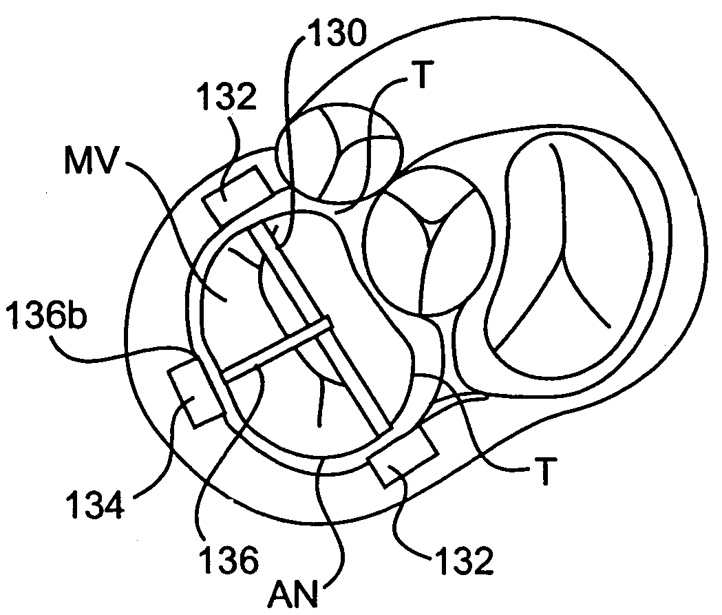
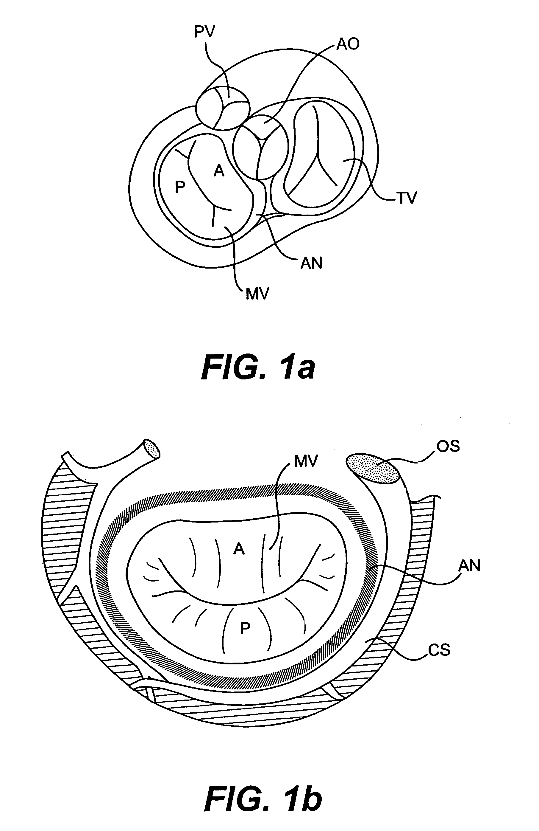
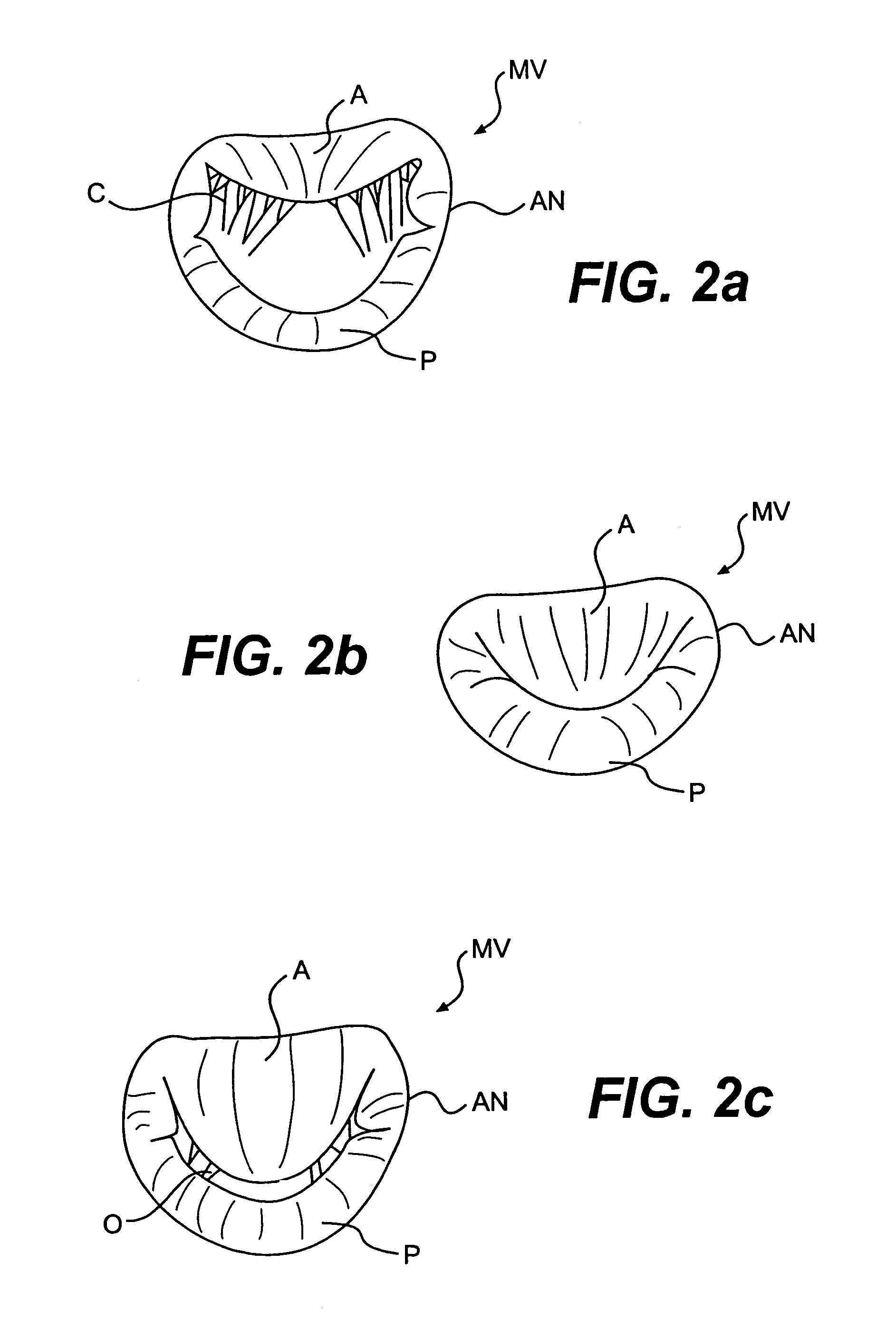
[0106]Certain aspects of the invention that will be discussed herein generally pertain to devices and methods for treating valve insufficiency arising from heart conditions, including, for example, ventricle dilatation, valve incompetencies, congenital defects, and other conditions. The various devices to be described may operate passively in that, once implanted in the heart, they do not require an active stimulus, either mechanical, electrical, or otherwise, to function. Implanting one or more of the devices of the present invention may assist in closing a valve to prevent regurgitation by, for example, assisting in the proper coaptation of the heart valve leaflets, either against one another or independently against another surface. Assisting this coaptation may be accomplished by directly geometrically altering the shape of the dysfunctional mitral valve annulus, by repositioning one or both of the papillary muscles to a more normal state, and / or by otherwise facilitating annula...


PUM


Abstract

Devices and methods for treating heart valves include members that assist the valve in closing during at least a portion of the cardiac cycle. Such devices include members configured to alter the shape of a valve annulus, reposition at least one papillary muscle, and / or plug an orifice of the valve so as to provide a coaptation surface for the valve leaflets.

Description

[0001]This is a continuation of application Ser. No. 10 / 040,784, filed Jan. 9, 2002, now U.S. Pat. No. 6,764,510, which is incorporated herein by reference.BACKGROUND OF THE INVENTION[0002]1. Field of the Invention[0003]The present invention relates to devices and related methods for treating and improving the function of dysfunctional heart valves. More particularly, the invention relates to devices and related methods that passively assist to close a heart valve to improve valve function of poorly functioning valves.[0004]2. Description of the Related Art[0005]Various etiologies may result in heart valve insufficiency depending upon both the particular valve as well as the underlying disease state of the patient. For instance, a congenital defect may be present resulting in poor coaptation of the valve leaflets, such as in the case of a monocusp aortic valve, for example. Valve insufficiency also may result from an infection, such as rheumatic fever, for example, which may cause a...

Claims


Application Information

Patent Timeline
Patent Type & Authority Patents(United States)
IPC IPC(8): A61F2/24
CPCA61B17/00234A61B17/0401A61F2/2487A61F2/2451A61F2/2454A61F2/246A61B17/0487A61B2017/00243A61B2017/0417A61B2017/0464A61B2017/06052A61F2/2445A61F2/2481A61B2017/0496
Inventor VIDLUND, ROBERT M.KALGREEN, JASON E.MORTIER, TODD J.SCHWEICH, JR., CYRIL J.SCHROEDER, RICHARDKUSZ, DAVID
Owner EDWARDS LIFESCIENCES LLC
Why Eureka
Try Eureka