Bronchoscopic lung volume reduction method

a lung volume and bronchial tube technology, applied in the field of lung disease treatment, can solve the problems of reducing lung function and efficiency, reducing the supportive structure of the airway, and losing the ability to remain open during exhalation, so as to reduce the risk of reinflation and achieve effective lung volume reduction

Inactive Publication Date: 2006-09-05
GYRUS ACMI INC (D B A OLYMPUS SURGICAL TECH AMERICA)
View PDF230 Cites 137 Cited by
  • Summary
  • Abstract
  • Description
  • Claims
  • Application Information

AI Technical Summary

Benefits of technology

[0011]In one application of the present method, the distal end of a bronchoscope is used to guide the catheter to a diseased lung section, where an inflammation-causing substance is injected into the diseased lung section. In one variation, the present method comprises using the distal end of a bronchoscope to guide a multilumen catheter to a position adjacent the target region, inserting a puncturing tip through a section of diseased lung tissue and into the target region, and equilibrating the target region with atmospheric air. The target region air is equilibrated through a lumen extending from a distal opening of the lumen to the atmosphere. With the target region at atmospheric pressure, one or more occlusion devices may then be deployed in airways leading into the target region, thereby isolating the target region from inspiratory air flow. In one application, the obstruction is a one-way valve, such as those described in the '951 and '100 Alferness patents, or those described in pending U.S. patent application Ser. No. 09 / 951,105 to Alferness et al. filed on Sep. 11, 2001, Ser. No. 10 / 104,487 to Alferness et al. filed on Mar. 30, 2002, and Ser. No. 10 / 124,790 to DeVore filed on Apr. 16, 2002. The entire disclosures of the above patents and applications are incorporated herein by reference and made part of the present disclosure. The advantage of an obstruction device is reduced risk of reinflation during the adhering process, resulting in more effective lung volume reduction. Another advantage, among others, is precluding migration of the injected substance to non-targeted regions of the lung.

Problems solved by technology

Patients with chronic obstructive pulmonary disease or emphysema have reduced lung function and efficiency, typically due to the breakdown of lung tissue.
Lung tissue and alveoli are destroyed, reducing the supportive structure of the airways.
This reduction in strength of the airway walls allows the walls to become “floppy,” thereby losing their ability to remain open during exhalation.
In this diseased state, the patient suffers from the inability to get the air out of their lungs due to the loss of elasticity of the lungs as well as the collapse of the airways during exhalation.
Within the confines of the chest cavity, this hyper-inflation restricts the in-flow of fresh air and the proper function of healthier tissue, resulting in significant breathlessness.
With, severe emphysema, when patients take in as much air as their chest cavity can accommodate, they still have insufficient gas exchange because their chest is full of non-functional air filling large cavities in the lungs.
A wide variety of drugs are available for treating the symptoms of chronic obstructive pulmonary disease, but none are curative.
A large number of patients are not responsive to these medications.
This surgery has substantial risks of serious post-operative complications, such as pneumonia, and requires an extended convalescence.

Method used


Image

  • Bronchoscopic lung volume reduction method
  • Bronchoscopic lung volume reduction method
  • Bronchoscopic lung volume reduction method

Examples


Embodiment Construction

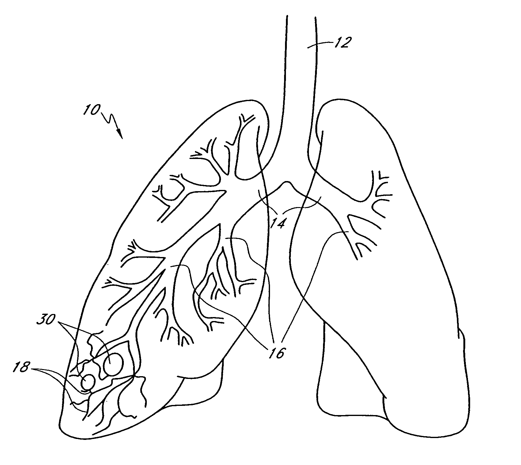
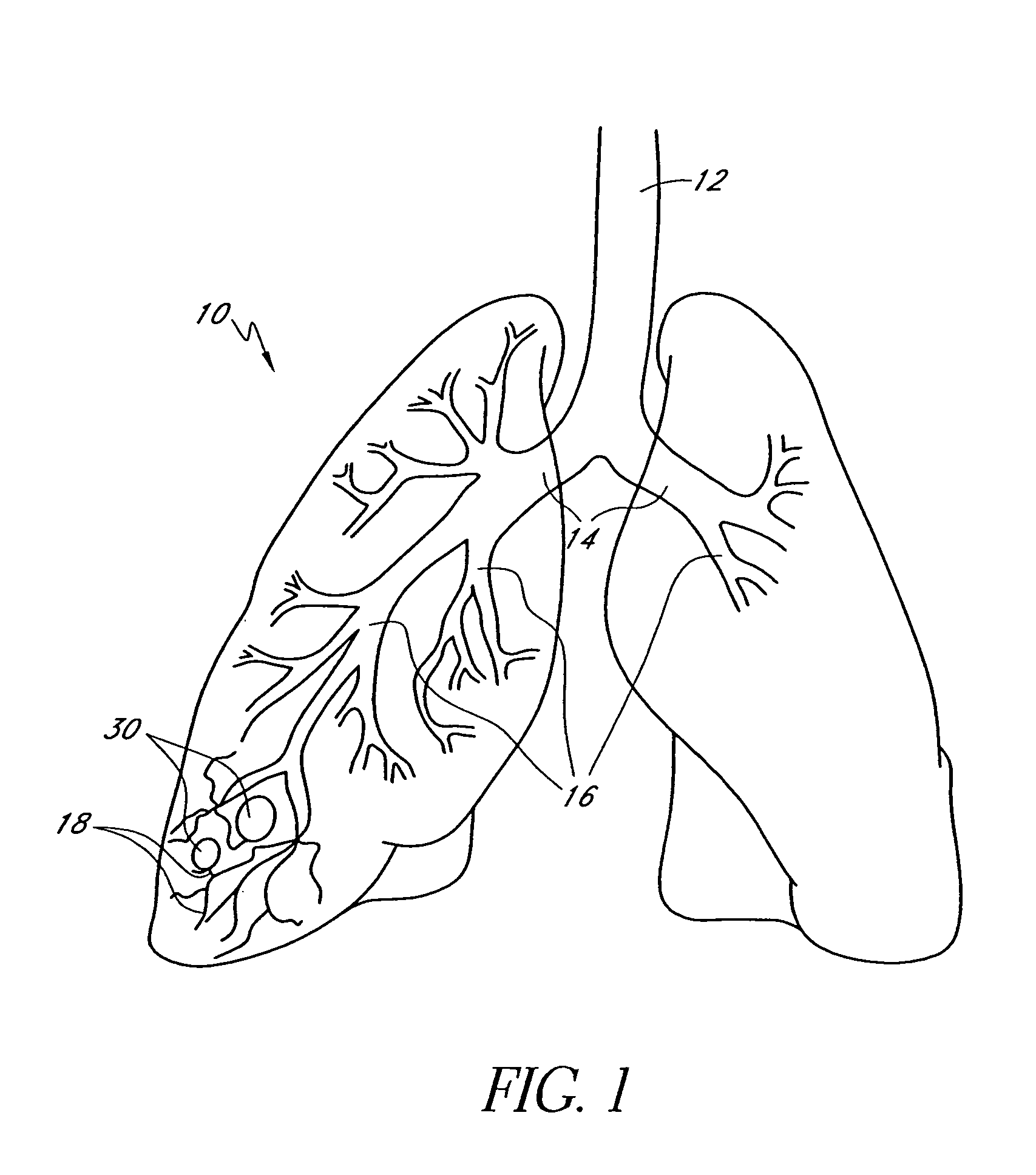
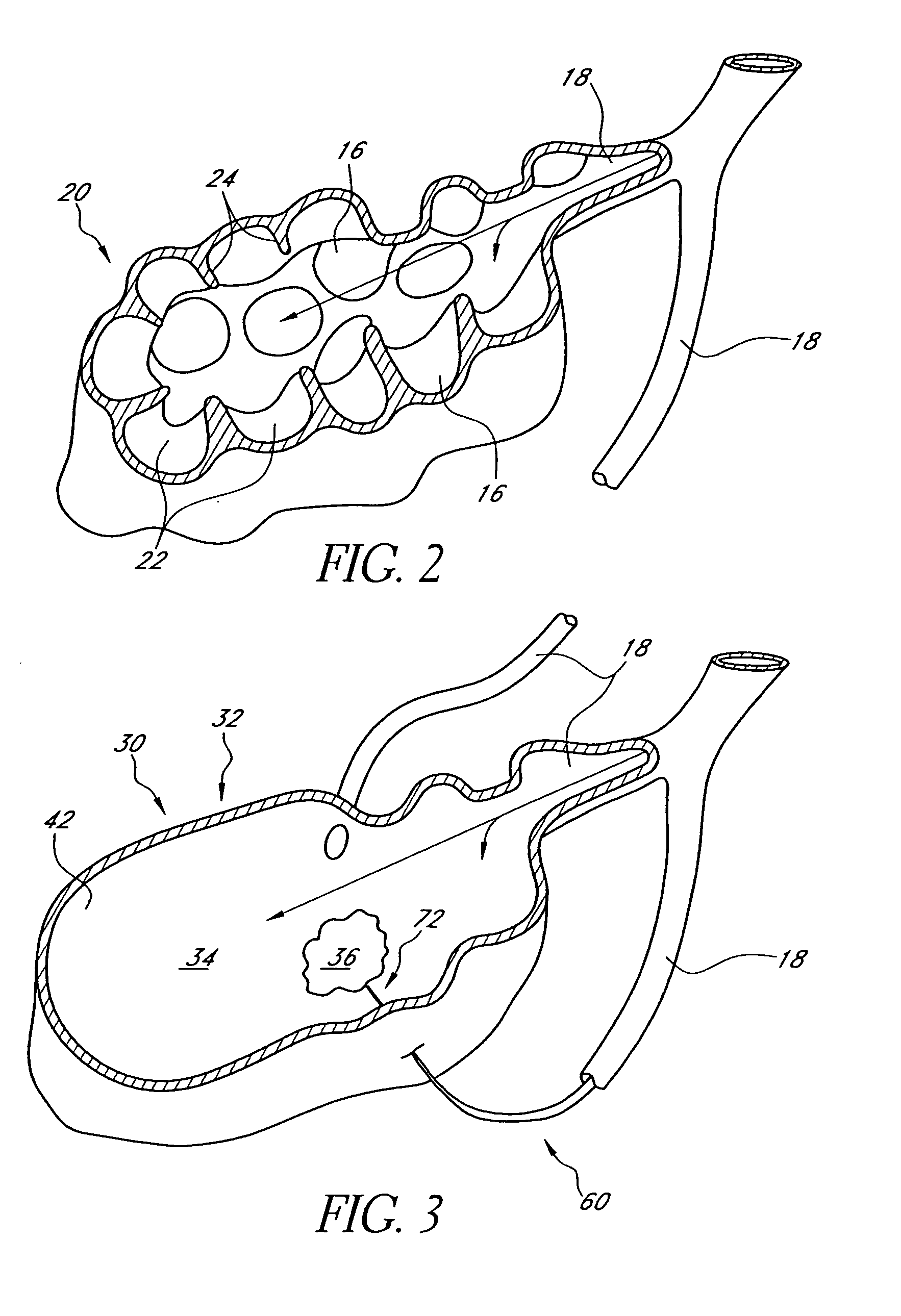
[0020]The methods described herein involve minimally invasive procedures for reducing the volume of a diseased target region of a patient's lung. Referring to FIGS. 1 and 2, airflow in a lung 10 to be treated generally follows a path from the trachea 12, through the main branch bronchial tubes 14, then through the sub-bronchial tubes 16 to the numerous tiny bronchioles 18. The bronchioles 18 lead to alveolar sacs 20 which include multiple alveoli 22 separated by alveolar walls 24 for the exchange of oxygen and carbon dioxide. In lungs suffering from emphysema, the alveolar walls 24 are deteriorated, thus leaving a number of large open spaces or cavities 30. Often, the largest of such cavities 30 will be chosen as a target region 32 for volume reduction first, although the specific regions to be reduced will be chosen by a clinician based on a number of factors. A target region 32 of diseased lung to be reduced in volume will generally include at least one enlarged target cavity 34.

[...


PUM


Abstract

A method of minimally invasively reducing a volume of a hyper-inflated target section of diseased lung comprising the steps of introducing a bronchoscope into a patient's airway to a position adjacent the target section and equilibrating air within the target section with atmospheric air to at least partially deflate the target lung section; injecting an inflammation-causing substance into the target section to precipitate adhesion of the walls within the target lung section, preventing substantial re-inflation of the target section by occluding an airway upstream of the target section for a period of time, and removing the airway occlusion after the target section has substantially permanently been reduced in volume. The injected substance can be autologous blood or a constituent thereof.

Description

BACKGROUND[0001]1. Field of the Invention[0002]The invention relates in general to the field of pulmonary disease treatments, and specifically to a bronchoscopic method of performing lung volume reduction surgery using an obstruction or one-way valve.[0003]2. Description of the Related Art[0004]The lungs deliver oxygen to the body by directing air through numerous air passageways that lead from the trachea to respiratory bronchiole to small sacs called alveoli. These alveoli are generally grouped together in a tightly packed configuration called an alveolar sac, and surround both alveolar ducts and respiratory bronchiole throughout the lung. The alveoli are small, polyhedral recesses composed of a fibrillated connective tissue and surrounded by a few involuntary muscular and elastic fibers. These alveoli inflate and deflate with the respiration of air. Oxygen and carbon dioxide are exchanged through the ultra-thin walls between alveoli and capillary sized blood vessels.[0005]During ...

Claims


Application Information

Patent Timeline
Patent Type & Authority Patents(United States)
IPC IPC(8): A61B19/00A61B17/12A61F2/04
CPCA61B17/12022A61B17/12104A61M16/00A61B17/12186A61F2002/043
Inventor SPRINGMEYER, STEVEN C.
Owner GYRUS ACMI INC (D B A OLYMPUS SURGICAL TECH AMERICA)
Why Eureka
Try Eureka