Endometriosis-specific secretory protein
a secretory protein and endometriosis technology, applied in the field of endometriosis diagnosis and diagnosis, can solve the problems of limited information available regarding endometriosis-specific markers, protein synthesis, secretion, regulation and expression in endometriotic tissue, and not useful in the development of endometriosis-specific markers for the diseas
- Summary
- Abstract
- Description
- Claims
- Application Information
AI Technical Summary
Benefits of technology
Problems solved by technology
Method used
Image
Examples
example 1
Isolation and Characterization of Glycoprotein
Materials and Methods
[0069]Endometrial and Endometriotic Tissue: Human tissues were obtained from randomly selected, informed volunteer patients routinely presenting to the physicians in the Department of Obstetrics and Gynecology at the University of Missouri Medical School as approved by the Institutional Review Board. Patients presented for a variety of routine diagnostic and therapeutic examinations including diagnosis of endometrial function, endometriosis, tubal ligation for sterilization, routine gynecological care and gamete intrafallopian transfer.
[0070]Endometrial tissue was obtained using a Pipelle (Unimar, Wilton, Conn.) endometrial suction curette. Endometriotic tissue was obtained at the time of laparoscopic examination. Peritoneal endometriotic implants, including red petechia and reddish-brown lesions, were elevated with biopsy forceps and the area circumscribed by either laser or sharp dissection. Powder-burn implants an...
example 2
The Synthesis and Release of Endometriotic Secretory Proteins Differs from that of the Uterine Endometrium
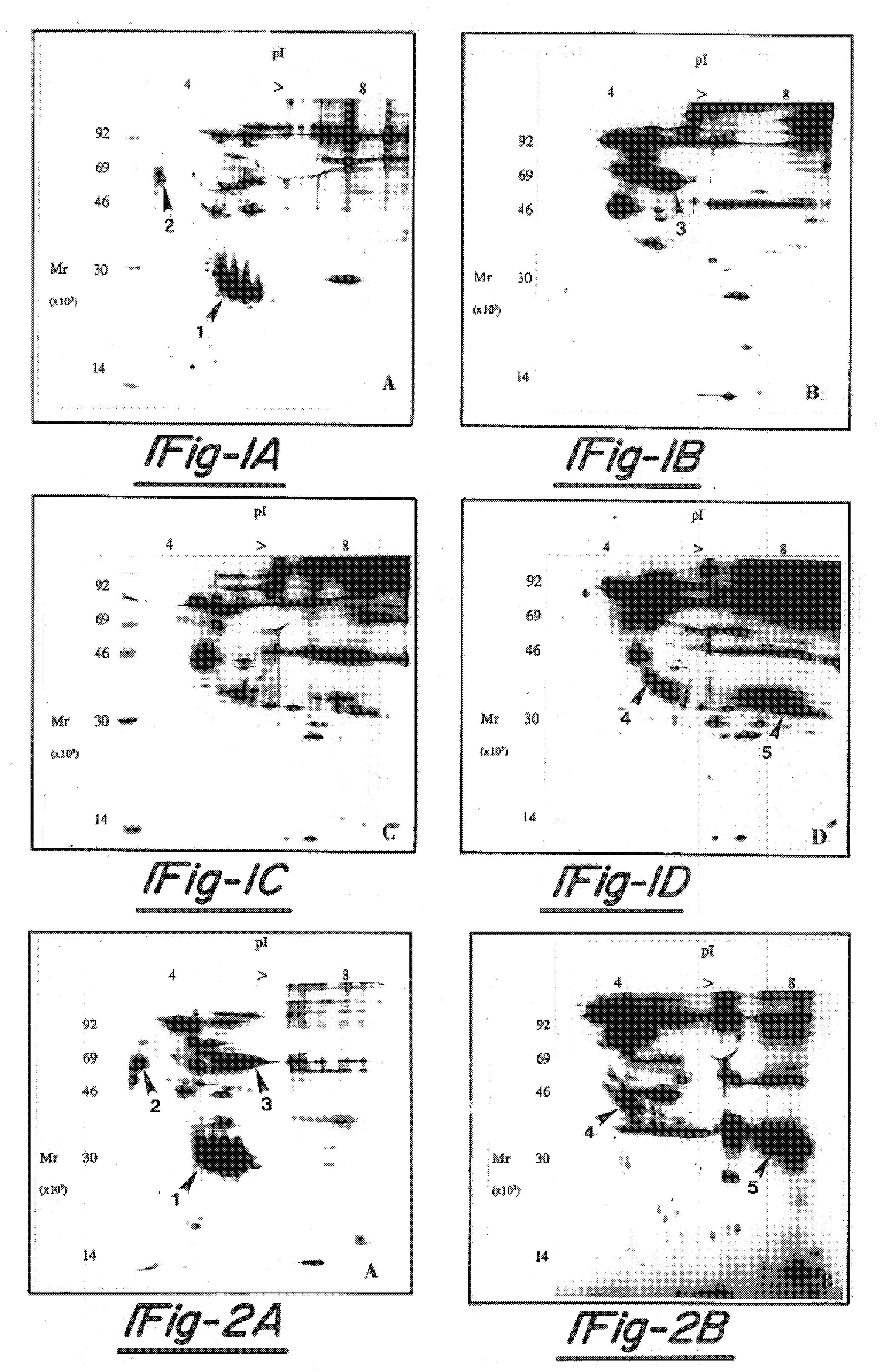
[0097]To assess the ability of the endometriotic lesion to synthesize and secrete endometrial proteins, in vitro protein production by uterine endometrium and endometriotic tissues was examined. Matched biopsy specimens of uterine and endometriotic tissues were collected at the time of laparoscopic diagnosis for endometriosis. Menstrual cycle stage (n=5 follicular {cycle day 4–12} and 7 secretory {cycle day 19–27}) and the presence of endometriosis was documented histologically. Tissue explants plus isolated, purified 90% confluent epithelial and stromal cells were cultured for 24 hours in minimal essential medium containing 36S-methionine (100 mCi / ml). Tissue and cell culture media containing the de novo synthesized proteins was centrifuged, dialyzed and lyophilized and the proteins separated and visualized by two-dimensional gel electrophoresis and fluorography.
[0098]Although ...
example 3
[0101]Applicants determined the partial amino acid sequence for the rat ENDO-I glycoprotein as set forth in SEQ ID No: 3. Specifically, rat ENDO-I was given to the Protein Core Facility at the University of Missouri for amino acid sequencing. Partially purified, wheat germ lectin fractionated ENDO-I protein from stromal cell culture media were separated by 2D-PAGE and electrophoretically transferred to polyvinylidene difluoride (PVDF) membranes. A minimum of 50 pmol quantities of protein for amino acid sequencing “off blot” is required. Transfer to PVDF membranes overnight at 4° C. provided samples free of contaminants such as Tris, glycine, sodium dodecyl sulfate or acrylamide. Transferred proteins were visualized by Commassie blue staining and cut from the membrane for sequencing. NH2-terminal sequence analysis was carried out by automated Edman degradation on an Applied Biosystems 470A gas phase sequencer with a Model 120A on-line phenylthiohydantion analyzer.
[0102]The partial am...
PUM
| Property | Measurement | Unit |
|---|---|---|
| Mass | aaaaa | aaaaa |
| Mass | aaaaa | aaaaa |
Abstract
Description
Claims
Application Information
 - R&D
- Intellectual Property
- Life Sciences
- Materials
- Tech Scout
- Unparalleled Data Quality
- Higher Quality Content
- 60% Fewer Hallucinations
Browse by: Latest US Patents, China's latest patents, Technical Efficacy Thesaurus, Application Domain, Technology Topic, Popular Technical Reports.
© 2025 PatSnap. All rights reserved.Legal|Privacy policy|Modern Slavery Act Transparency Statement|Sitemap|About US| Contact US: help@patsnap.com


