

Antibodies to a fibrinogen binding protein of staphylococcus epidermidis
a technology of fibrinogen binding protein and staphylococcus epidermidis, which is applied in the directions of antibody ingredients, bacterial antigen ingredients, antibody ingredients, etc., can solve the problem of difficult to obtain homogeneous and reproducible products
- Summary
- Abstract
- Description
- Claims
- Application Information
AI Technical Summary
Problems solved by technology
Method used
Image

Examples
example 1
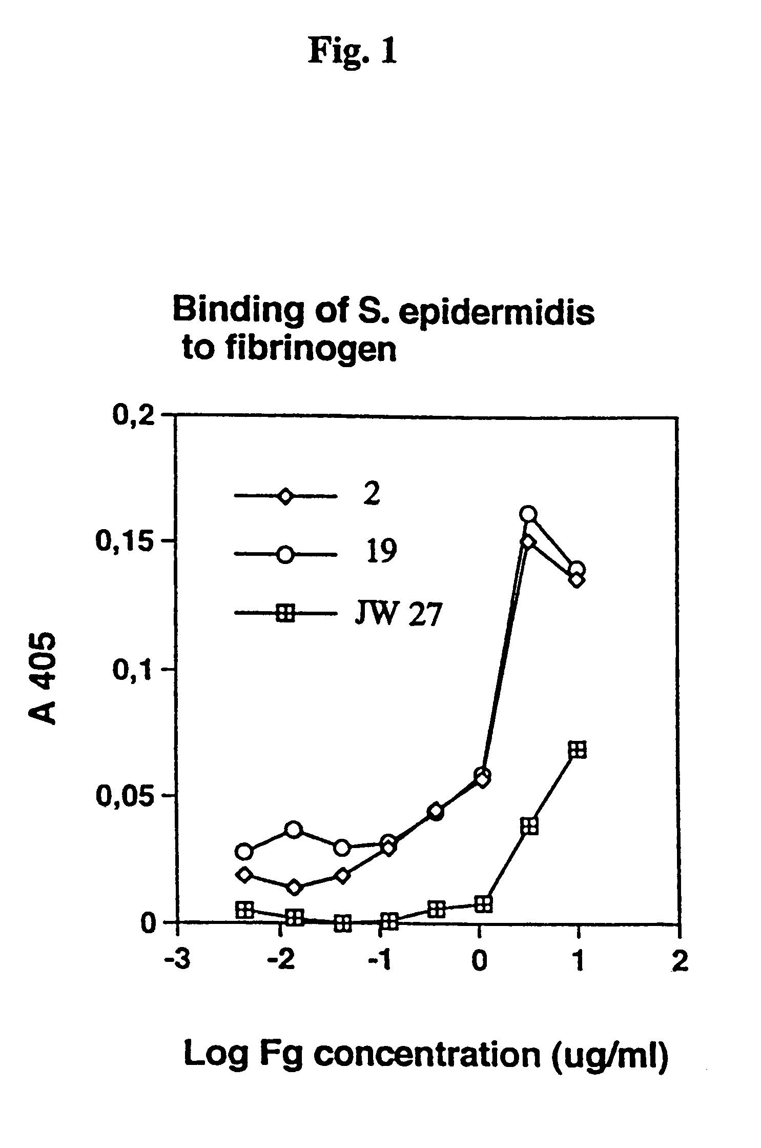
The Adherence of Staphylococcus epidermidis to Immobilised Fibrinogen and Investigation of the Nature of the Binding Mechanism (A–E)
[0039]Strains of Staphylococcus epidermidis isolated from cases of peritonitis were grown on Blood agar plates at 37° C. overnight. The bacteria from one plate was harvested with 5 ml phosphate buffered saline (PBS), washed once, and the optical density (OD) was adjusted to 1.0.
(A) Bacterial Adherence
[0040]Fibrinogen was dissolved in PBS at 10 mg / ml and added in serial 3-fold dilution to microtiter wells (Nunc), from top to bottom. THe plates were incubated overnight at room temperature (RT). To cover uncoated plastic sites the plates were coated with 2% bovine serum albumin for 1 hour at 37° C. The plates were washed with PBS with 0.05% TWEEN 20 (PBST). Next, bacteria were added in serial 2-fold dilution in PBST, from left to right, to the fibrinogen coated microtiter plates. Bacterial adherence was allowed for 2 hours at 37° C. or at 4° C. overnight. ...
example 2
Isolation of a Clone Expressing Fibrinogen Binding Activity
[0045]A gene library of S. epidermidis strain HB was produced in a manner as described by Jacobsson and Frykberg (1996). Staphylococcal DNA was randomly fragmented by sonication. The library resulted in 4×10⁷ independent clones, which after amplification had a titer of 2×10¹⁰ cfu / ml. Two hundred microlitres of the library were added to each of three fibrinogen coated wells and incubated for 4 hour at room temperature (RT). The wells were washed extensively with PBS-T and once with 50 mM Na-citrate / 140 mM NaCl, pH 5.4. Finally, the bound phages were eluted stepwise in the same buffer with decreasing pH (3.4 and 1.8). The eluates from the three wells were neutralised with 2 M Tris-HCl, pH 8.6. Aliquots of the eluates were used to infect E. coli TG1 cells, which thereafter were grown overnight on LA plates containing glucose and ampicillin. The colonies (obtained after infection of TG1 cells with the phage and eluted at pH 3.4 ...
example 3
DNA Sequencing and Sequence Analysis
[0047]Eight colonies coming from the second panning (pH 3.4) against fibrinogen described in Example 2 were chosen for further studies. Phagemid DNA from these colonies was prepared and partially sequenced. Seven of the clones seemed to contain the same insert. One of these seven clones called pSE100 was chosen for further studies. Purified phagemid DNA from the clone pSE100 was analysed by restriction mapping which revealed that the phagemid contained an insert of ˜1.8 kilo base pair (kb). The nucleotide (nt) sequences of the complete inserts of pSE100 were determined and the nt and deduced amino acid (aa) sequences were analysed using the PC-gene program. This analysis revealed that the insert of pSE100 contains an open reading frame of 1.745 nt (sequence list). Thus the insert encodes a 582 aa protein, termed protein FIG (and the corresponding gene termed fig), with a calculated molecular mass of ˜65 kDa (sequence list). Furthermore, the sequen...
PUM
 Abstract
Description
Claims
Application Information